3672
Analysis of Generation of Arbitrary Spectral Profiles (GASP) for Water-Fat Separation1Bioengineering, Imperial College London, London, United Kingdom, 2Biomedical Engineering, University of Utah, Salt Lake City, UT, United States, 3Electrical and Computer Engineering, Boise State University, Boise, ID, United States
Synopsis
Keywords: Fat & Fat/Water Separation, Fat
Motivation: This research explores using multiple bSSFP images to generate arbitrary spectral profiles for use in water-fat separation
Goal(s): Our goal is to analyze the generation of synthetic spectral profiles and use this technique for water-fat separation.
Approach: We use computational simulations to analyze the effectiveness of generating arbitrary spectral profiles for filtering different tissues and validate our technique with an in-vivo knee experiment.
Results: Our experiments demonstrate that the banding artifacts from multiple bSSFP acquisitions with varying TRs and phase cycles can be used to spectrally isolate water from fat signals.
Impact: Multiple bSSFP images can be linearly combined to generate arbitrary spectral profiles and isolate tissues for water-fat separation
Introduction
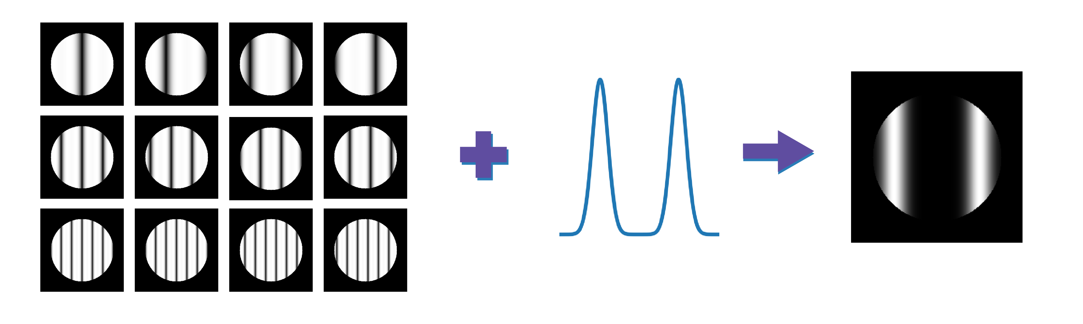
Balanced Steady State Free Precession (bSSFP) MRI is often used for its high signal-to-noise (SNR), however is limited by banding artifacts caused from off-resonance dependence1. Many techniques have been developed to mitigate or exploit these banding artifacts, often visualized as spectral profile responses2-6. For large flip angles, bSSFP images have sinusoidal-like spectral profiles for certain tissues, containing a pass-band region and region of signal null2. As the bSSFP profiles are similar to sines and cosines, we use multiple phase-cycled acquisitions to generate a series of basis functions similar to the Fourier series. These basis functions can be used to generate arbitrary spectral profiles in off-resonance space. Spectral profiles can be used to create spectral filters for suppressing undesired signals. Spectral filters can be created to suppress off-resonance signal peaks corresponding to water and fat signals to generate separate fat and water images, respectively. In this work we propose the use of the GASP7 technique for the application of water-fat separation8.Theory
The technique for generating arbitrary spectral profiles, known as GASP, uses a set of linear RF phase-cycled bSSFP to generate a basis set of functions to arbitrarily generate any set of desired spectral profiles. These spectral profiles, $$$S$$$, are approximated using the following basis expansion $$S = \sum_{n}^N (G_n * M_n)$$ where $$$G_n$$$ denotes a complex weighting (GASP) coefficient and $$$M_n$$$ denotes the magnetization for a set of $$$N$$$ phase-cycles. A linear combination of these basis combinations are used after calculating the corresponding GASP coefficients using linear least squares regression. In previous works3, the GASP coefficients were calculated using an orthonormalization operation to generate an orthonormal basis for function estimation. In this work, we relax this constraint to allow for general application by using a least squares regression to calculate the coefficients. If the desired function is approximately representable in the span of the basis, then voxel-wise spectral profiles can be generated to approximate the desired function.Methods
Data from simulated and physical water phantoms were collected to validate and verify the ability of GASP to generate desired spectral profiles. Real data was collected using a 3T Siemens Verio scanner [Erlangen, Germany] with an 8-channel knee coil. No accelerated techniques were used. For each phantom, 16 evenly spaced phase-cycled images at three TR values were acquired as training datasets to calculate GASP coefficients for a variety of spectral profiles. To spatially represent the bSSFP spectral profile physically, a linear gradient was applied across the x-axis of the phantoms to project a single period of the bSSFP spectrum across the FOV. In-vivo knee images were collected from a healthy volunteer to validate the use of GASP for water-fat separation. Knee images were acquired for a 2D axial slice of the knee at 16 evenly-spaced linear phase-cycles at three TR values (TE = TR / 2 = 3ms, 6ms, 12ms). The spatial resolution was 1.0x1.0x.2.0 mm with a field-of-view of 256 mm and flip angles of 20° and 90°. Separate water-fat images were reconstructed using the GASP technique.Results
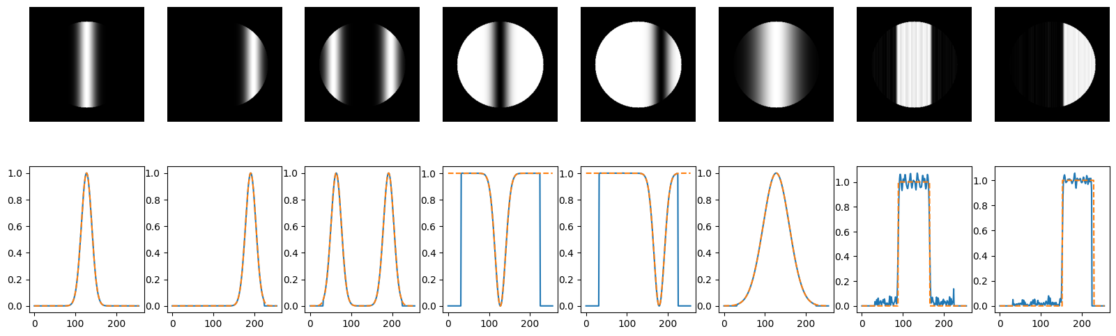
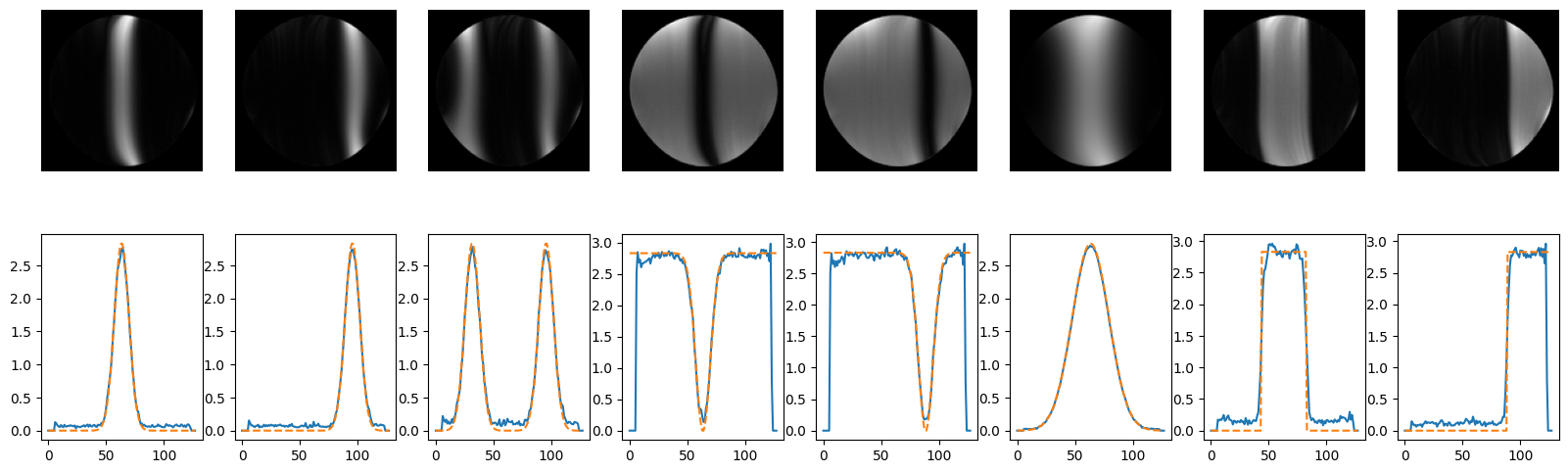
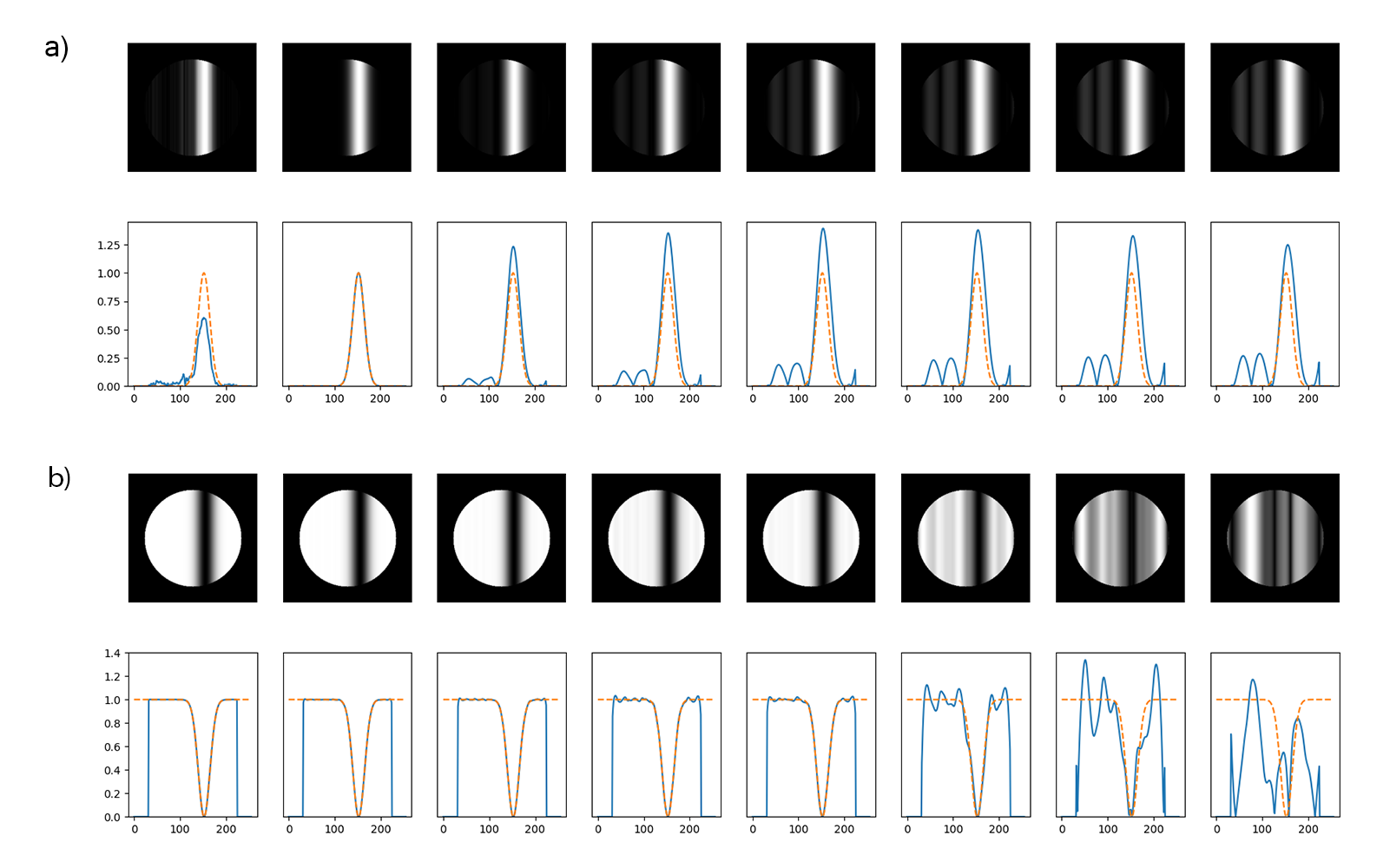
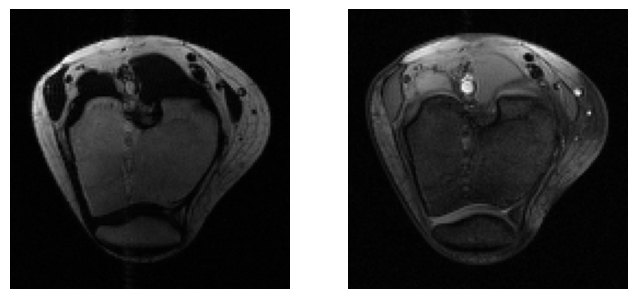
A numerical Bloch simulation was used to compute the GASP coefficients for a variety of desired spectral profiles (Figure 2). Simulations showed that the quality of the images improves with an increased number of GASP coefficients (Figure 3b). Data was collected from a doped-water phantom in a 3T scanner to validate and GASP coefficients were calculated for the same set of spectral profiles using flip angles of 20° and 90° (Figure 4). Water-fat separated images were generated from in-vivo knee acquisitions (Figure 5).Discussion
GASP can be used to generate a variety of desired spectral profiles, however GASP has limitations in generating these profiles. The first limitation is caused by the number of coefficients used in the GASP reconstruction. Using a large number of basis coefficients, GASP can be used to reconstruct any arbitrary spectral profile. As the number of coefficients is reduced the ability to represent a function is reduced (Figure 3a). A possible mitigation is optimization of scanning parameters over known tissues and desired profiles. The second limitation is caused by variation in the shape of bSSFP off-resonance profiles due to changes in tip angle, T2, and T1. As the shape changes from the assumed basis, the effectiveness of the reconstruction is reduced (Figure 3b). This method can be extended to any spectrally-selective experiment [e.g. multi-peak fat isolation, spectroscopy] and spatially-selective experiment [e.g. segmentation, reduced field-of-view].Conclusion
In this work we analyzed the use of GASP to generate desired spectral profiles which were then used for fat suppression, successfully creating water-fat separated images.Acknowledgements
We acknowledge generous support from The Wellcome Trust (220473/Z/20/Z), The Edmond J Safra Foundation, UK Dementia Research Institute, NIHR Imperial Biomedical Research Centre, and National Institutes of Health (R01EB002524).References
1. Carr, H. Y. "Steady-state free precession in nuclear magnetic resonance." Physical Review 112.5 (1958): 1693.
2. Bangerter, et al. “Analysis of Multiple-Acquisition SSFP”, Magnetic Resonance in Medicine, 2004.
3. Vasanawala, et al. “Linear Combination Steady-State Free Precession MRI”, Magnetic Resonance in Medicine, 2000
4. Xiang, Hoff. "Banding Artifact Removal for bSSFP Imaging with an Elliptical Signal Model, Magnetic Resonance in Medicine", 2014
5. Hargreaves, et al. “Fat-suppressed steady-state free precession imaging using phase detection”, Magnetic Resonance in Medicine, 2003
6. Quist, et al. “Simultaneous fat suppression and band reduction with large-angle multiple-acquisition balanced steady-state free precession”, MagneticResonance in Medicine, 2012
7. Mendoza M, McKibben N, Bangerter NK. "Generation of arbitrary spectral profiles using orthonormal basis combinations of bSSFP MRI." Proceedings of the 26th Annual Meeting of the ISMRM.
8. Bley, T. A., Wieben, O., François, C. J., Brittain, J. H., & Reeder, S. B. (2010). Fat and water magnetic resonance imaging. Journal of Magnetic Resonance Imaging, 31(1), 4-18.
Figures