3662
Tensor PCA denoising improves reproducibility of quantitative Multi-Parameter Mapping.1Berlin Center for Advanced Neuroimaging, Charité Medical University, Berlin, Germany
Synopsis
Keywords: Multi-Contrast, Contrast Mechanisms
Motivation: Quantitative biomarkers such as proton density, longitudinal- and transverse relaxation rates, and magnetization transfer saturation measured by multi-parameter mapping (MPM) reflect microstructural tissue characteristics. However, resolution and reproducibility of MPM are constrained by SNR limits of the imaging process resulting in long acquisition times.
Goal(s): We investigate tMPPCA denoising performance for improved SNR and reproducibility of quantitative maps at different voxel volumes for a clinically optimized protocol.
Approach: Denoising of repeated MPM acquisitions at different voxel volumes and its effects on model-based SNR and reproducibility of parameter maps.
Results: Denoising increase SNR of quantitative maps up to sixfold while scan-rescan fluctuations were halved.
Impact: Tensor-based MPPCA denoising of multi-contrast images enhances SNR of quantitative Multi-Parameter Mapping which results in improved reproducibility with greater benefits for high-resolution applications. Consequently, tMPPCA denoising could be considered for future studies and for retrospectively enhancing sensitivity of MPM studies.
Introduction
Quantitative biomarkers such as proton density (PD), longitudinal- (R1) and transverse (R2*) relaxation rates, and semi-quantitative magnetization transfer saturation (MT) measured by multi-parameter mapping (MPM)[1] reflect microstructural tissue characteristics with physiological relevance.[2] However, spatial resolution and reproducibility of MPM are constrained by signal-to-noise ratio (SNR) limits of the imaging process resulting in long acquisition times.Retrospective PCA based denoising promises SNR improvements which allow higher resolutions or shorter scan times without stability loss of quantitative maps. Tensor MPPCA (tMPPCA) denoising[3] leverages data redundancy of multi-contrast acquisitions to optimally separate noise from signal.[4]
On the example of a clinically optimized MPM protocol[5], we investigate tMPPCA denoising performance for improved SNR and reproducibility of quantitative maps at different voxel volumes and doubled acceleration factors.
Methods
3D-FLASH volumes for MPM quantification were acquired twice in one subject (male, 44 years) at different isotropic resolutions at a 3.0-Tesla scanner (Siemens-Prisma, 20-channel head coil) with vendor sequences. Twelve equidistant echo times from 1.60 to 15.9 ms (steps of 1.30 ms, bandwidth: 1400 Hz/pixel) with varying flip angles for PD-weighting (PDw, 4°, TR=18 ms) and T1-weighting (T1w, 25°, TR=18 ms) and additional pre-pulse induced magnetization transfer weighting (MTw, 6°, TR=37 ms) were sampled. Given a field-of-view of 256×224x176 mm3, we achieved different isotropic resolutions of (2.67, 2.00, 1.60, 1.33, 1.00) mm by adapting the matrix size accordingly (96×84×64, 128×112×88, 160×140×112, 192×168×128, 256×224×176). GRAPPA acceleration factor 2 was used in both phase-encoding directions. Total scan time per resolution was (1.6, 2.4, 3.5, 4.6, 7.9) min plus 2 min for transmit field estimation.Magnitude images were denoised with the tMPPCA algorithm using a [3×3×3] window.[3] Denoised and original data were processed using the hMRI toolbox for R2*, R1, PD, and MT calculation[6] including bias correction[7] and model-based SNR (mSNR) estimation[8]. Quantitative maps were normalized to MNI space to calculate voxel-wise differences between repeated measurements (scan-rescan fluctuation in %). White matter (WM) averages for MPM values, mSNR and voxel-wise differences using original and denoised data were determined.
Results
Figure 1 presents maps for R2*, MT, R1 and PD based on original and denoised data at 1 mm isotropic resolution. The denoising performance can be appreciated in the enlarged sections. A substantial noise reduction was apparent in all quantitative maps, while anatomical details were preserved without noticeable blurring.Figure 2 illustrates maps of proton density and respective scan-rescan fluctuations at different isotropic resolutions based on original and denoised data. Spatial noise and scan-rescan fluctuations increased with resolution, also benefits from denoising increased with resolution. Mean values were affected minorly (<1%) whereas standard deviations across WM were reduced.
Figure 3 shows WM averages of mSNR and scan-rescan fluctuations at different isotropic resolutions using original and denoised data. As seen previously, mSNR increased with resolution, whereas scan-rescan fluctuations reduced with resolution. Denoising markedly improved mSNR, which in turn reduced scan-rescan fluctuations by 50%.
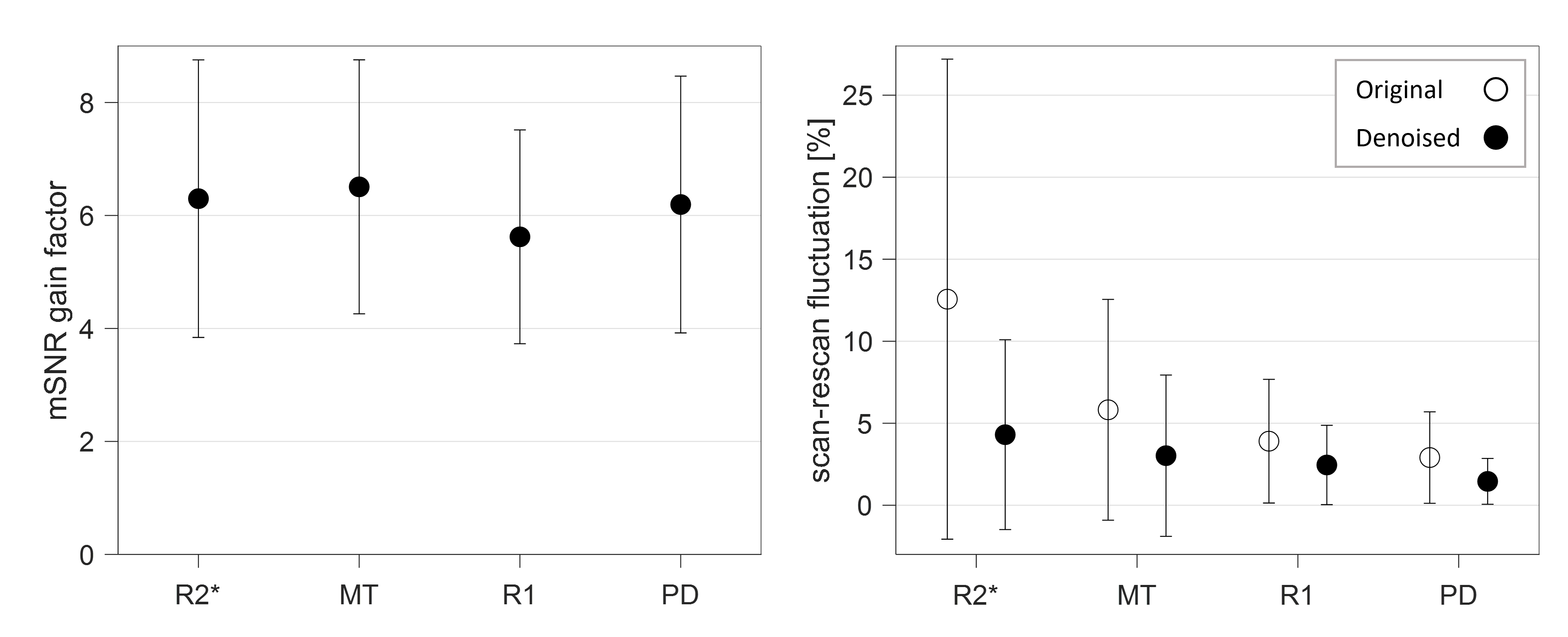
mSNR gain and scan-rescan fluctuations in WM for all parameters at 1 mm resolution are given in Figure 4. Denoising gains were similar for all quantitative maps (R2*: 6.3±2.5, MT: 6.5±2.2, R1: 5.6±1.9, PD: 6.2±2.3), whereas reproducibility of R2* improved almost threefold using denoising. (R2*: 12.6% to 4.3%, MT: 5.8% to 3.0%, R1: 3.9% to 2.5%, PD: 2.9% to 1.5%).
Discussion
tMPPCA denoising was applied to repeated MPM acquisitions for reducing image noise as well as increasing model-based SNR and ultimately enhance reproducibility of quantitative maps at different resolutions.mSNR of MPM quantification was increased up to sixfold without introducing bias in mean values. This resulted in considerably improved reproducibility for all quantitative maps. After denoising our results at 1 mm isotropic resolution exhibited a similar reproducibility as previously at 1.6 mm which was already close to the physiological noise limit.
PCA denoising was previously applied to diffusion and relaxometry for spinal cord[9] and mouse brain[10], but never in the context of MPM quantification for high-resolution human brain imaging or regarding reproducibility of clinically optimized MPM protocols.
tMPPCA was derived from Veraart et al.[11] and shares common limitations such as unwanted image blurring when mathematical assumptions are violated.[3, 4] Although practical applications are likely at risk, we believe that the presented benefits prevail.
Conclusion
In conclusion, we applied tMPPCA denoising to MPM acquisitions and demonstrated substantial noise reduction which resulted in improved MPM model fitting and high-fidelity quantitative maps with up to sixfold SNR and halved scan-rescan fluctuations. This study verified the potential of tMPPCA denoising to compensate SNR and stability loss from high-resolution imaging. Denoising can not only be considered for future study designs but also retrospectively increase MPM stability.Acknowledgements
The authors would like to thank Olesen et al.[3], Mohammadi et al.[8] and Tabelow et al.[12] for code sharing.
References
[1] N. Weiskopf, J. Suckling, G. Williams, M.M. Correia, B. Inkster, R. Tait, C. Ooi, E.T. Bullmore, A. Lutti, Quantitative multi-parameter mapping of R1, PD*, MT, and R2* at 3T: a multi-center validation, Frontiers in Neuroscience 7 (2013) 95.
[2] P. Tofts, Quantitative MRI of the Brain : Measuring Changes Caused by Disease, Wiley, Chichester, 2010.
[3] J.L. Olesen, A. Ianus, L. Østergaard, N. Shemesh, S.N. Jespersen, Tensor denoising of multidimensional MRI data, Magn Reson Med 89(3) (2023) 1160-1172.
[4] J.P. Manzano-Patron, S. Moeller, J.L.R. Andersson, E. Yacoub, S.N. Sotiropoulos, Denoising Diffusion MRI: Considerations and implications for analysis, bioRxiv (2023).
[5] G. Cooper, S. Hirsch, M. Scheel, A.U. Brandt, F. Paul, C. Finke, P. Boehm-Sturm, S. Hetzer, Quantitative Multi-Parameter Mapping Optimized for the Clinical Routine, Front Neurosci 14 (2020) 611194.
[6] N. Weiskopf, M.F. Callaghan, O. Josephs, A. Lutti, S. Mohammadi, Estimating the apparent transverse relaxation time (R2*) from images with different contrasts (ESTATICS) reduces motion artifacts, Frontiers in Neuroscience 8 (2014).
[7] A. Lutti, J. Stadler, O. Josephs, C. Windischberger, O. Speck, J. Bernarding, C. Hutton, N. Weiskopf, Robust and fast whole brain mapping of the RF transmit field B1 at 7T, PloS one 7(3) (2012) e32379.
[8] S. Mohammadi, T. Streubel, L. Klock, L.J. Edwards, A. Lutti, K.J. Pine, S. Weber, P. Scheibe, G. Ziegler, J. Gallinat, S. Kühn, M.F. Callaghan, N. Weiskopf, K. Tabelow, Error quantification in multi-parameter mapping facilitates robust estimation and enhanced group level sensitivity, Neuroimage 262 (2022) 119529.
[9] F. Grussu, M. Battiston, J. Veraart, T. Schneider, J. Cohen-Adad, T.M. Shepherd, D.C. Alexander, E. Fieremans, D.S. Novikov, C.A.M. Gandini Wheeler-Kingshott, Multi-parametric quantitative in vivo spinal cord MRI with unified signal readout and image denoising, Neuroimage 217 (2020) 116884.
[10] M.D. Does, J.L. Olesen, K.D. Harkins, T. Serradas-Duarte, D.F. Gochberg, S.N. Jespersen, N. Shemesh, Evaluation of principal component analysis image denoising on multi-exponential MRI relaxometry, Magn Reson Med 81(6) (2019) 3503-3514.
[11] J. Veraart, D.S. Novikov, D. Christiaens, B. Ades-Aron, J. Sijbers, E. Fieremans, Denoising of diffusion MRI using random matrix theory, Neuroimage 142 (2016) 394-406.
[12] K. Tabelow, E. Balteau, J. Ashburner, M.F. Callaghan, B. Draganski, G. Helms, F. Kherif, T. Leutritz, A. Lutti, C. Phillips, E. Reimer, L. Ruthotto, M. Seif, N. Weiskopf, G. Ziegler, S. Mohammadi, hMRI - A toolbox for quantitative MRI in neuroscience and clinical research, Neuroimage 194 (2019) 191-210.
Figures
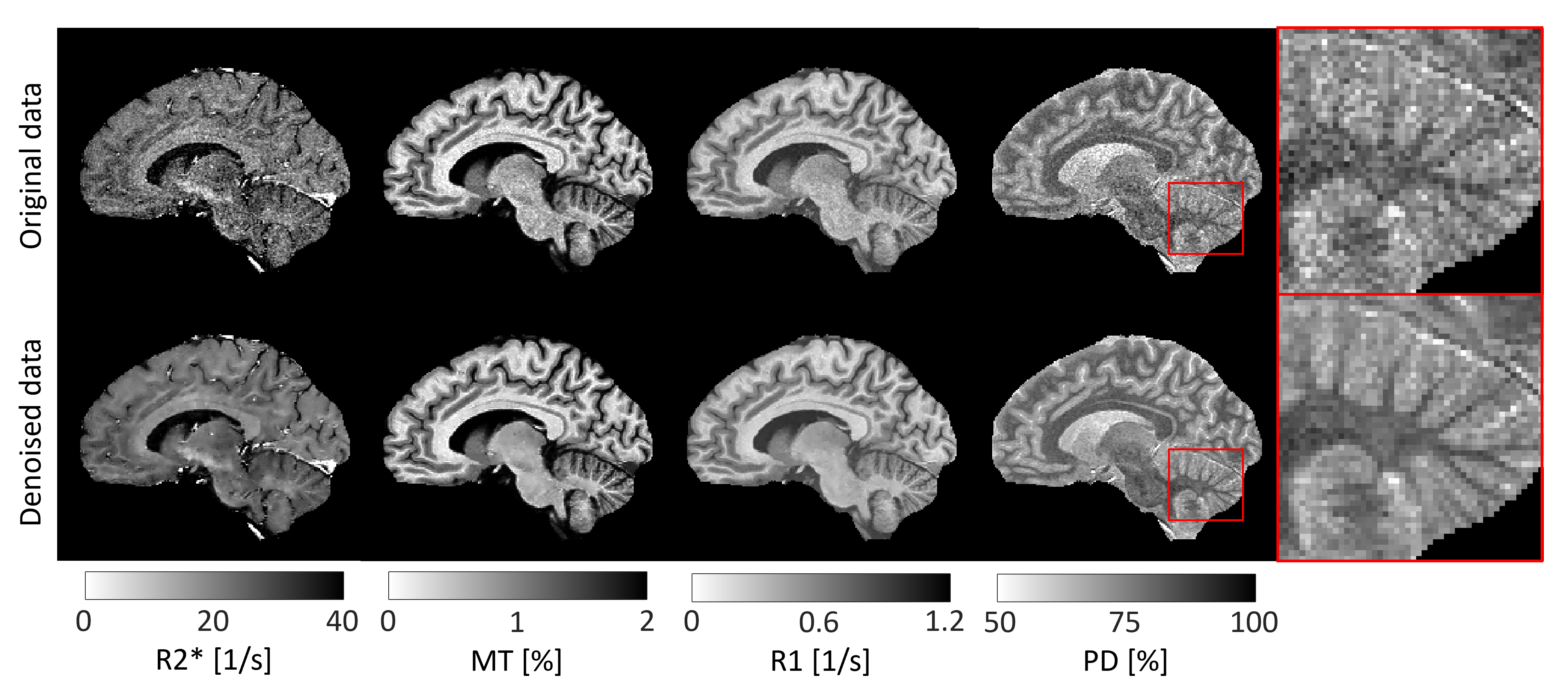
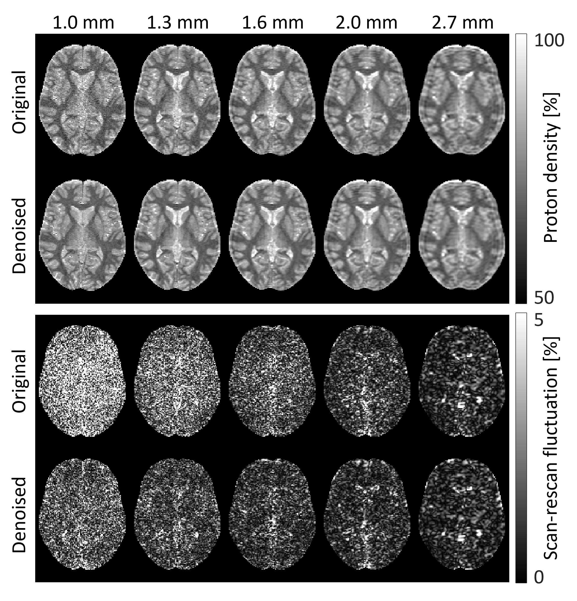
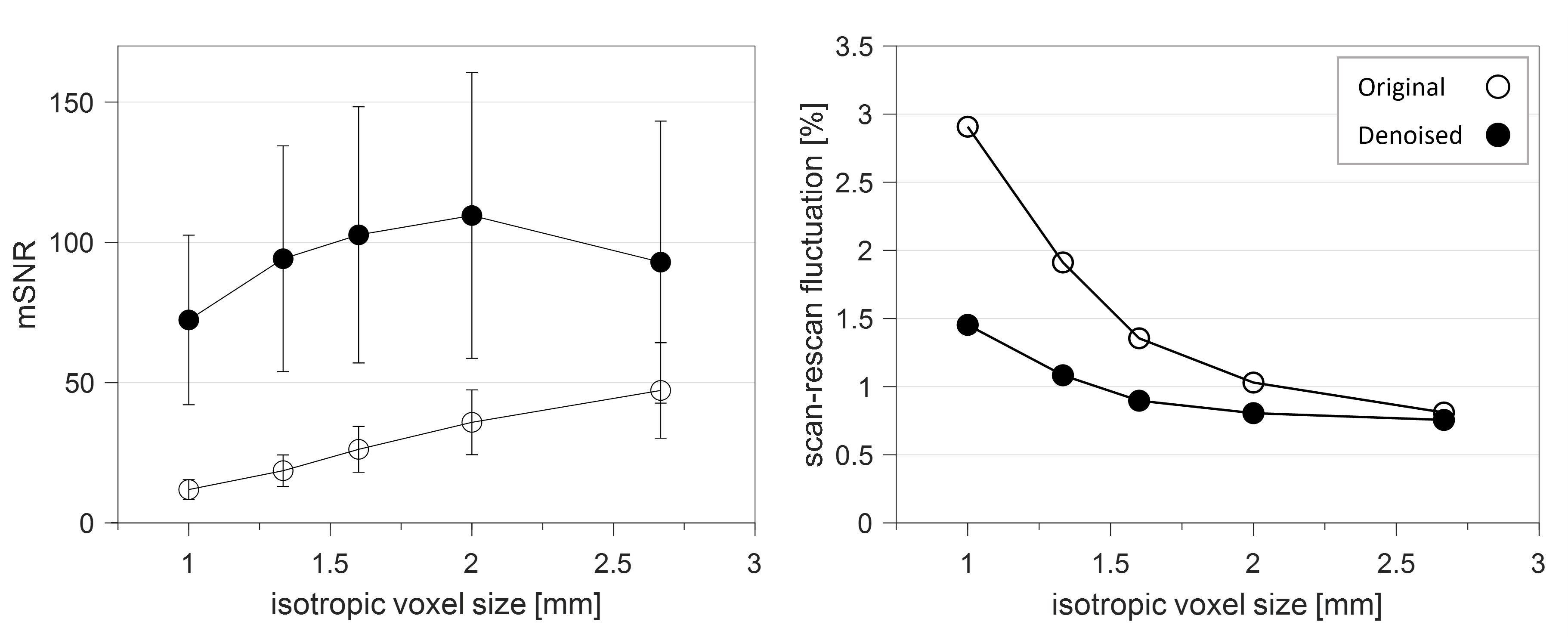
Figure 3: Model-based SNR (mSNR) (left) and scan-rescan fluctuations (right) at different isotropic resolutions based on original and denoised raw data. Results are based on proton density maps in MNI space averaged across WM. Error bars for scan-rescan fluctuations were omitted for clarity.