3652
Evaluation of deep learning HASTE sequence for liver MRI at 3.0 Tesla: a qualitative and quantitative prospective study1Zhongshan hospital, Fudan university, Shanghai, China, 2Shanghai Institute of Medical Imaging, Shanghai, China, 3Zhongshan Hospital, Fudan University, Shanghai, China, 4MR Collaboration, Siemens (Shenzhen) Magnetic Resonance Ltd., Shenzhen, China, 5MR Application Predevelopment, Siemens Healthineers AG, Erlangen, Germany
Synopsis
Keywords: Liver, Liver
Motivation: Liver T2-weighted imaging usually requires a long scan time. A faster sequence with adequate image qualities is essential in clinical practice.
Goal(s): To evaluate deep-learning reconstruction accelerated T2-weighted HASTE sequence in liver application on the image quality and diagnostic confidence.
Approach: One hundred and five patients were imaged using both sequences. Images were reviewed independently by two blinded observers.
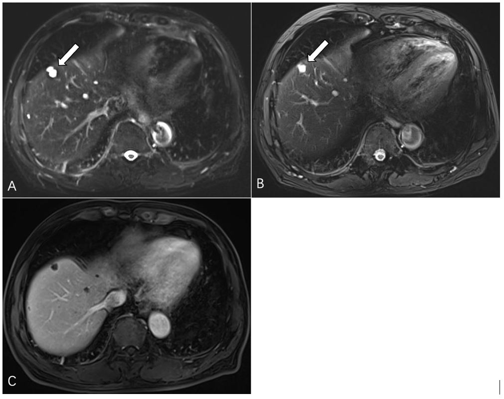
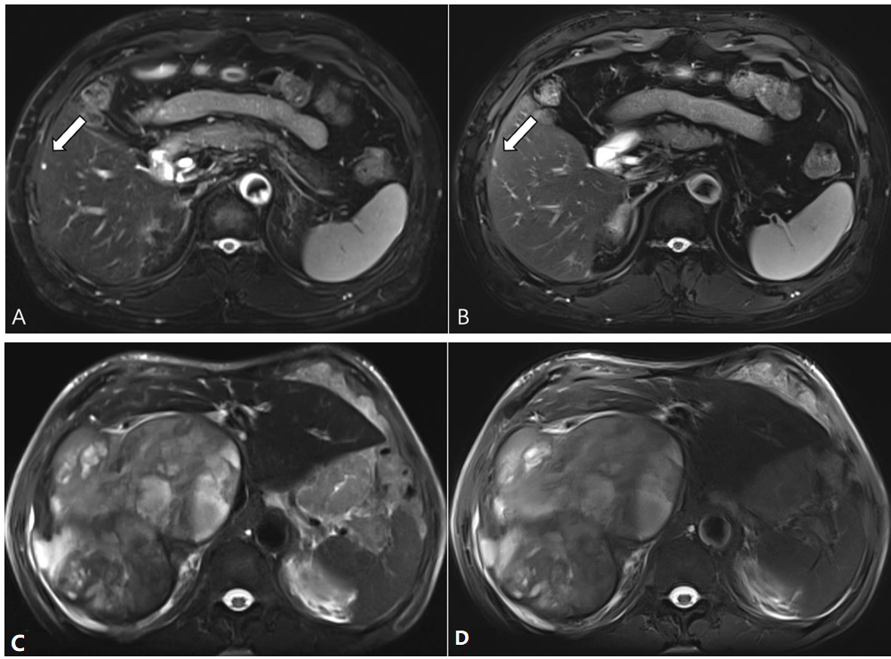
Results: The DL HASTE sequence can detect more liver lesions and improve the CNR of the lesion compared to the conventional T2-weighted BLADE sequence, with a 2.5-fold reduction in acquisition time.
Impact: DL HASTE sequence has the potential to replace the conventional BLADE sequence in routine clinical liver MRI, reducing the scan time and detecting more liver lesions.
Introduction
In clinical liver MRI, T2-weighted imaging (T2WI) is critical important for the detection and characterization of lesions1-2. But it has been plagued by long scanning times and motion artifacts for a long time. The fast half-Fourier single-shot turbo spin echo sequence with deep learning reconstruction (DL HASTE), as an emerging T2WI sequence, has significantly shorter acquisition time than that of T2-weighted BLADE sequence. The purpose of the study was to compare the image quality and diagnostic confidence of DL HASTE with BLADE sequence for liver T2WI at 3T MRI.Methods
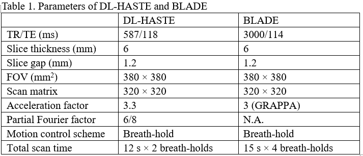
From July 2023 to October 2023, one hundred and five consecutive patients with suspected liver lesion were prospectively included. Liver MRI was performed on a single clinical 3.T MR imaging system (MAGNETOM Prisma; Siemens Healthineers, Erlangen, Germany) for all patients. The scan protocols included a research application T2- weighted DL HASTE sequence with 2 breath-holds and a conventional T2-weighted BLADE sequence with 4 breath-holds. Other parameters were shown in table 1. Images were reviewed independently by two blinded observers. The chi-squared and McNemar tests were used to assess qualitative imaging features, such as overall image quality, artifacts, sharpness of liver margin, hepatic vessel margin, pancreatic duct margin, homogeneity of fat suppression, strength of fat suppression, conspicuity of the smallest lesion and imaging interlayer. The paired Wilcoxon signed-rank test was used to assess quantitative parameters, such as acquisition time, number of liver lesions, size of the smallest lesion, and the contrast-to-noise ratio (CNR) of the lesion; Intra-class correlation coefficient (ICC) and kappa coefficients were used to assess agreement between the two readers.Results
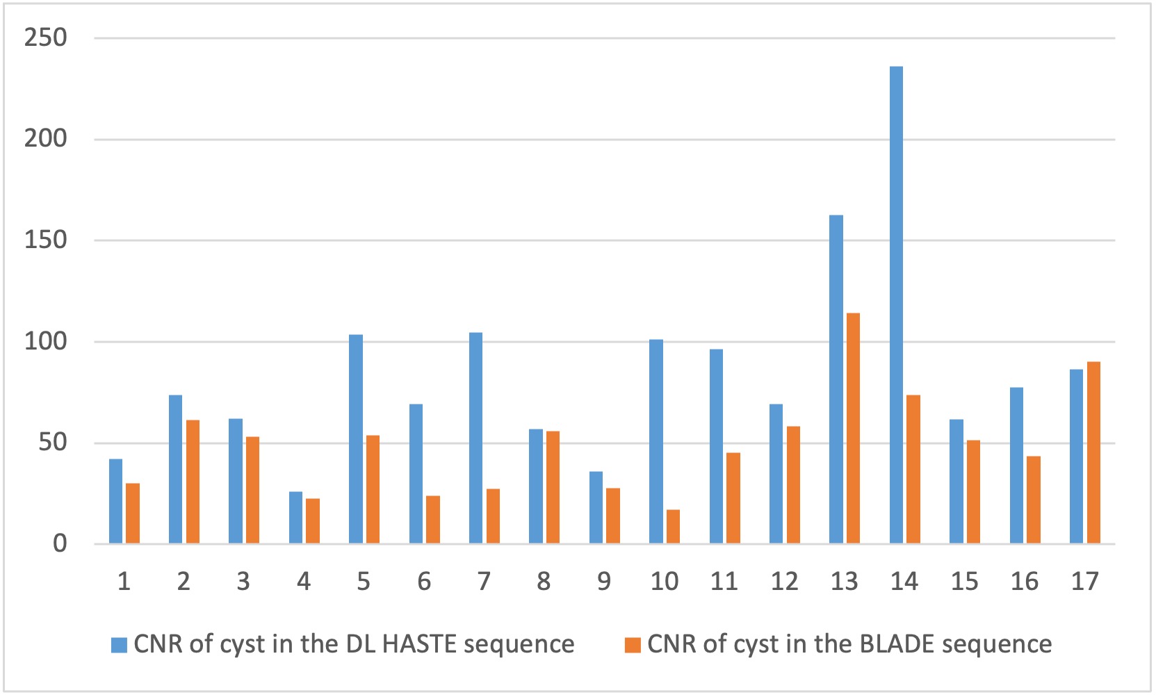
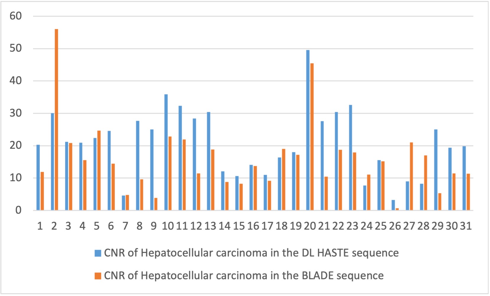
Acquisition time for DL HASTE sequence was 2 x12 s, significantly (p <0.001) shorter than BLADE sequence (4 x15 s). For qualitative imaging feature, there was no statistical difference (p >0.05) between the two sequences. For quantitative imaging feature, significantly more liver lesions were detected with the DL HASTE sequence (243 lesions) than with BLADE sequence (219 lesions; p <0.001), especially in the liver lesions ≤10mm (159 lesions VS. 138 lesions; p <0.001). In the liver lesions >10mm, CNR of cyst and hepatocellular carcinoma (HCC) were significantly higher for the DL HASTE sequence (p =0.001; p =0.004). Inter-reader agreement was moderate to excellent depending on the sequence. Of the 24 supernumerary lesions visible only on the DL HASTE sequence, 23 (95.8%) were true positives.Discussion
In our study, the qualitative and quantitative performances of the DL HASTE sequence and BLADE sequence of liver 3.0T MRI were assessed. The results suggest that the DL HASTE sequence can detect more liver lesions and improved CNR of the lesion compared with the conventional BLADE sequence. One possible explanation for these results is that, firstly, DL HASTE sequence was performed within only 2 short breath-holds, which will help reduce motion artifacts and slice discontinuity and thus improve the lesion detection rates. Secondly, the strong T2 weighting of the HASTE sequence linked to the filling of the majority of k-space with long-TE echoes enabled excellent contrast to be obtained, thus resulting in greater conspicuity and detection of small lesions. Thirdly, the application of DL techniques has the potential to improve the image qualities, significantly reduce the total acquisition time and improve the CNR of the lesions.Conclusion
The 2-breath-hold DL HASTE sequence can detect more liver lesions and improve the CNR of the lesions compared to the conventional 4 breath-hold BLADE sequence, with a 2.5-fold reduction in acquisition time.Acknowledgements
Thank you to those who helped with this study.References
1. Marrero, J.A., et al., Diagnosis, Staging, and Management of Hepatocellular Carcinoma: 2018 Practice Guidance by the American Association for the Study of Liver Diseases. Hepatology, 2018. 68(2): p. 723-750.
2. Girardet, R., et al., The combination of non-contrast abbreviated MRI and alpha foetoprotein has high performance for hepatocellular carcinoma screening. Eur Radiol, 2023. 33(10): p. 6929-6938.
Figures