3648
Automated Respiratory Pattern Analysis for Dynamic MRI of the Lung in Post COVID-19 patients at 0.55 T1Radiology, Bernard and Irene Schwartz Center for Biomedical Imaging, Department of Radiology, New York University Grossman School of Medicine, New York, NY, United States, 2Department of Electrical and Computer Engineering, NYU Tandon School of Engineering, New York, NY, United States, 3Center for Advanced Imaging Innovation and Research (CAI2R), Department of Radiology, New York University Grossman School of Medicine, New York, NY, United States
Synopsis
Keywords: Lung, Lung
Motivation: There is a lack of non-invasive approaches for quantitatively analyzing the patterns of respiration motion in proton MRI in patients with lung diseases such as post-COVID symptoms.
Goal(s): The goal of this work is to determine whether post-COVID-19 patients can be classified as having either long COVID or no symptoms by analyzing dynamic MRI motion fields within various regions of lungs.
Approach: A deep learning-assisted framework was developed for automatically analyzing localized respiratory motion in lung MRI.
Results: The framework was able to successfully categorize patients into different categories based on their degrees of no symptoms using the proposed image analysis framework.
Impact: This work develops an automated framework that can aid radiologists in quickly determining not only the presence but also the severity of long COVID. It can also be extended for applications in other lung diseases.
Introduction
After recovering from COVID, some patients continue to experience lingering symptoms, such as dyspnea, a condition commonly associated with long COVID. Patients with long COVID exhibit different breathing patterns, which can be analyzed to characterize the severity of the long COVID patients.A study previously examined the breathing patterns of long-COVID patients by introducing a new concept of incoherence for quantitatively measuring the periodicity of the area change within the lungs [1]. It has been shown that incoherence can be used to categorize post-COVID patients into different groups reflecting the severity of symptoms. However, the area-based breathing pattern analysis is not sufficient to capture the regional breathing pattern, as direct segmentation of the lungs into different sub-regions is challenging.
In this study, we propose a deep learning-assisted framework for automated analysis of localized respiratory motion in lung MRI to allow for efficient and automated segmentation of the lungs into, calculation of respiratory motion fields, and then estimation of regional incoherence of respiratory motion patterns. This would enable analysis of localized breathing patterns to study disease heterogeneity.
Method
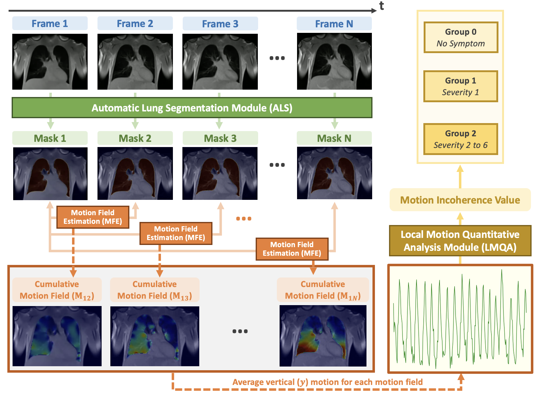
A total of 36 dynamic 2D lung MRI datasets were acquired on a ramped-down 0.55T MRI scanner (Aera, Siemens) using a real-time 2D bSSFP sequence. Each dataset has 250 motion frames with a matrix size of 256x256 or 92x112.The proposed framework consists of the Automatic Lung Segmentation (ALS) module, the Motion Field Estimation (MFE) module, and the Local Motion Quantitative Analysis (LMQA) module, as shown in Figure 1.
The ALS module employs a UniverSeg [2] model, which is a pre-trained universal medical image segmentation model to perform segmentation tasks. It has been adapted for our study by including a small number of lung MRI images and labels for fine-tuning. The ALS module was utilized to generate lung masks for every 2D frame in dynamic MRI.
In the MFE module, Symmetric Diffeomorphism [3] was used to estimate the mapping (e.g. transformation matrix or grid in Figure 3) between the reference image frame (e.g. frame 1) and other image frames. Based on these mappings, motion curves can then be generated for different regions of the lungs.The vertical(Y) motion curves for different segmented regions are the average displacement of all Y coordinate pixels between the reference frame and other frames.
In the LMQA module, the incoherence of a specific curve for a specific region of the lung is computed as follows:
$$Incoherence(t)=\frac{\sum_{i=0}^{Nsetps}|C(i\times step)-C(i\times step+t)|}{Nsteps\times Avg(C)} $$
where C represents the curve. Nsteps represents the total number of frames. 'step' is set to 1. ‘t’ represents the time period at which incoherence is minimal, which was found by an exhaustive search over a range of values.
For comparison, the total lung area-based incoherence was also computed for each patient. All patients were categorized into three groups based on their self-reported severity.
Results and Discussion
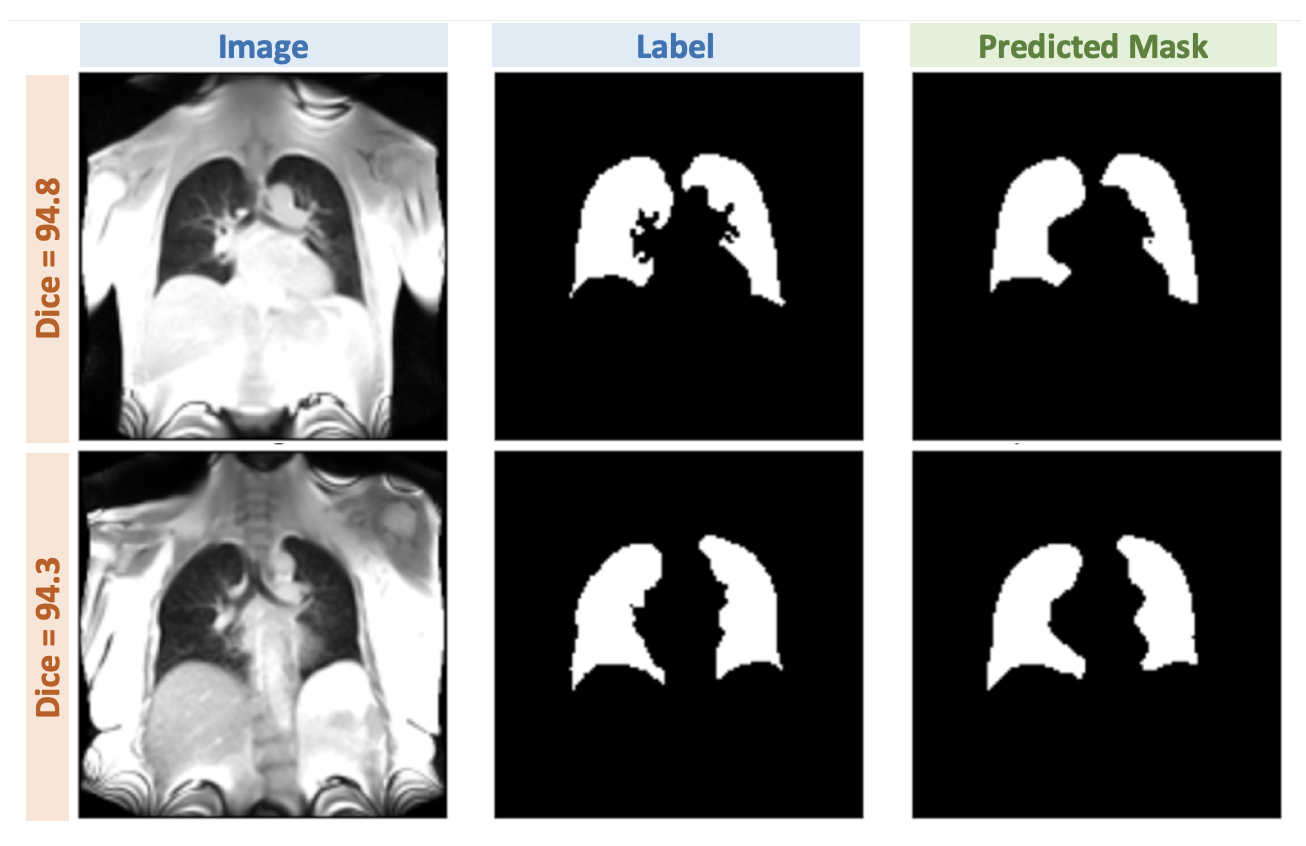
Figure 2 shows the lung segmentation result of two cases. The DICE score was calculated between the predicted mask and the ground truth mask. The results indicate that the ALS module can produce accurate segmentation results based on the DICE score (~0.90 for a total of 60 frames).Figure 3 shows the results for motion field estimation. The assessment of the motion fields was carried out by warping the mask from frame 1 to align with the mask from frame N. The RMSE/DICE between warped masks and reference masks was calculated across all patients and was found to be 0.03206/0.979.
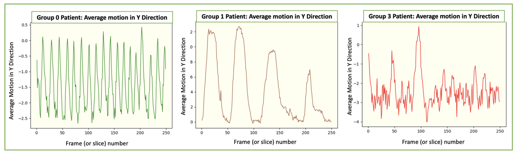
As shown in Figure 4, the irregularity of the Y motion curves increases with severity. This demonstrates that the Y motion allows for the identification of the irregularity of the motion pattern for patients with different severity.
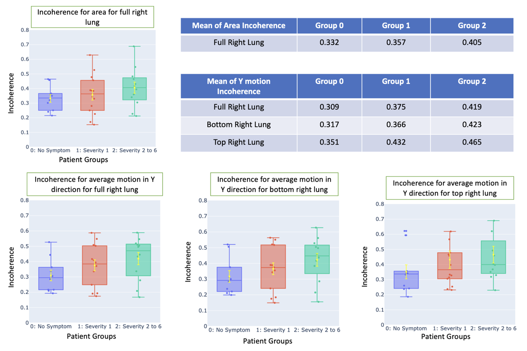
Figure 5 compares the mean incoherence calculated based on the area (total right lung only) and Y motion (both total right and regional right lung). The standard error bars for groups 0 and 1 do not overlap for Y motion incoherence but they overlap significantly for the area-based incoherence, indicating Y motion could be a better marker than area. Incoherence calculated from the bottom lung distinguishes between various severities, while the upper portion suggests that similar measurement.
Conclusion
This study proposed a framework for automated analysis of localized motion in dynamic lung MRI, which enables automated segmentation of the lung to different regions of interest and then the calculation of regional incoherence to analyze motion patterns. Our results have shown that regional motion analysis may provide additional information of value compared to that based on the total lung.Acknowledgements
This work was supported by the NIH (R01EB031083 and and P41EB017183) and was performed under the rubric of the Center for Advanced Imaging Innovation and Research (CAI2R), an NIBIB Biomedical Technology Resource Center.References
1. Lea Azour, et al. " Dysfunctional Respiratory Patterns in Symptomatic Post-Acute Covid-19 Patients on Dynamic High Temporal Resolution Free-Breathing Lung MRI."
2. Butoi*, V. I., Ortiz*, J. J. G., Ma, T., Sabuncu, M. R., Guttag, J., & Dalca, A. V. (2023). UniverSeg: Universal Medical Image Segmentation. International Conference on Computer Vision.
3. Avants, Brian B., et al. "Symmetric diffeomorphic image registration with cross-correlation: evaluating automated labeling of elderly and neurodegenerative brain." Medical image analysis 12.1 (2008): 26-41.
Figures
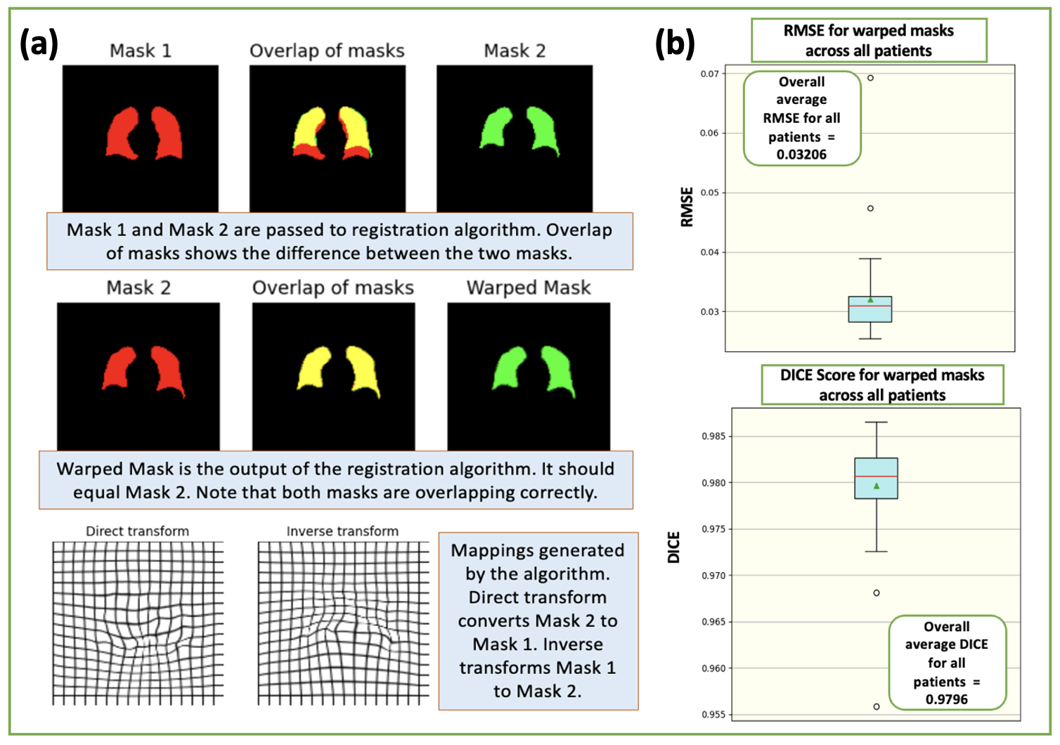
(a) The results for motion field estimation. The result shows that the estimated motion field map enables an accurate warp between mask1 and mask2.
(b) Quantitative analysis of the accuracy of motion field estimation. The assessment of the motion fields was carried out by warping the mask from frame 1 to align with the mask from frame N. The RMSE/DICE between warped masks and reference masks was calculated across all patients and was found to be 0.03206/0.979.