3647
Deep learning reconstructed 3D zero echo time MRI for lung imaging: a preliminary study1Department of Radiology, the Second Xiangya Hospital, Central South University, Changsha 410011, China, chang sha, China, 2GE Healthcare, MR Research China, Beijing, China, Bei jing, China, 3GE Healthcare, MR Research China, Beijing, China, BEI Jing, China, 4Department of Radiology, the Second Xiangya Hospital, Central South University, Changsha 410011, China, Chang sha, China
Synopsis
Keywords: Lung, Lung, Zero echo time, pulmonary ventilation
Motivation: Lung MRI using UTE and ZTE techniques is limited by its SNR and tissue interface blurring. Deep learning based reconstruction (DLR) technique has been used to improve MRI image quality via noise reduction.
Goal(s): To evaluate potentially clinical applications of breath-hold DLR ZTE lung MRI in ventilation function.
Approach: DLR and conventional reconstructed ZTE lung images of thirty patients with pulmonary nodules were compared for image quality and image-based pulmonary ventilation estimation.
Results: Compared to conventional reconstructed results, DLR ZTE images demonstrated improved image quality and better correlation with clinical measurements for ventilation estimation.
Impact: This preliminary study demonstrated the feasibility of DLR ZTE technique in lung MRI. DLR ZTE images showed improved image quality and better correlation with clinical measurements for ventilation estimation.
Introduction
Pulmonary MRI is challenging due to low proton density and high inhomogeneity of magnetic susceptibility in lung. With progress in MRI techniques, ultra-short echo time (UTE) and zero echo time (ZTE) sequences have been used for lung imaging to provide both morphological and functional information for various pulmonary diseases in the past decades [1-3]. However, spatial resolution and image quality of pulmonary MRI is still limited by signal-to-noise ratio (SNR). Recently, deep learning-based reconstruction (DLR) technique was proposed to increase SNR and improve sharpness of MRI images [4-6]. In this preliminary study, we aim to evaluate the quality of DLR 3D ZTE images and to investigate whether DLR based pulmonary ventilation estimation could be in better coordinate with clinical measurements for patients with lung nodules.Methods
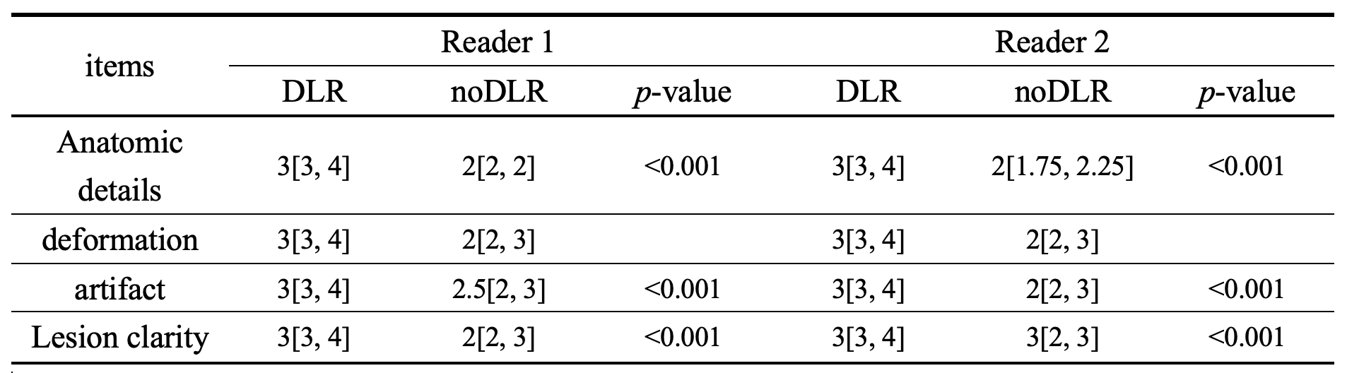
Thirty patients diagnosed with lung nodules were included in this study with IRB approval and written informed consents. All patients were scanned with a 3T MRI scanner (SIGNA Premier, GE Healthcare) using a single inspiration- and expiration breath-hold 3D radial ZTE sequence. The scan parameters were as follows: FOV = 38 cm, frequency = 128, slice thickness = 1.5 mm, number of excitations = 1.5, scan time = 17 second). For pulmonary ventilation estimation, lung images of both end-inspiration and expiration were scanned for all patients. Acquired k-space data was reconstructed by conventional method and DLR, respectively. The overall image quality was independently evaluated by two radiologists using four-point Likert scale assessment (Anatomical details :1= poor, 2= medium, 3= good, 4= excellent; deformation: 1= severe, 2= moderate, 3= mild, 4= missing; artifacts: 1= severe, 2= moderate, 3= mild, 4= missing; Lesion clarity: 1= poor, considered unrecognized, 2= medium, most poorly defined, 3= good, a few poorly defined, 4= excellent, clearly defined). Both lungs were semi-automatically segmented for all patients to measure their volumes. Based on a previous study [7], signal intensity of both lungs parenchyma and background were measured to assess SNR with manually drawn region-of-interest (ROI) in anterior, middle and posterior parts of lung (see Fig. 1). Additional ROIs were placed on trachea to calculate contrast-to-noise ratio (CNR) between parenchyma and trachea. Whole lung averaged fractional ventilation (FV) and relative FV (rFV) values were generated using the following equations [8]:FV=(〖SI〗exp−〖SI〗insp)/〖SI〗exp [1] rFV=FV/((〖Vol〗insp−〖Vol〗exp)/〖Vol〗insp) [2] SI and Vol means signal intensity and volume, respectively. All measurements were generated for both DLR and conventional reconstructed images. Paired t-tests or Wilcox signed rank tests were conducted to assess statistical differences between results of different reconstruction methods. Moreover, forced expiratory volume in the first second (FEV1) and forced vital capacity (FVC) were tested for all patients and correlated with ZTE based pulmonary ventilation values to investigate the effect of DLR.
Resuls
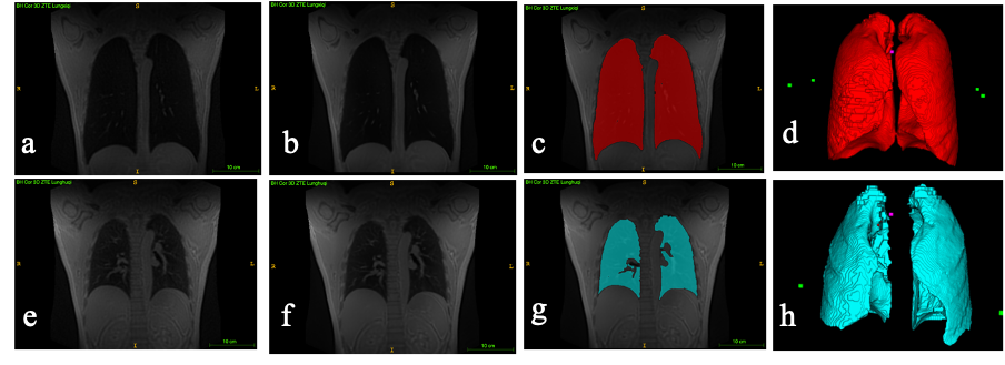
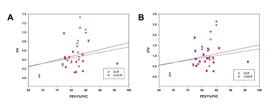
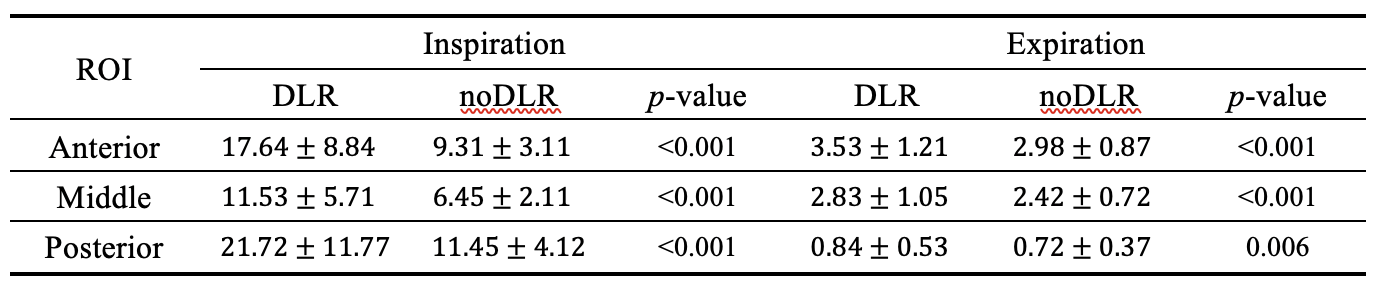
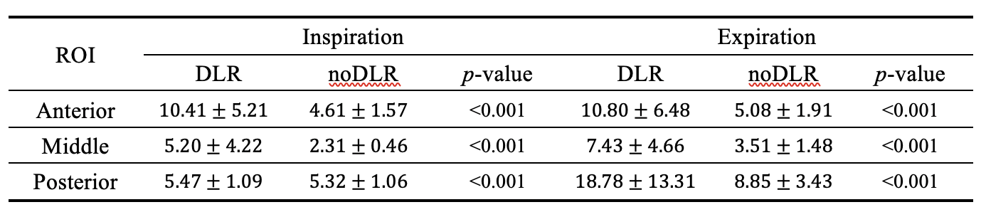
DLR and conventional reconstructed ZTE lung images of a typical patient are demonstrated in Fig. 1. As it is shown, elevated SNR and improved sharpness can be found in DLR images. In quantification, significantly increased SNR and CNR values of DLR images can be seen in both end-inspiration and end-expiration phases, respectively (see Table 1 and Table 2). Moreover, the quality scores of DLR images are significantly higher than those of conventional reconstructed ones for both observers (p<0.001, Table 3). In clinical, the percentage between FEV1 and FVC (FEV1%FVC ) is used to reflect restrictive or obstructive ventilation. And the whole lung averaged FV and rFV values are obtained from ZTE MRI images. The correlation between FV, rFV values and FEV1%FVC for both DLR and conventional reconstructed images (noDLR) are illustrated in Fig. 2. Significant correlation can be found between FV, rFV and clinical measurements for both DLR and noDLR images. Besides, elevated correlation values can be found for DLR compared to noDLR results.Discussion and Conclusion
This is a preliminary study for deep learning reconstructed ZTE lung MRI. Similar to previous DLR related studies, image quality of ZTE lung images were significantly improved through deep learning reconstruction with both increased SNR and CNR as well as elevated subjective evaluation at anatomical details, deformation, artifacts and lesion clarity. In addition, the improved image quality of DLR images result in better correlation between image-derived pulmonary ventilation estimation and clinical measurements. Furthermore, the posterior SNR, as well as CNR, was constantly higher than the anterior values for all patients, consistent with previous studies of the vertical gravity gradient as the air density distribution in lung. The findings of this study may facilitate the potential clinical applications of deep learning reconstructed ZTE technique in lung imaging.Acknowledgements
No acknowledgement found.References
1. Ohno, Y., Takenaka, D., Yoshikawa, T., Yui, M., Koyama, H., Yamamoto, K., ... & Toyama, H. (2022). Efficacy of ultrashort echo time pulmonary MRI for lung nodule detection and Lung-RADS classification. Radiology, 302(3), 697-706. 2. Bae, K., Jeon, K. N., Hwang, M. J., Lee, J. S., Ha, J. Y., Ryu, K. H., & Kim, H. C. (2019). Comparison of lung imaging using three-dimensional ultrashort echo time and zero echo time sequences: preliminary study. European radiology, 29, 2253-2262. 3. Heidenreich, J. F., Weng, A. M., Metz, C., Benkert, T., Pfeuffer, J., Hebestreit, H., ... & Veldhoen, S. (2020). Three-dimensional ultrashort echo time MRI for functional lung imaging in cystic fibrosis. Radiology, 296(1), 191-199. 4. Zhu, B., Liu, J. Z., Cauley, S. F., Rosen, B. R., & Rosen, M. S. (2018). Image reconstruction by domain-transform manifold learning. Nature, 555(7697), 487-492. 5. Kim, M., Kim, H. S., Kim, H. J., Park, J. E., Park, S. Y., Kim, Y. H., ... & Lebel, M. R. (2021). Thin-slice pituitary MRI with deep learning–based reconstruction: diagnostic performance in a postoperative setting. Radiology, 298(1), 114-122. 6. Almansour, H., Herrmann, J., Gassenmaier, S., Afat, S., Jacoby, J., Koerzdoerfer, G., ... & Othman, A. E. (2022). Deep learning reconstruction for accelerated spine MRI: prospective analysis of interchangeability. Radiology, 306(3), e212922. 7. Zeimpekis, K. G., Geiger, J., Wiesinger, F., Delso, G., & Kellenberger, C. J. (2021). Three-dimensional magnetic resonance imaging ultrashort echo-time cones for assessing lung density in pediatric patients. Pediatric radiology, 51, 57-65. 8. Metz, C., Weng, A. M., Heidenreich, J. F., Slawig, A., Benkert, T., Köstler, H., & Veldhoen, S. (2023). Reproducibility of non-contrast enhanced multi breath-hold ultrashort echo time functional lung MRI. Magnetic Resonance Imaging, 98, 149-154.Figures