3646
Feasibility study of stack-of-spirals free breathing ultrashort echo time for lung MRI in patients with malignant tumors at 1.5T1Union Hospital, Tongji Medical College, Huazhong University of Science and Technology, wuhan, China, 2MR Collaborations, Siemens Healthineers Ltd., Chengdu, China, 3MR Application Predevelopment, Siemens Healthcare GmbH, Erlangen, Germany, Erlangen, Germany
Synopsis
Keywords: Lung, Lung
Motivation: Ultrashort echo time (UTE) MR imaging has been developed to visualize lung tissues, but is limited by long acquisition time and capacity for displaying pulmonary tissues.
Goal(s): 3D free breathing stack-of-spirals UTE (spiral-UTE) could provide a higher readout efficiency, but its comprehensive performance with and without contrast material for follow-up of patients with identified malignant tumors at 1.5T has not been reported.
Approach: Aim to investigate clinical feasibility of spiral-UTE compared to enhanced-VIBE with CT as the reference.
Results: Spiral-UTE was superior with improved image qualities for depicting bronchi and lung parenchyma than enhanced-VIBE in lung screening during the oncology patient follow-ups
Impact: Spiral-UTE provides a potential alternative to CT for lung follow-up with a significantly superior quality visualization of the pulmonary anatomy than routine enhanced-VIBE
Introduction/Purpose:
Very low proton density and fast signal decay of lung tissues makes it a big challenge for MR imaging, and hasn’t been widely accepted in clinical practice. For the lung follow-up of patients with malignant tumors, CT screening is still regarded as the first-choice, but the repetitive x-ray ionizing radiation doses may increase the risk of developing cancer, which has aroused great concerns. Ultrashort echo time (UTE) MR imaging makes lung imaging better than the usual sequences with a very short echo time (TR). However, the long acquisition time is a main outcome. Three-dimensional free breathing stack-of-spirals UTE method (Spiral-UTE) could provide a higher readout efficiency and has been applied in 3T for oncology patients for metastasis and nodule evaluations. However, the comprehensive performance of spiral-UTE with and without contrast material has not been reported. Aim of this study is to investigate the clinical feasibility of Spiral-UTE lung MRI compared to enhanced- volumetric interpolated breath-hold examination (VIBE) for follow-up of patients with identified malignant tumors at 1.5T, with CT as the reference standard.Materials and methods
This study was approved by local ethics committee, and all subjects provided written informed consent. All the examinations were performed by a 1.5T MR scanner (MAGNETOM AERA, Siemens Healthcare, Erlangen, Germany). A total of 17 oncology patients (10 males, 7 females, mean ± SD: 57.29±10.76 years old) completed MRI and CT with a maximum of 7 days between the two studies. These patients had various types of cancer including lung cancer (n=7), breast cancer (n=3), gastrointestinal cancer (n=4) and cervical cancer (n=3). Gd-DOTA enhanced and unenhanced spiral-UTE sequence during free-breathing (research application) and enhanced VIBE sequence during breath-holding were scanned at 1.5T for lung follow-ups (Table 1). Enhanced spiral-UTE was scanned 3~4 minutes after contrast injection following the completion of routine clinical examination. Two experienced radiologists evaluated image quality in consensus with respect to the visibility of fissures, airways and vessels, signal homogeneity, motion artifacts related with thorax and heart, lesion conspicuity and overall image quality with a 5-point scale (4, excellent; 0, poor) regarding CT as the reference standard. Quantitative comparison of image quality was performed using SNR and CNR. Kruskal-Wallis one-way ANOVA and Bonferroni correction were used for statistical analyses.Results:
Spiral-UTE outperformed VIBE in terms of visibility of fissures, airways, vessels, signal homogeneity, motion artifacts, lesion conspicuity and overall image quality with a statistically significant difference (p<0.05, Table 2, Fig 1-3). Notably, there was no statistical difference in vessel visualization between unenhanced and enhanced spiral-UTE. Bronchi were consistently visible up to fourth-level in spiral-UTE, while VIBE only provided level-one visibility. Additionally, enhanced spiral-UTE was found to be the best for depicting pulmonary vessels, outperforming unenhanced spiral-UTE and enhanced VIBE. Regarding apparent SNR and CNR, there were significant difference among enhanced and unenhanced UTE and VIBE (SNR:1.95±0.43, 1.46±0.39 and 0.41±0.13, p<0.001; CNR: 0.08±0.03, 0.06±0.04, -0.03±0.01, p<0.001). Enhanced spiral-UTE provided the best SNR, while spiral-UTE obtained the highest CNR, with no statistical difference between enhanced and unenhanced spiral-UTE methods (p=0.498).Conclusions:
Spiral-UTE provides a free-breathing technique with superior image quality compared to routine enhanced-VIBE with high quality visualization of the pulmonary anatomy. This technique offers a potential alternative to CT for lung lesion follow-up.Clinical Relevance/Application
For patients with malignant tumors, CT scans are routinely used for lung follow-ups, but this can result in a high cumulative radiation dose. Our study showed that spiral-UTE enables free-breathing lung imaging of bronchi and lung parenchyma with superior SNR and CNR compared to routine enhanced-VIBE sequence for oncology patients in their lung screening during the follow-ups. Further analysis comparing lesion characterization and measurement is now required to determine the necessity for contrast material.Acknowledgements
NoneReferences
1. Cha MJ, Park HJ, Paek MY, et al. Free-breathing ultrashort echo time lung magnetic resonance imaging using stack-of-spirals acquisition: A feasibility study in oncology patients. Magnetic Resonance Imaging. 2018;51:137–143. doi: 10.1016/j.mri.2018.05.002.
2. Cha MJ, Ahn HS, Choi H, et al. Accelerated Stack-of-Spirals Free-Breathing Three-Dimensional Ultrashort Echo Time Lung Magnetic Resonance Imaging: A Feasibility Study in Patients With Breast Cancer. Front Oncol. 2021;11:746059. doi: 10.3389/fonc.2021.746059.
3. Dournes G, Grodzki D, Macey J, et al. Quiet Submillimeter MR Imaging of the Lung Is Feasible with a PETRA Sequence at 1.5 T. Radiology. 2015;276(1):258–265. doi: 10.1148/radiol.15141655.
Figures
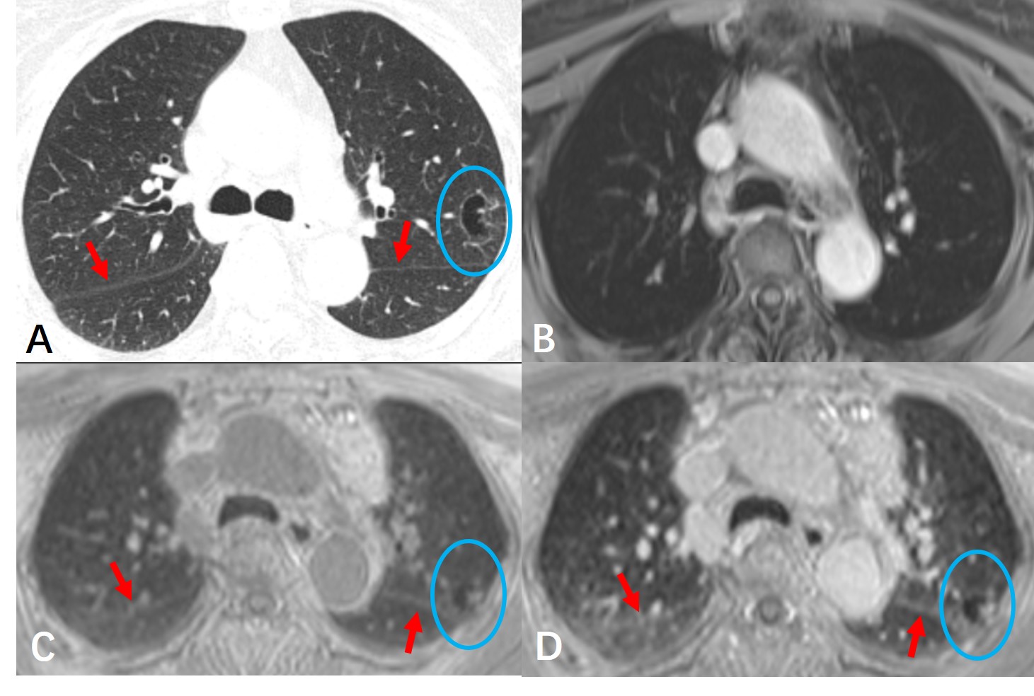
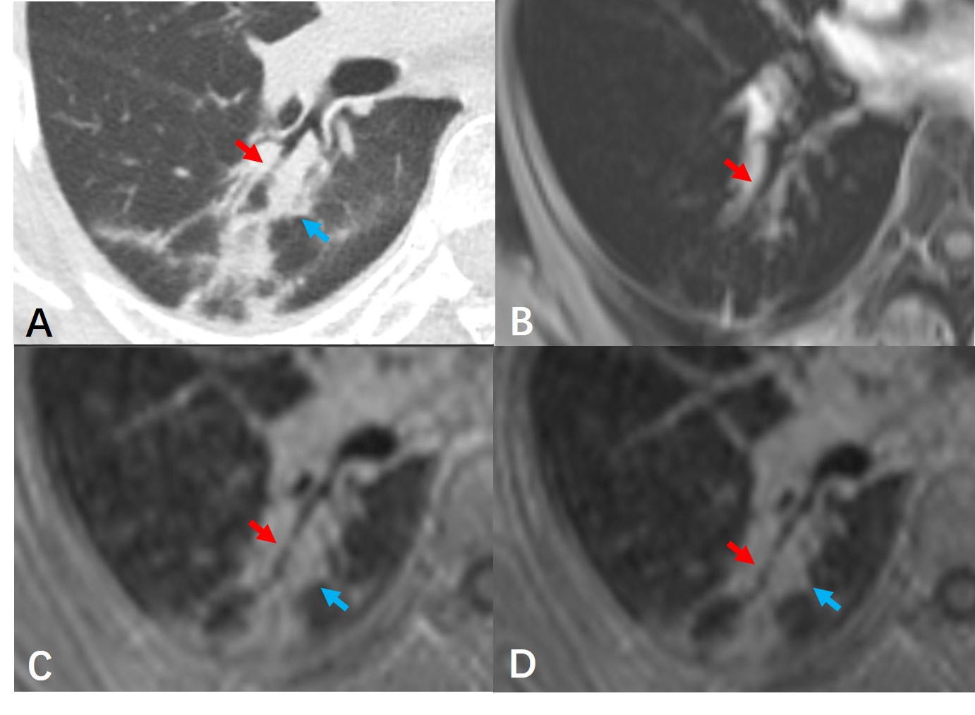
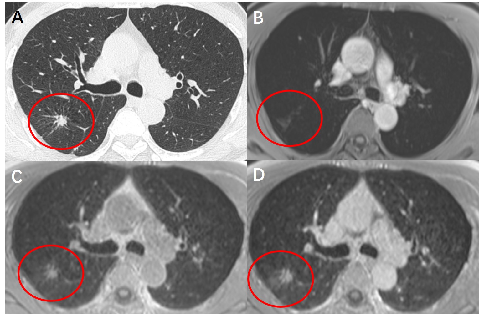
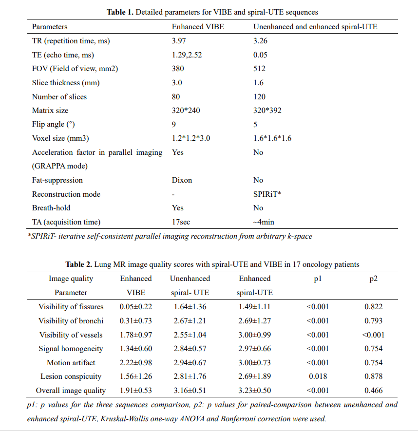
Table 1. Detailed parameters for VIBE and spiral-UTE sequences
Table 2. Lung MR image quality scores with spiral-UTE and VIBE in 17 oncology patients