3645
Accelerated Spiral Ultrashort Echo Time (Spiral-UTE) MRI of the Lung Using Deep Learning1Bernard and Irene Schwartz Center for Biomedical Imaging, Department of Radiology, New York University Grossman School of Medicine, New York City, NY, United States, 2Center for Advanced Imaging Innovation and Research (CAI2R), Department of Radiology, New York University Grossman School of Medicine, New York City, NY, United States, 3Department of Electrical and Computer Engineering, NYU Tandon School of Engineering, New York City, NY, United States
Synopsis
Keywords: Lung, Lung
Motivation: Spiral-UTE MRI has been proposed for more efficient lung imaging to permit breath-hold ultra-short echo time acquisition of the lung. It is more valuable to further accelerate the acquisition of the spiral-UTE MRI of lung images, thus enabling shorter breath-holds and higher spatial resolutions.
Goal(s): This work presents a deep learning based method to enable the reconstruction of spiral-UTE MRI of lung images from accelerated spiral k-space.
Approach: An unrolled network was developed for reconstructing images from the accelerated non-cartesian k-space.
Results: The unrolled network allows for higher reconstruction quality for spiral-UTE MRI of lungs compared to a standard U-Net.
Impact: The proposed unrolled network tailored for spiral MRI reconstruction enables reconstruction of accelerated spiral-UTE of lung images to allow shorter breath-holds and higher spatial resolutions. This reconstruction technique can also extended to other multi-coil non-cartesian accelerated MRI reconstructions.
Introduction
MRI is a promising radiation-free imaging modality for lung imaging compared to Computed Tomography (CT). While MRI has traditionally not been employed for lung imaging, recent technological advancements in MRI have enabled its utilization in both research and clinical settings1,2. For instance, the use of ultra-short echo time acquisition in MRI(UTE-MRI) has made it feasible to visualize short T2* structures within the lungs3,4. Recently, a new UTE-MRI sequence based on stack-of-spirals sampling, called spiral-UTE, has been proposed for more efficient lung imaging to permit breath-hold ultra-short echo time acquisition of the lung5. Nevertheless, further acceleration of spiral UTE would be useful to enable short breath-holds and/or higher spatial resolutions. In recent years, deep learning-based approaches for accelerated MRI acquisition have gained popularity due to their ability to outperform traditional model-based methods like compressed sensing(CS)6, 7. In this study, we proposed a deep learning based non-Cartesian multi-coil MRI reconstruction method, which is demonstrated to further accelerate the acquisition of spiral-UTE MRI of the lungs.Methods
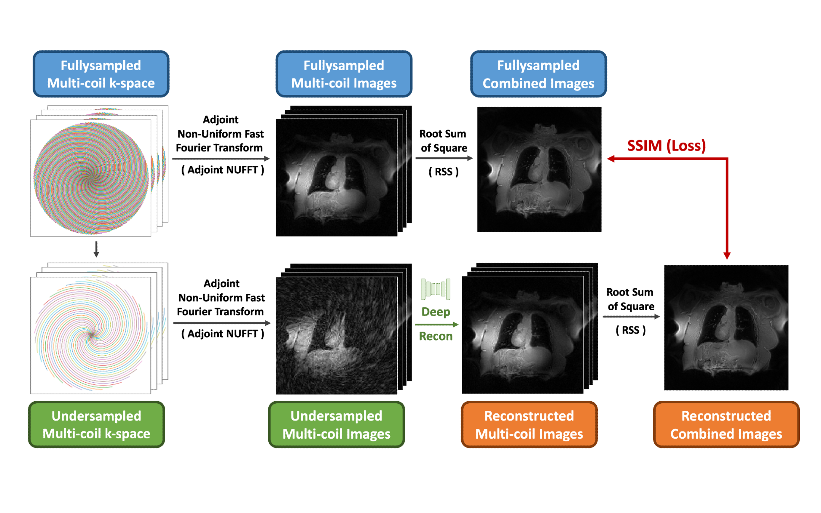
The overall training pipeline is shown in Figure 1. The deep learning-based reconstruction network was built to reconstruct the undersampled non-Cartesian multi-coil images with aliasing artifacts, which were generated by applying the adjoint non-uniform Fast Fourier Transform (NUFFT) to the undersampled multi-coil k-space. The network was optimized by enforcing a structural similarity index measure (SSIM) loss between the reconstructed and fullysampled coil combined images that were acquired from the root sum of square(RSS) combination of the reconstructed and fullysampled multi-coil image, separately.The cost function for reconstructing the accelerated non-Cartesian MRI can be formulated as follows:
$$x=argmin\frac{1}{2}\ \left|\left|Ax-b\right|\right|_2^2+\lambda\Phi(x)$$
Where $$$A=\ {DF}_NC(·)$$$, $$$C(·)$$$ represents coil expansion operation to expand the coil combined image to multi-coil images. $$$b$$$ is the measured undersampled multi-coil k-space. $$$F_N$$$ denotes NUFFT. $$$D$$$ is the pre-defined downsample operation to downsample the non-cartesian k-space to make it consistent with the measured undersample k-space. $$$x$$$ is the reconstructed image. $$$\Phi$$$ is a regularization function.
This above cost function can be solved using iterative gradient descent methods, which involve updating the image estimate in each iteration until convergence is achieved. In the t-th step, the image is updated from $$$x^t$$$ to $$$x^{t+1}$$$ using the following formula:
$$x^{t+1}=x^t-\eta\left(A^H\left(Ax-b\right)\right)-\lambda\ \nabla\Phi(x^t)$$
A convolutional neural network was employed to estimate the gradient of the regularization function:
$$x^{t+1}=x^t-\eta^t\left(A^H\left(Ax-b\right)\right)-CNN(x^t)$$
Where $$$A^H=C^H(F_N^HD)$$$ is the conjugate transpose of the forward operator $$$A$$$. $$$C^H$$$ represents coil combination operation to combine the multi-coil images into coil-combined images. $$$F_N^H$$$ denotes the Adjoint of the NUFFT. $$$\eta^t$$$is the decay rate that can be learned from data.
To reconstruct the multi-coil images, we apply a coil expansion operator $$$C(·)$$$ to both sides of the formula:
$$x_{mc}^{t+1}=x_{mc}^t-\eta^t\left(F_N^HD\left({DF}_NC\left(x\right)-b\right)\right)-C(CNN\left(C^H(x_{mc}^t)\right))$$
Where $$$x_{mc}$$$ is the multi-coil images. In practice, the downsampling operation $$$D$$$ is implemented with NUFFT or adjoint of NUFFT with downsampled k-space trajectory. Therefore, the final formula to build the unrolled network is:
$$x_{mc}^{t+1}=x_{mc}^t-\eta^t\ \left(F_N^H\left(F_NC\left(x\right)-b\right)\right)-C(CNN\left(C^H(x_{mc}^t)\right))$$
The final reconstructed image was acquired from the RSS combination of the final reconstructed multi-coil image $$$x_{mc}$$$.
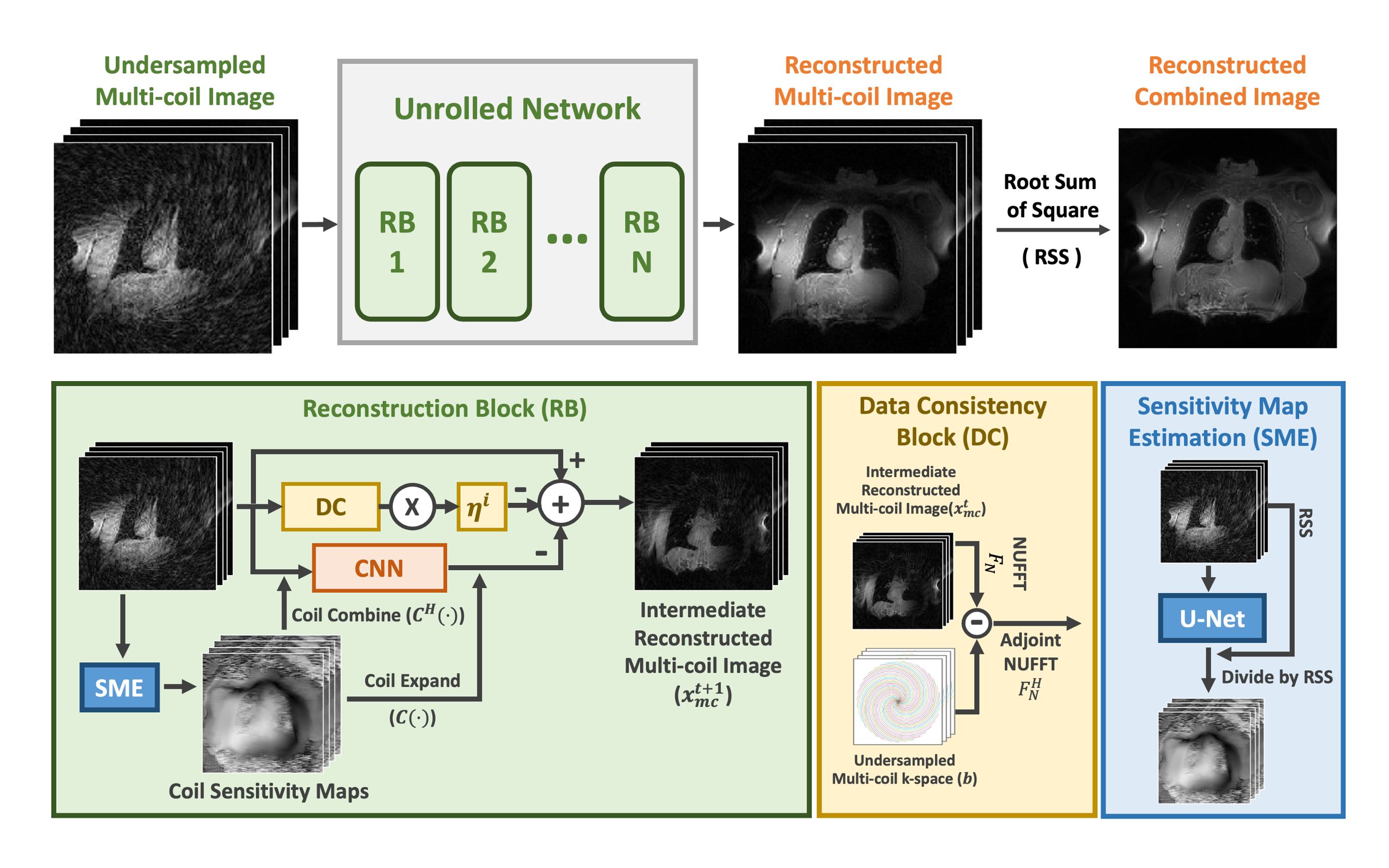
Based on the above equation, the unrolled network was developed with multiple blocks to model the model iterative gradient descent updates as shown in Figure 2. The coil sensitivity estimation module (SME)7 employs a small U-Net to estimate coil sensitivity maps employed in the reconstruction process from undersample multi-coil images.
47(30/5/12 training/validation/testing) spiral UTE lung MRI datasets(48 slices, 140 spiral lines/slice/sample) were used to evaluate the unrolled network with two acceleration factors(R=2/4 with 70/35 spiral lines). All the data were previously acquired on a 3T MRI scanner (Skyra, Siemens) during a single breath hold. For comparison, a standard U-Net8 was trained to directly reconstruct images from the coil-combined undersampled images.
Results
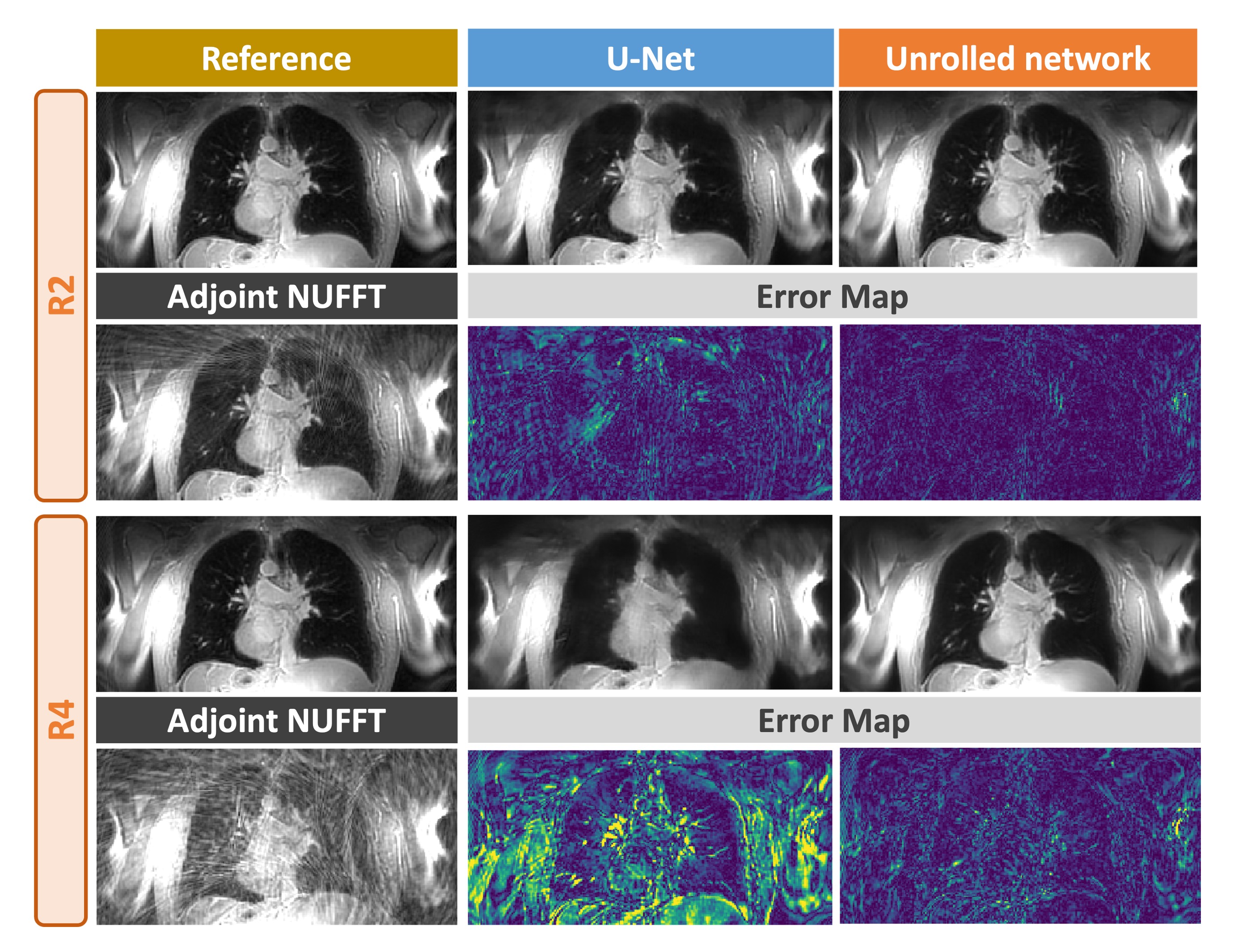
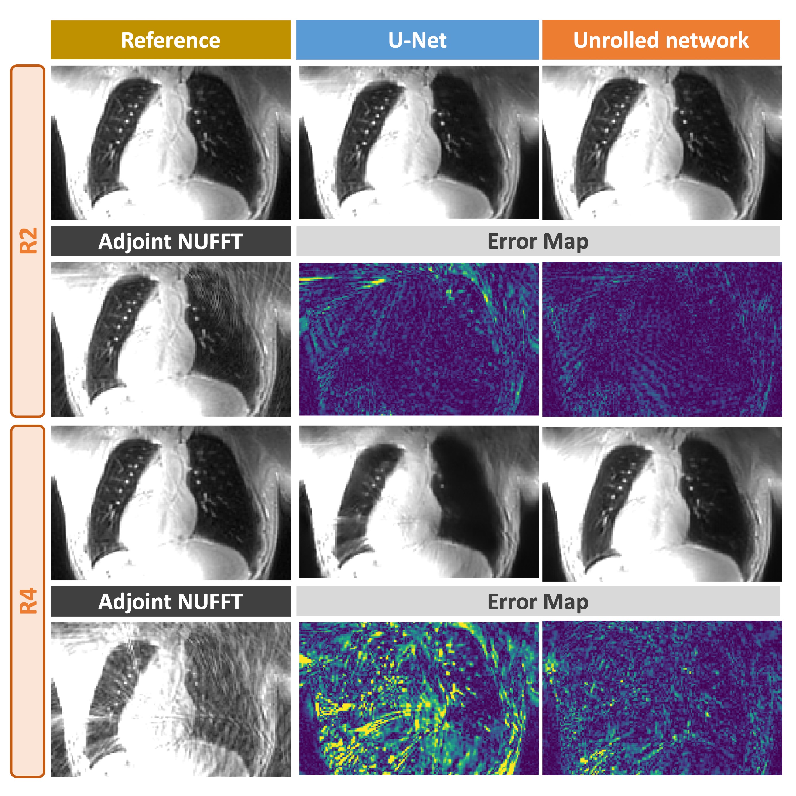
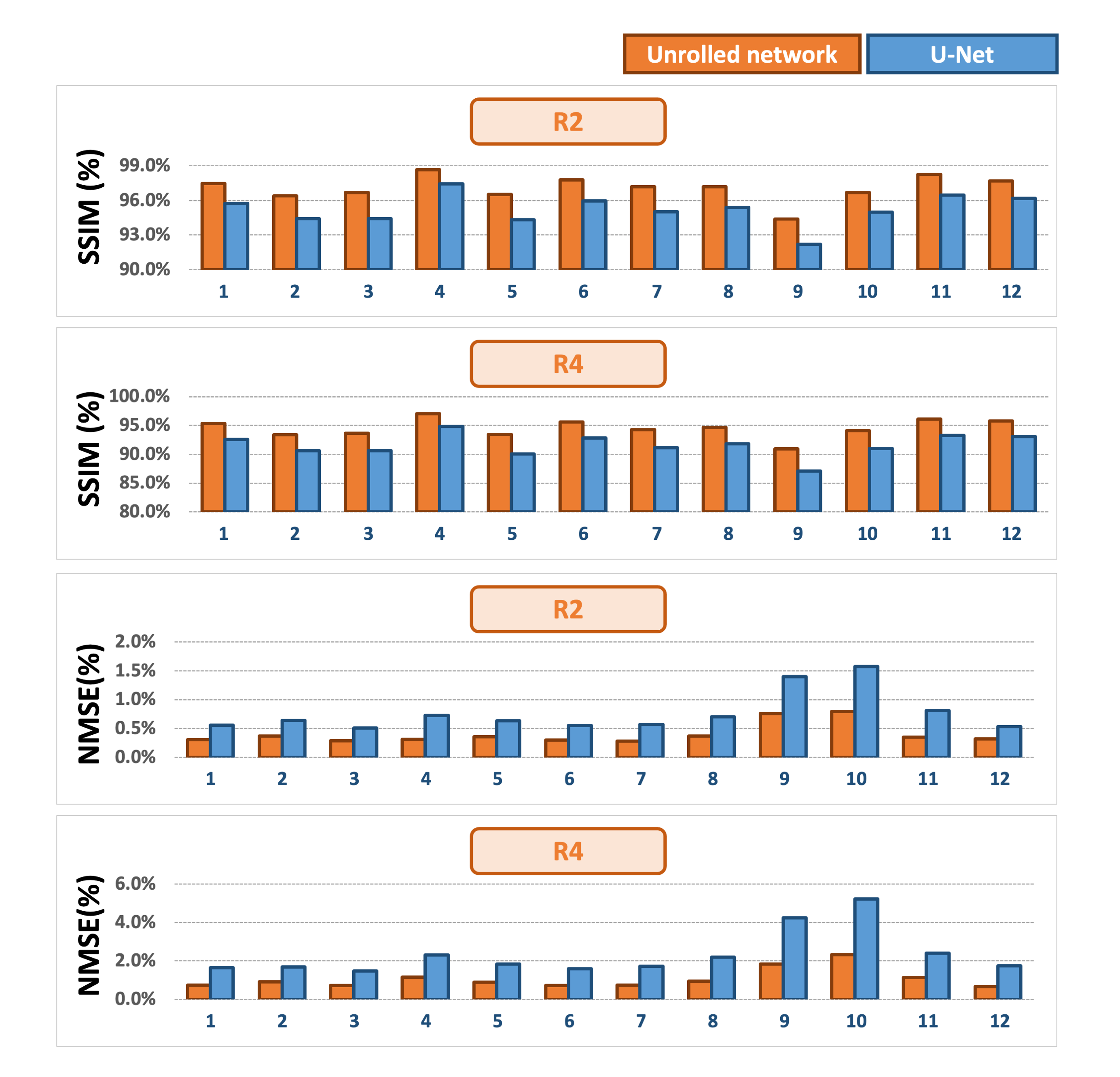
Figures.3 and 4 show two cases comparing reconstructed lung images using the unrolled network and U-Net with different acceleration factors. The error maps indicate that the unrolled network can reconstruct higher-quality images with less errors compared to the U-Net. When the acceleration factors increased to 4, the U-Net failed to recover the fine-grained structure inside of the lung.Figure.5 summarizes the quantitative comparison (SSIM and normalized mean squared error(NMSE)) of the unrolled network and U-Net with different acceleration factors for all 12 testing cases. The results demonstrate that the unrolled network enables high-quality reconstruction of lung images compared to U-Net reconstruction across all test cases in terms of SSIM and NMSE.
Conclusion
In this study, we proposed a new deep learning based method to perform the accelerated multi-coil non-cartesian MRI reconstruction, which has demonstrated high-performance reconstruction for spiral-UTE MRI the lung to allow shorter breath-holds and higher spatial resolutions in clinical settings.Acknowledgements
This work was supported by the NIH (R01EB031083, R01EB030549 and P41EB017183) and was performed under the rubric of the Center for Advanced Imaging Innovation and Research (CAI2R), an NIBIB Biomedical Technology Resource Center.References
1. Wild J.M., Marshall H., Bock M., Schad L.R., Jakob P.M., Puderbach M., et al. MRI of the lung (1/3): methods. Insights Imaging. 2012;3:345–353. doi: 10.1007/S13244-012-0176-X/FIGURES/10.2. Biederer J., Beer M., Hirsch W., Wild J., Fabel M., Puderbach M., et al. MRI of the lung (2/3). Why… When … How? Insights Imaging. 2012;3:355–371. doi: 10.1007/S13244-011-0146-8.
3. Delacoste J., Chaptinel J., Beigelman-Aubry C., Piccini D., Sauty A., Stuber M. A double echo ultra short echo time (UTE) acquisition for respiratory motion-suppressed high resolution imaging of the lung. Magn Reson Med. 2018;79:2297–2305. doi: 10.1002/mrm.26891.
4. Zhang J., Feng L., Otazo R., Kim S.G. Rapid dynamic contrast-enhanced MRI for small animals at 7T using 3D ultra-short echo time and golden-angle radial sparse parallel MRI. Magn Reson Med. 2019;81:140–152. doi: 10.1002/mrm.27357.
5. John P., Mugler I., Meyer C.H., Pfeuffer J., Stemmer A., Kiefer B. (ISMRM 2017) Accelerated Stack-of-Spirals Breath-hold UTE Lung Imaging. https://archive.ismrm.org/2017/4904.html
6. Aggarwal, Hemant K., Merry P. Mani, and Mathews Jacob. "MoDL: Model-based deep learning architecture for inverse problems." IEEE transactions on medical imaging 38.2 (2018): 394-405.
7. Sriram, Anuroop, et al. "End-to-end variational networks for accelerated MRI reconstruction." Medical Image Computing and Computer Assisted Intervention–MICCAI 2020: 23rd International Conference, Lima, Peru, October 4–8, 2020, Proceedings, Part II 23. Springer International Publishing, 2020.
8. Ronneberger, Olaf, Philipp Fischer, and Thomas Brox. "U-net: Convolutional networks for biomedical image segmentation." Medical Image Computing and Computer-Assisted Intervention–MICCAI 2015: 18th International Conference, Munich, Germany, October 5-9, 2015, Proceedings, Part III 18. Springer International Publishing, 2015.
Figures