3642
Radiomics based on tumor and multiple body components on MRI to predict outcome in rectal Cancer1The First Hospital of Jilin University, Changchun, China, 2Dalian Municipal Central Hospital, Dalian, China
Synopsis
Keywords: Cancer, Radiomics
Motivation: In rectal cancer, the existing stratification of prognosis is mainly based on the established TNM tumor staging system, which limits clinical decision-making.
Goal(s): Construct a model to provides prognostic information preoperatively.
Approach: This study established radiomics models to predict the disease-free survival (DFS) in rectal cancer, based on tumor, and multiple body components (including mesenteric fat and pelvic skeletal muscles) in MRI.
Results: The radiomics model based on tumor and multiple body components of MRI in rectal cancer, have good predictive value for DFS in rectal cancer patients at 2 years after surgery.
Impact: The model that provides prognostic information at the time of cancer diagnosis would be useful for optimizing treatment and monitoring.
Introduction
In Rectal cancer (RC), the existing preoperative stratification of prognosis is mainly based on the established TNM tumor staging.1 However, TNM is not an optimal risk assessment tool because of the clinical heterogeneity of patients at the same stage. Therefore, a model that provides more precise preoperative prognostic information would be useful for optimizing treatment and monitoring. Recently, as part of body composition, fat and skeletal muscles are gaining more attention which shown to be associated with morbidity and mortality in caners and following abdominal surgery.2,3 MRI is recommended for RC staging, further, fat and pelvic muscles can also be clearly shown.1,4 Currently, radiomics5 may improve the accuracy of prognosis prediction by extracting and analyzing the first-order and high-order image features of medical images.Therefore, the purpose of this study was to build a radiomics model using tumor and multiple body components (fat and skeletal muscles) based on MRI to predict DFS so as to differentiate the underlying poor outcome patient in RC.Purpose
To establish radiomics models based on tumor and multiple body components , including mesenteric fat (MF) and pelvic skeletal muscles (PSM) in MRI to predict the disease-free survival (DFS) in RC.Methods
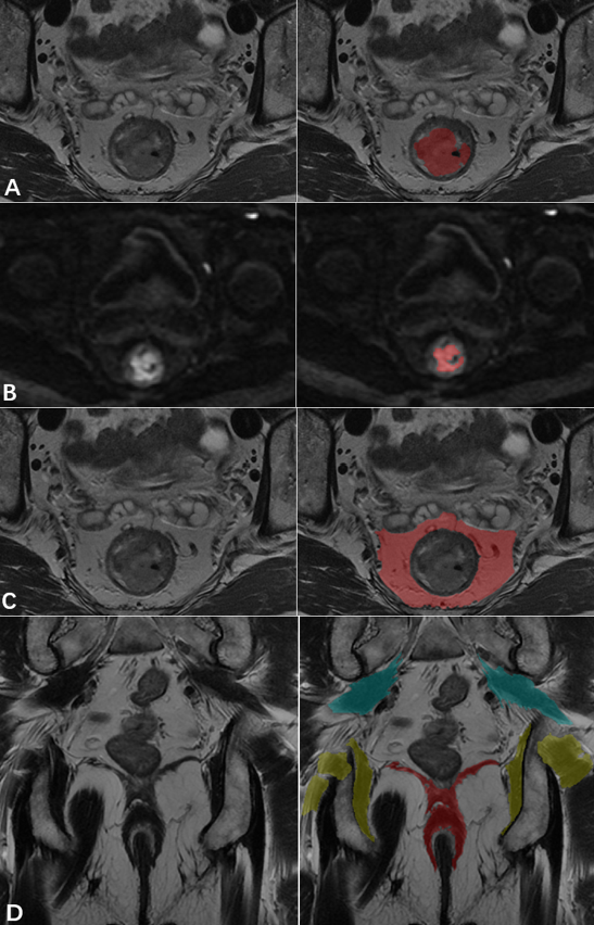
A retrospective analysis was conducted on 244 patients with RC confirmed by postoperative pathology at the First Hospital of Jilin University from January 2016 to June 2020. They were randomly divided into a training set (170 cases) and a testing set (74 cases) in a 7:3 ratio. The patients’ clinical data and preoperative rectal MRI were collected. After conducting univariate and multivariate COX regression analysis on clinical features, the strongest correlation with rectal cancer prognosis was obtained, and a clinical model was established based on the selected features. Use the ITK-SNAP software to delineate the primary lesion of rectal cancer in the axial T2WI and DWI, the MF in the axial T2WI, and the PSM (piriformis muscle, obturator muscle, anal sphincter complex) in the coronal T2WI sequence. Subsequently, the radiomics features were extracted from above regions of interest (ROIs). Then, we used single factor COX regression and autocorrelation algorithm Corr_ Group, LASSO COX regression, and multivariate COX regression to select features. Three radiomics models were established based on the tumors (R1-model), tumors combined with MF (R2-model), tumors combined with MF and PSM (R3-model), respectively. A integrated model was established based on clinical features and radiomics features used in R3-model. Finally, we verified the relationship between radiomics features and DFS using K-M survival curve.Results
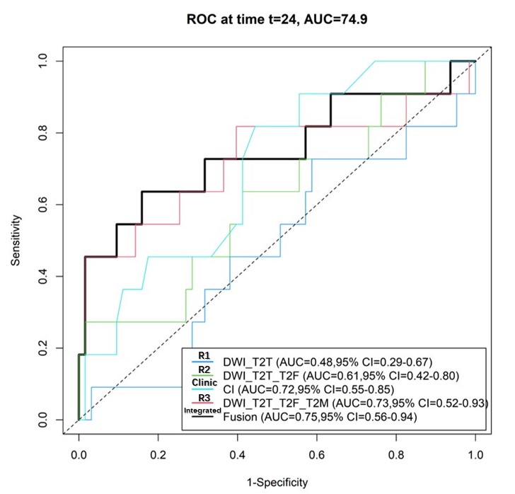
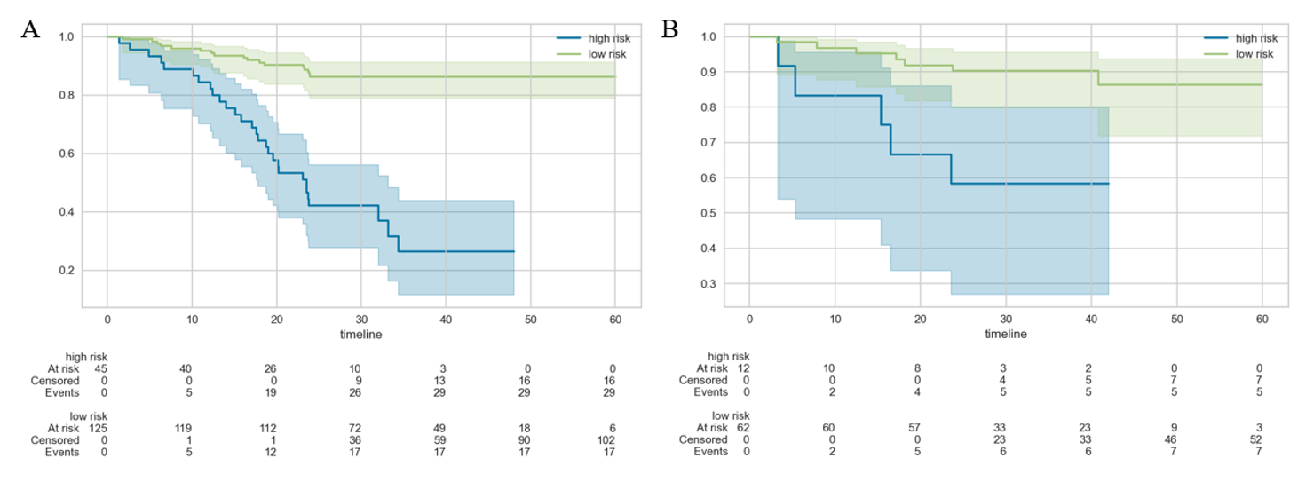
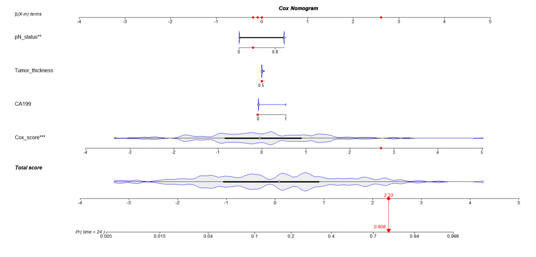
The pN stage, CA-199 and depth of tumor invasion were identified as clinical factors significantly related to the prognosis of RC (P<0.05). After the radiomics features selecting, eight radiomic features based on tumor, five radiomic features based on tumor combined with MF, and eight radiomic features based on tumor combined with MF and PSM were obtained, then, three radiomics models were established. All three radiomics models were significantly associated with 2-year DFS in the training cohort (P<0.001), while R1-model and R2-model were not significantly associated with 2-year DFS in the testing cohort (P=0.42). The R3-model was significantly associated with 2-year DFS (P=0.0037), and with a higher prediction performance (C-index: 0.697, 95%CI: 0.470-0.846; AUC = 0.73, 95%CI: 0.52-0.93) in the test set. Further, the integrated model incorporating R3 and clinical features had the highest performance (C-index=0.724, 95%CI: 0.493-0.875; AUC=0.75, 95%CI: 0.56-0.94) in the test set. K-M survival curves were plotted in the training and testing sets, there was a significant statistical difference in DFS between the high-risk and low-risk groups (Ptrain<0.0001,Ptest=0.0037) in integrated model.Discussion
We successfully established a radiomics model using tumor and multiple body components (PSM and MF) to predict DFS in RC. The above objectively results would help clinicians to screen the bad outcome patients and ultimately improve the treatment decision making. Muscle and fat are the distinct entities from other markers of physiological reserve, which are considered markers of overall health. 6 And in our study, we constructed radiomics models based on tumor and multiple body components, which maybe new biomarkers to make a novel risk stratification for RC. However, our data is biased, which is consistent with clinical reality. Further, we will increase the sample size, hoping to build a more robust and generalized model.Conclusion
The MRI radiomic model based on the primary tumor, MF and PSM have good predictive value for DFS of patients with RC. The integrated model incorporating clinical feature, tumor and multiple body components radiomics signaturs has the best performance, which can predict the prognosis of RC before operation and help to guide the individualized treatment of patients.Acknowledgements
This work was supported by National Natural Science Foundation of China (NSFC)(12226003, U22A20351), Jilin Province Science and Technology Department (2YDZJ202201ZYTS679), Jilin Provincial International Joint Research Center for Medical Artificial Intelligence Precision Diagnosis and Treatment (020210504008GH), and Youth Development Fund of the First Hospital of Jilin University (JDYY13202204).References
1. Glynne-Jones R, Wyrwicz L, Tiret E, et al. Rectal cancer: ESMO Clinical Practice Guidelines for diagnosis, treatment and follow-up. Annals of oncology : official journal of the European Society for Medical Oncology 2017;28(suppl_4):iv22-iv40.
2. Lee CM, Kang J. Prognostic impact of myosteatosis in patients with colorectal cancer: a systematic review and meta-analysis. Journal of Cachexia Sarcopenia and Muscle 2020;11(5):1270-1282. 3. Weerink LBM, van der Hoorn A, van Leeuwen BL, de Bock GH. Low skeletal muscle mass and postoperative morbidity in surgical oncology: a systematic review and meta-analysis. Journal of Cachexia Sarcopenia and Muscle 2020;11(3):636-649.
4. Benson AB, Venook AP, Al-Hawary MM, et al. NCCN Guidelines Insights: Rectal Cancer, Version 6.2020. J Natl Compr Canc Netw 2020;18(7):806-815.
5. Lambin P, Rios-Velazquez E, Leijenaar R, Carvalho S, Aerts HJWL. Radiomics: Extracting more information from medical images using advanced feature analysis. European Journal of Cancer 2012;43(4):441-446.
6. Body S, Ligthart MAP, Rahman S, et al. Sarcopenia and Myosteatosis Predict Adverse Outcomes After Emergency Laparotomy A Multi-center Observational Cohort Study. Annals of Surgery 2022;275(6):1103-1111.
Figures