3638
Improving Abdominal MR Image Quality at 0.55T Using Deep Learning Reconstruction: A Comparative Study with Commercial 0.55T and High-Field Scans1Department of Radiology, University of Michigan, Ann Arbor, MI, United States, 2Siemens Healthineers, Erlangen, Germany
Synopsis
Keywords: Hepatobiliary, Low-Field MRI, Abdomen, deep-learning reconstruction
Motivation: Deep-learning reconstruction may overcome two shortcomings of 0.55T, low SNR and extended scan time, without compromising lesion conspicuity.
Goal(s): To demonstrate that image quality and SNR of deep-learning reconstructed 0.55T images are at least similar to 1.5T/3T images, while maintaining visibility of pathologies.
Approach: 23 patients imaged at 0.55T using standard and deep-learning HASTE and DWI. Three radiologists rated IQ and SNR at 0.55T and HF. Pathologies were evaluated in deep-learning images.
Results: Deep-learning reconstructed HASTE and DWI 0.55T images were of same or better quality and SNR than 1.5T/3T images. All pathologies were visible on deep-learning 0.55T images. DL reduced HASTE scan-time.
Impact: Deep-learning reconstruction algorithms of select sequences at 0.55T can help overcome low SNR and extended scan times of current 0.55T abdominal imaging, making it comparable or superior to standard-of-care 1.5/3T, thereby expanding global use of a more accessible MRI system.
Introduction
The recent introduction of a general-use 0.55T commercial scanner offers potential to improve MR access through a combination of reduced purchase price, installation requirements (smaller weight/footprint, lack of quench pipe) and maintenance costs (reduced power consumption and cooling needs)1. The system also offers some advantages over 1.5/3T scanners, including a more comfortable patient-experience via acoustic noise reduction and larger bore size (80 cm), reducing anxiety2. However, the image quality of 0.55T systems remain uncertain compared to conventional high-field scanners due to the lower signal-to-noise ratio (SNR) inherent to lower field-strengths, and the need to prolong scan times to acquire images suitable for clinical diagnosis. In response to these challenges, deep-learning (DL) reconstruction techniques have been introduced. These approaches work by using an iterative k-space-to-image reconstruction with interleaved, neural network-based regularization.The purpose of this study is to assess the impact of deep-learning reconstruction on the overall image quality (IQ) and SNR of scans acquired at 0.55T compared to those acquired at higher field strengths in the same patient cohort, and to evaluate the effect of the DL reconstruction on conspicuity of pathologies amongst abdominal organs.
Methods
In this IRB-approved prospective study, 23 clinical patients underwent abdominal MR imaging on a commercial 0.55T MRI system (Free.Max, Siemens Healthineers) upon providing informed-consent. In addition to “standard” product sequences, limiting matching sequences (coronal and axial T2w HASTE, and DWI b50 and b800) with work-in progress deep-learning reconstruction algorithms (WIP-DL) were acquired. Of the 23 patients, 16 had prior scans at higher field (HF) strengths (1.5T or 3T) and the same sequences were used for comparison. Three raters with 24, 11, and 18 years of experience in abdominal MR imaging independently reviewed sets of the individual sequences that consisted of randomized standard 0.55T, 0.55T WIP-DL, and HF images. The raters were blinded to field-strength, type of image, and patient history. Raters assessed overall image quality (IQ) and SNR on a 1-4 Likert scale (1=non-diagnostic/poor SNR – 4=excellent). Wilcoxon signed-rank tests were applied to determine differences between the paired ratings with p-value<0.05 considered to be statistically significant. Kendall’s coefficient of concordance (W) was calculated to determine inter-rater agreement.In patients with prior high-field scans, visibility of pathology mentioned in the HF and standard 0.55T radiology reports was compared to an independent assessment of the 0.55T WIP-DL sequences.
Results
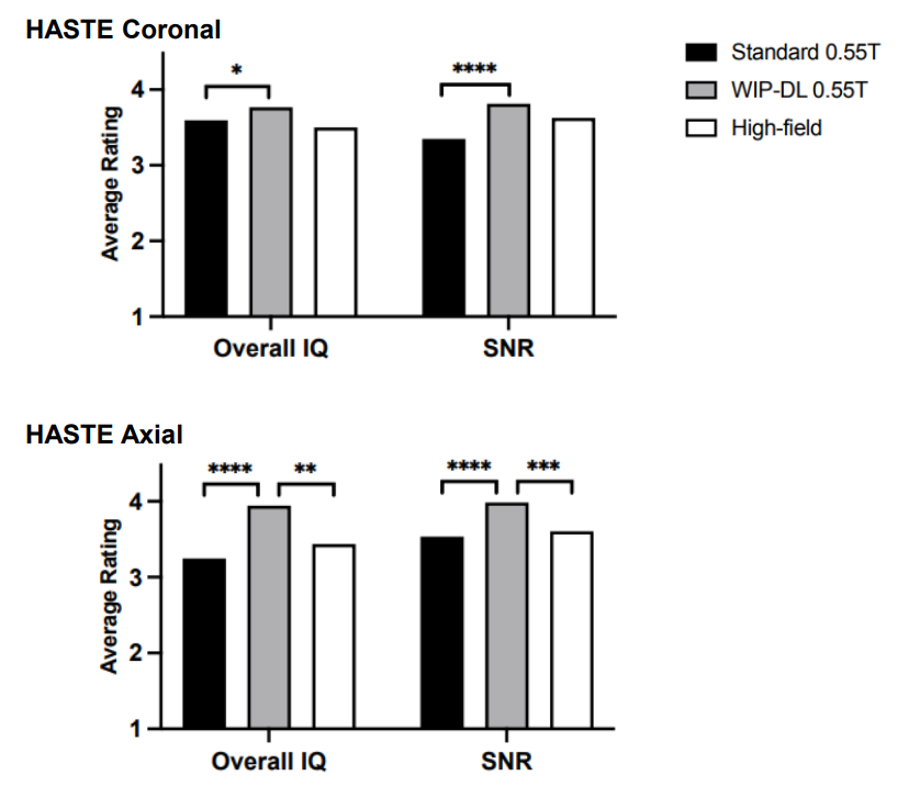
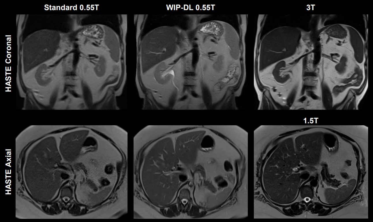
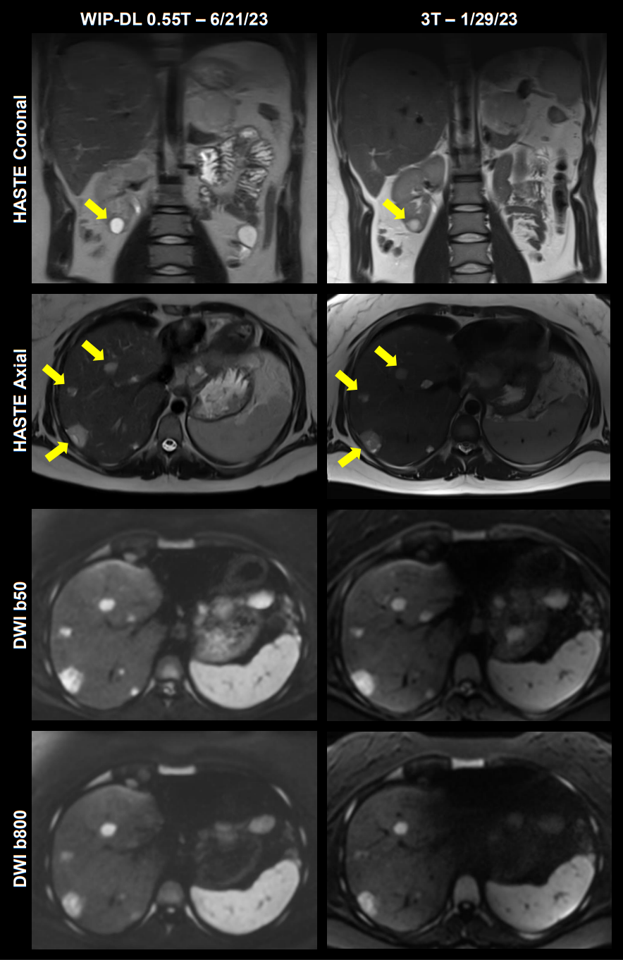
Analysis included 23 sets of coronal and axial T2w HASTE images and 22 sets of b50 and b800 DWI images, collected using standard and WIP-DL sequences at 0.55T. The HASTE and DWI sets also included 16 and 13 additional paired HF sequences, respectively.For coronal HASTE, the 0.55T WIP-DL had similar IQ and SNR scores compared to HF (p=0.1484; p=0.3828), but scored significantly higher than standard 0.55T (p=0.0343; p<0.0001). Ratings of axial HASTE IQ and SNR were significantly higher for 0.55T WIP-DL compared to HF (p<0.0001; p<0.0001) and standard 0.55T (p=0.0022; p=0.001).
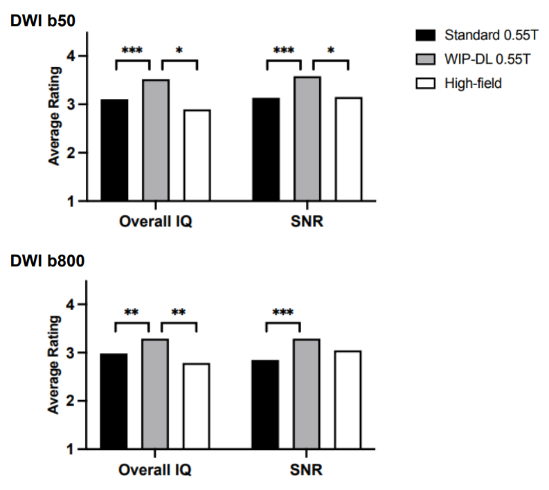
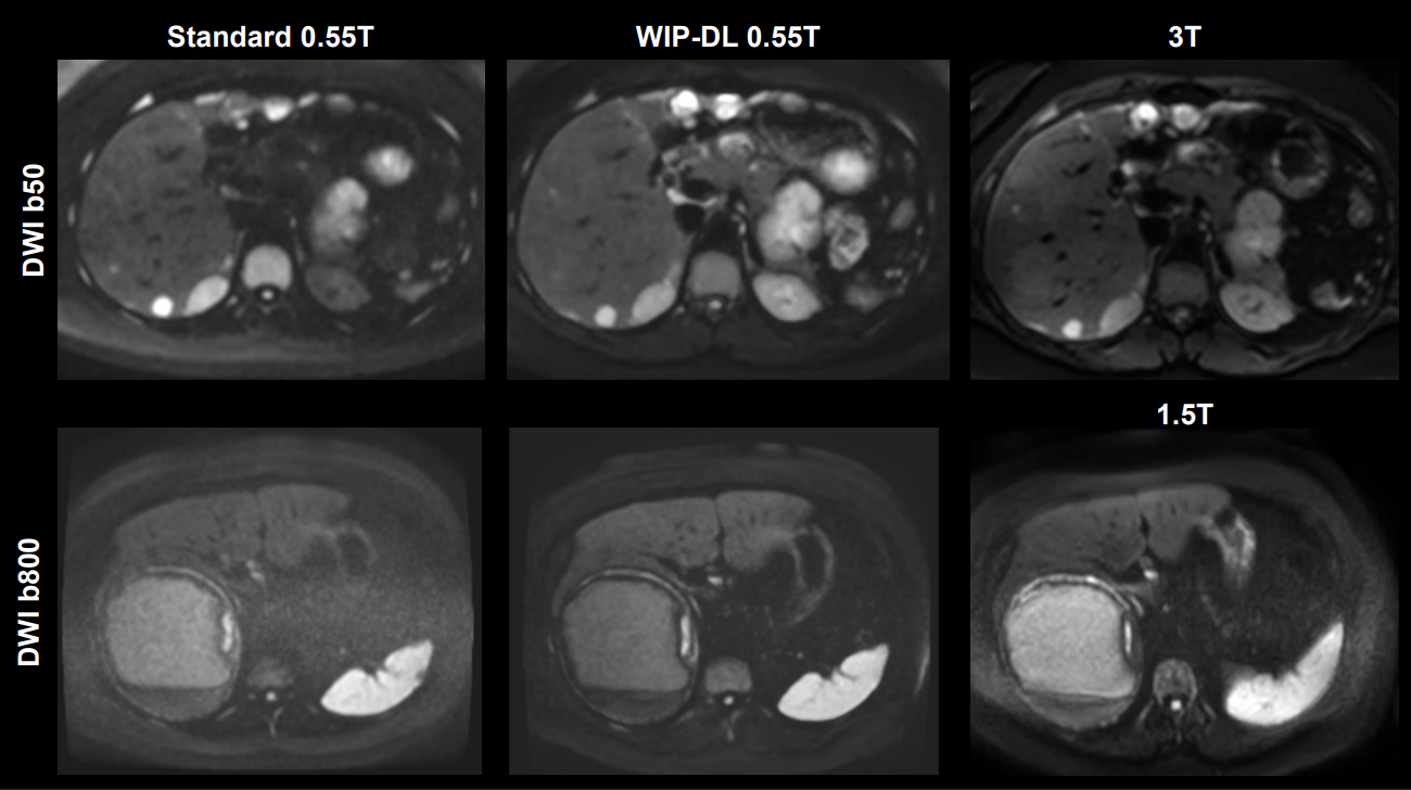
For DWI b50, the 0.55T WIP-DL scans scored significantly higher for IQ and SNR compared to HF (p=0.0107; p=0.0371) and standard 0.55T scans (p=0.001; p=0.0002). For DWI b800, the 0.55T WIP-DL scored similarly to the HF for SNR (p=0.1911) but scored significantly higher for IQ (p=0.0098) and for both features compared to standard 0.55T (p=0.0028; p=0.0003). Inter-rater agreement was moderate (W>0.30) to strong (W>0.60) for all features across the sequences.
The acquisition times of the WIP-DL sequences were shorter for coronal HASTE (54.0% decrease) and axial HASTE (39.5% decrease) and longer for DWI (11.7% increase) compared to standard 0.55T sequences.
All pathologies described on the official HF scan reports were clearly visible on the WIP-DL sequences.
Discussion
Comparing 0.55T scans with standard-of-care scans obtained at 1.5T or 3T is essential for assessing image quality and conspicuity of pathologies. Lower intrinsic SNR at 0.55T can create both a perception of suboptimal scans to radiologists, and also an actual diagnostic challenge, potentially obscuring small lesions. Our initial results suggest that the use of deep-learning algorithms significantly improves image quality and SNR of HASTE and DWI sequences, bringing them to the same or higher quality as higher field-strengths without compromising diagnostic efficacy. However, the WIP-DL helped reduce the acquisition time only for HASTE.Conclusion
Deep-learning algorithms can significantly improve the overall image quality and signal-to noise ratio of HASTE and DWI images collected at 0.55T, making them comparable or superior to higher field-strengths while maintaining diagnostic efficacy. Although the WIP-DL reduced acquisition time for HASTE imaging, it did not improve DWI. Addressing limitations of low field-strengths is essential to make low-field scanners a viable option alongside higher field-strengths for all MRI indications.Acknowledgements
No acknowledgement found.References
1 Vosshenrich, Jan, et al. "Economic aspects of low-field magnetic resonance imaging: Acquisition, installation, and maintenance costs of 0.55 T systems." Der Radiologe 62.5 (2022): 400-404.
2 Rusche, Thilo, et al. "More Space, Less Noise—New-generation Low-Field Magnetic Resonance Imaging Systems Can Improve Patient Comfort: A Prospective 0.55 T–1.5 T-Scanner Comparison." Journal of Clinical Medicine 11.22 (2022): 6705.
Figures