3637
Beyond the Tumor Region: Peritumoral Radiomics Reshapes Prognostic Accuracy in Rectal Cancer1Sun Yat-sen University Cancer Center, Guangzhou, China, 2Philips Healthcare, Guangzhou, China
Synopsis
Keywords: Digestive, Cancer, Rctal; Prognosis
Motivation: The prognostic value and importance of peritumoral radiomics remain underexplored in locally advanced rectal cancer (LARC).
Goal(s): To investigate the prognostic significance of peritumoral versus intratumoral radiomic features in LARC.
Approach: In a retrospective cohort of 409 patients with LARC, we extracted intratumoral and peritumoral radiomic features from pretreatment high-resolution small-field-of-view T2-weighted images. Various prognostic models incorporating clinicopathological and radiomic data were constructed and compared. Variable importance was analyzed.
Results: Peritumoral features demonstrated equivalent or superior prognostic value than intratumoral features, significantly enhanced accuracy of models relying on intratumoral or intratumoral-clinicpathological features. One peritumoral feature emerged as the leading predictor.
Impact: Peritumoral radiomics provides equal or even greater prognostic value compared to intratumoral radiomics, promising to enhance accuracy in prognostic estimation for locally advanced rectal cancer and thus facilitating personalized treatment strategies.
Introduction
Accurate prognostic predictions are crucial for patient management in locally advanced rectal cancer (LARC), which is marked by its less than 70% five-year disease-free survival (DFS) 1. The peritumoral region, being the tumor microenvironment with potential prognostic biomarkers 2-4, remains relatively underexplored in radiomics 5-8, where the focus has traditionally been on intratumoral region 9-11. This study aimed to investigate whether peritumoral radiomic features from baseline magnetic resonance images could provide additive value to enhance prognostic models that currently rely on intratumoral radiomics and clinicopathological features.Methods
This retrospective study included 409 biopsy-confirmed LARC patients treated with neoadjuvant chemoradiotherapy and surgery from January 2015 to July 2018 (median age, 59 years; 276 men,133 women). Both intratumoral and peritumoral radiomic features were extracted from pretreatment axial high-resolution small-field-of-view T2-weighted images. Patients were randomly allocated into training (n=273) and validation (n=136) sets at a 2:1 ratio. A series of multivariate Cox prognostic models for DFS were developed, each incorporating different types of features: only clinicopathological, only radiomic (intratumoral or peritumoral), and combined models. A comprehensive model integrated all features. Models were evaluated using the concordance index (C-index), calibration curves, and decision-curve analysis. Additionally, we analyzed the comprehensive model's risk stratification capability and used permutation testing to identify its most significant variables. Figure 1 shows the flowchart the study.Results
The comprehensive model that included seven peritumoral, three intratumoral, and four clinicopathological features demonstrated superior prognostic accuracy over other models (all p≤0.001), achieving a C-index of 0.836 (95% confidence interval [CI], 0.787-0.886) in the training set and 0.801 (95% CI, 0.722-0.879) in the validation set (Figure 2 and Figure 3). This comprehensive model showed robust calibration (Figure 4A-B). Decision-curve analysis underscored its superior clinical net benefit (Figure 4C-D). In addition, this model effectively distinguished high- from low-risk patients, showing significant differences in DFS rates (training: 97.2% vs. 67.6%; validation: 95.4% vs. 64.8%; both p<0.001; Figure 4E-F). The peritumoral model exhibited equal or superior discriminative performance compared to the intratumoral model, with C-index of 0.703 versus 0.669 in the validation set (p=0.316) and 0.754 versus 0.670 in the training set (p=0.015) (Figure 2 and Figure 3). Notably, combining peritumoral and intratumoral features significantly enhanced model performance (C-index 0.758; 95% CI, 0.670-0.847) (Figure 2 and Figure 3). Permutation testing identified that the three most important prognostic factors in the comprehensive model were, in order, a peritumoral feature, followed by an intratumoral and then a clinicopathological feature.Discussion
Our study has revealed the significant yet previously underexplored prognostic value of the peritumoral region in LARC. This is supported by three key findings: firstly, permutation testing identified a peritumoral feature as the primary prognostic factor; secondly, more peritumoral features (seven) than intratumoral features (three) were selected during the prognostic model development; and thirdly, peritumoral model displayed comparable or better discriminative abilities than intratumoral model, integrating peritumoral features significantly enhanced the predictive accuracy of models based solely on intratumoral features or intratumoral-clinicopatholocial features. These findings emphasize the importance of the peritumoral region and suggest future studies should include both the tumor and its surrounding region for more accurate prognostic assessments to improve patient management in LARC.Conclusion
This study confirms that peritumoral radiomics have prognostic value at least equal to, if not greater than, intratumoral radiomics in LARC. Future research should extent its use beyond prognostic estimation, exploring the role of peritumoral radiomics in guiding therapeutic choices and monitoring treatment responses, thereby enhancing patient management in LARC.Acknowledgements
No acknowledgement found.References
1. Oronsky B, Reid T, Larson C, Knox SJ. Locally advanced rectal cancer: The past, present, and future. Semin Oncol. 2020;47(1):85-92.2. Schneider S, Park DJ, Yang D, El-Khoueiry A, Sherrod A, Groshen S, et al. Gene expression in tumor-adjacent normal tissue is associated with recurrence in patients with rectal cancer treated with adjuvant chemoradiation. Pharmacogenet Genomics. 2006;16(8):555-63.
3. Yang D, Schneider S, Azuma M, Iqbal S, El-Khoueiry A, Groshen S, et al. Gene expression levels of epidermal growth factor receptor, survivin, and vascular endothelial growth factor as molecular markers of lymph node involvement in patients with locally advanced rectal cancer. Clin Colorectal Cancer. 2006;6(4):305-11.
4. Zhang S, Regan K, Najera J, Grinstaff MW, Datta M, Nia HT. The peritumor microenvironment: physics and immunity. Trends Cancer. 2023;9(8):609-23.
5. Zhao R, Wan L, Chen S, Peng W, Liu X, Wang S, et al. MRI-based Multiregional Radiomics for Pretreatment Prediction of Distant Metastasis After Neoadjuvant Chemoradiotherapy in Patients with Locally Advanced Rectal Cancer. Acad Radiol. 2023.
6. Jayaprakasam VS, Paroder V, Gibbs P, Bajwa R, Gangai N, Sosa RE, et al. MRI radiomics features of mesorectal fat can predict response to neoadjuvant chemoradiation therapy and tumor recurrence in patients with locally advanced rectal cancer. Eur Radiol. 2022;32(2):971-80.
7. Li H, Chen XL, Liu H, Lu T, Li ZL. MRI-based multiregional radiomics for predicting lymph nodes status and prognosis in patients with resectable rectal cancer. Front Oncol. 2022;12:1087882.
8. Delli Pizzi A, Chiarelli AM, Chiacchiaretta P, d'Annibale M, Croce P, Rosa C, et al. MRI-based clinical-radiomics model predicts tumor response before treatment in locally advanced rectal cancer. Sci Rep. 2021;11(1):5379.
9. Davey MS, Davey MG, Ryan É J, Hogan AM, Kerin MJ, Joyce M. The use of radiomic analysis of magnetic resonance imaging in predicting distant metastases of rectal carcinoma following surgical resection: A systematic review and meta-analysis. Colorectal Dis. 2021;23(12):3065-72.
10. Stanzione A, Verde F, Romeo V, Boccadifuoco F, Mainenti PP, Maurea S. Radiomics and machine learning applications in rectal cancer: Current update and future perspectives. World J Gastroenterol. 2021;27(32):5306-21.
11. Feng L, Liu Z, Li C, Li Z, Lou X, Shao L, et al. Development and validation of a radiopathomics model to predict pathological complete response to neoadjuvant chemoradiotherapy in locally advanced rectal cancer: a multicentre observational study. Lancet Digit Health. 2022;4(1):e8-e17.
Figures
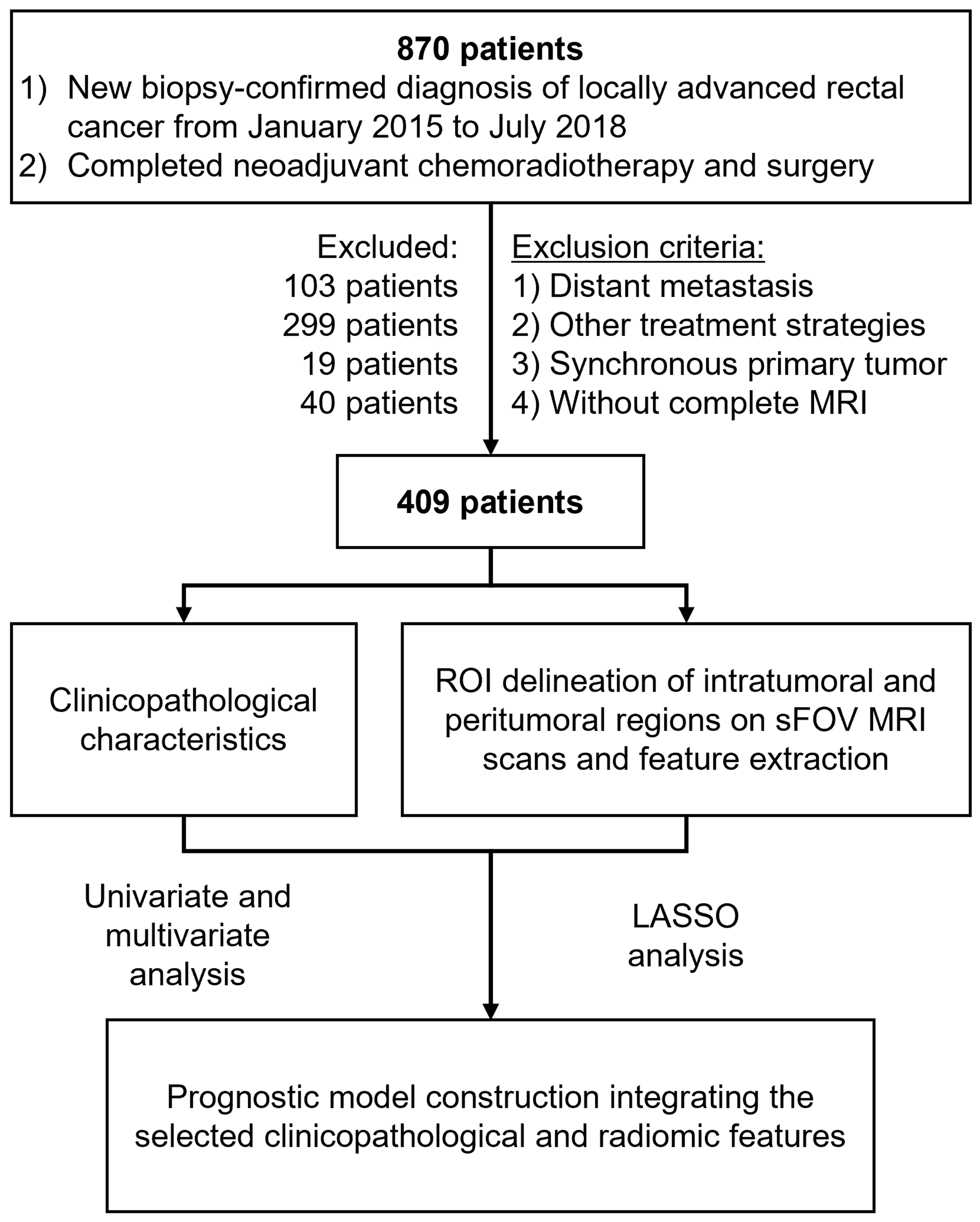
Abbreviations: MRI = magnetic resonance imaging; ROI = region of interest; sFOV = small field-of-view; LASSO = least absolute shrinkage and selection operator.
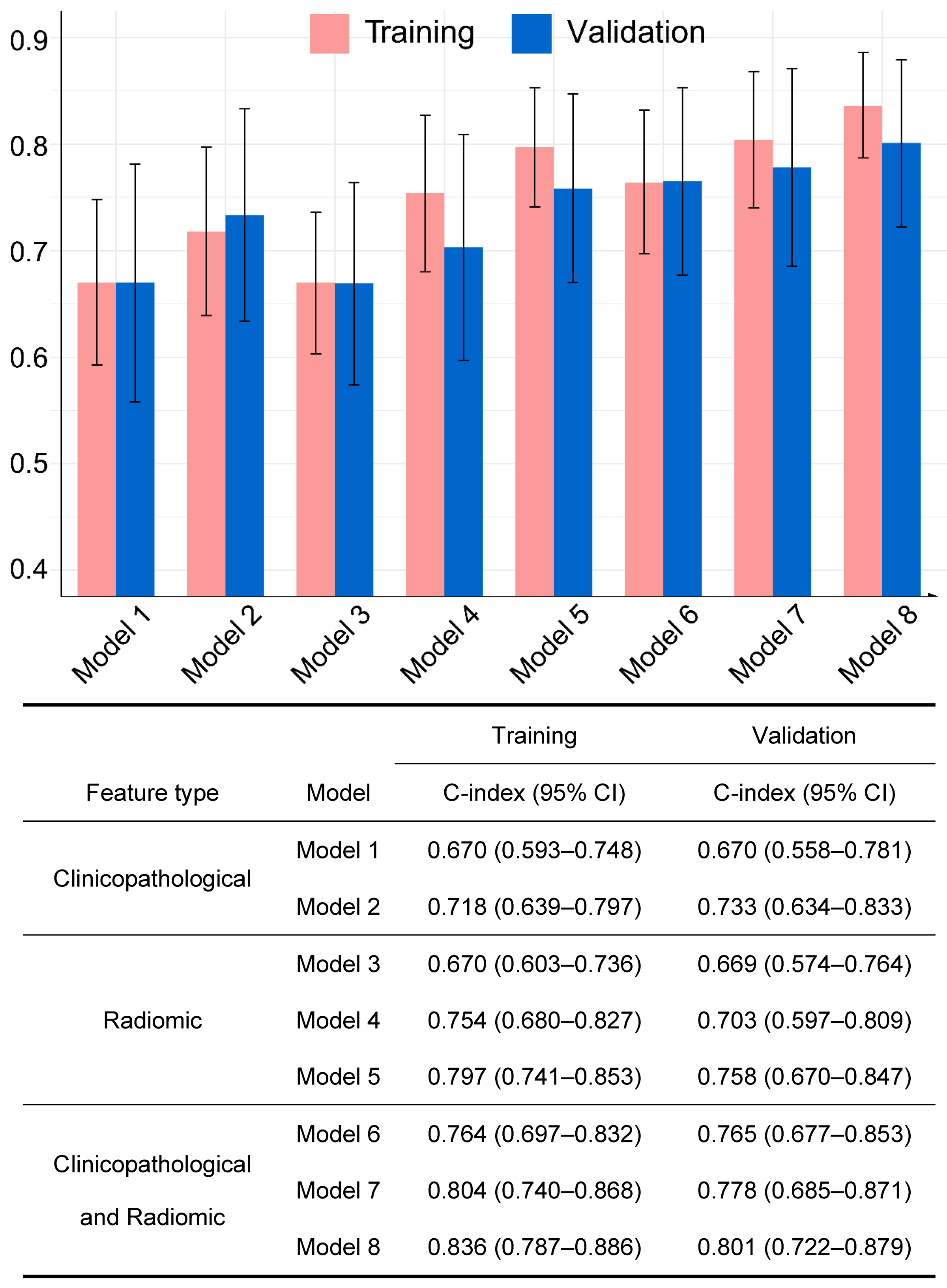
Figure 2. C-indexes of prognostic models for disease-free survival prediction for LARC
Prognostic models for LARC DFS prediction: Model 1: ypTN; Model 2: clinicopathologic features (BMI, ypN, TRG, MRF); Model 3: intratumoral radiomics; Model 4: peritumoral radiomics; Model 5: combined intratumoral-peritumoral radiomics; Model 6: clinicopathologic-intratumoral; Model 7: clinicopathologic-peritumoral; Model 8: clinicopathologic-intratumoral-peritumoral. C-index = Harrell's concordance index; CI = confidence interval.
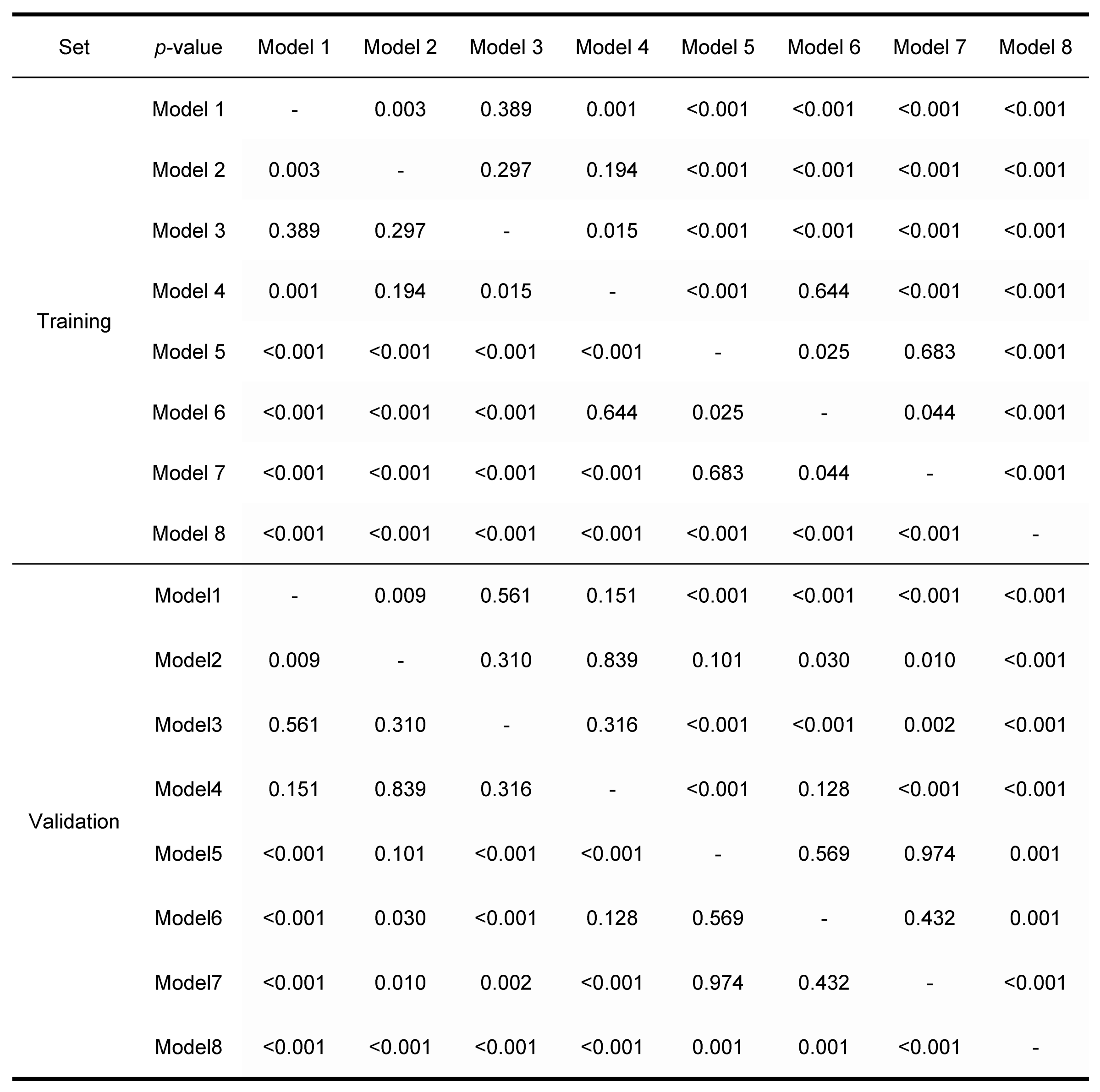
Figure 3. The p-values for C-index comparison between prognostic models for disease-free survival prediction in training and validation sets
Comparing C-indexes of prognostic models for DFS in LARC using DeLong test. Model 1: ypTN; Model 2: clinicopathologic features (BMI, ypN, TRG, MRF); Model 3: intratumoral radiomics; Model 4: peritumoral radiomics; Model 5: combined intratumoral-peritumoral radiomics; Model 6: clinicopathologic-intratumoral; Model 7: clinicopathologic-peritumoral; Model 8: clinicopathologic-intratumoral-peritumoral.
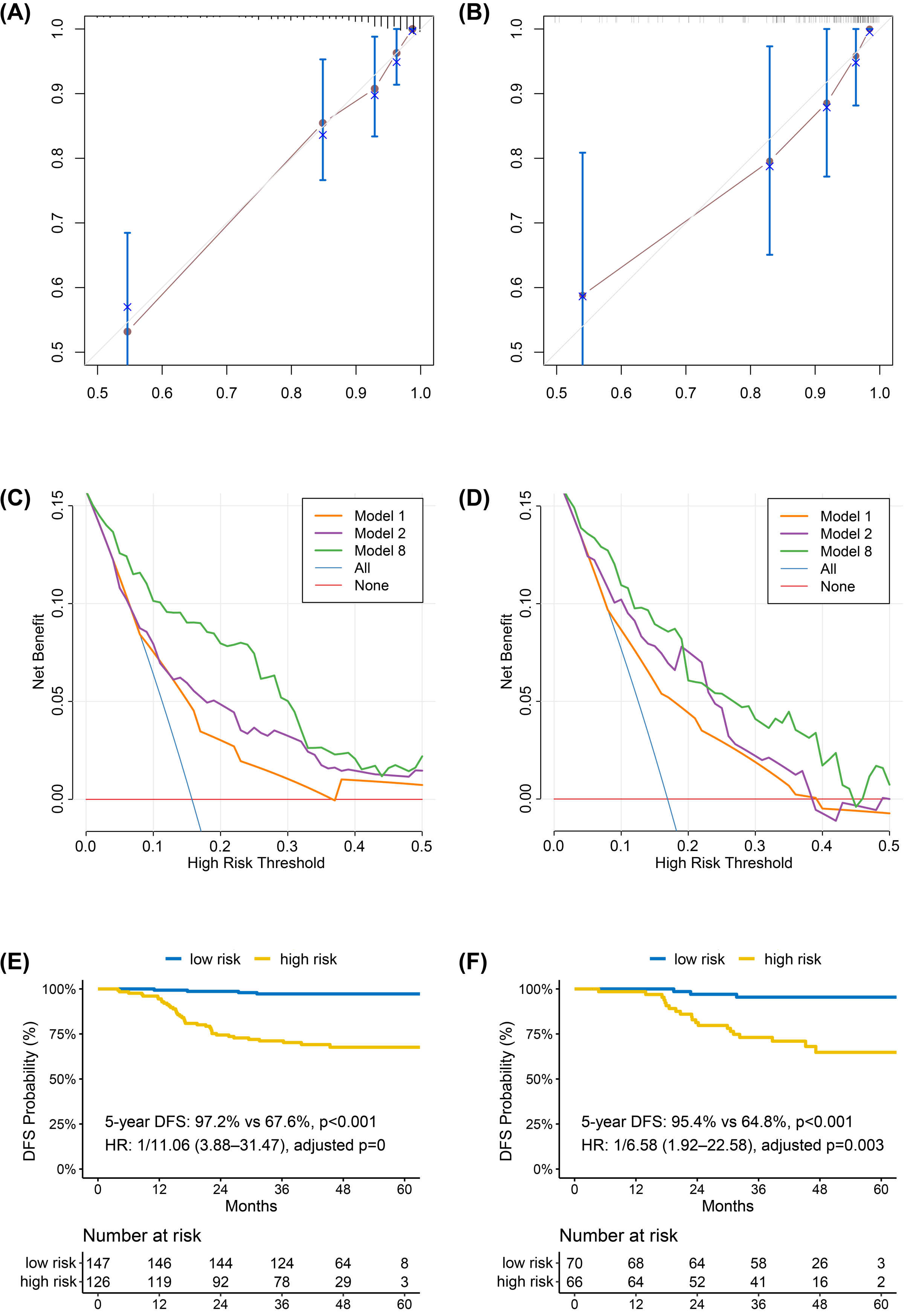
Figure 4. The calibration curves, decision curves, and Kaplan-Meier curves of the comprehensive prognostic model
The Model 8 is the comprehensive prognostic model integrating all available features. Calibration curves (A-B) demonstrate Model 8's accuracy for 3-year DFS in training and validation sets. Decision curves (C-D) for Models 1, 2, and 8 assess net benefits across threshold probabilities, with Model 8 showing superior performance. Kaplan-Meier curves (E-F) detail DFS risk stratification in both sets, as determined by Model 8.
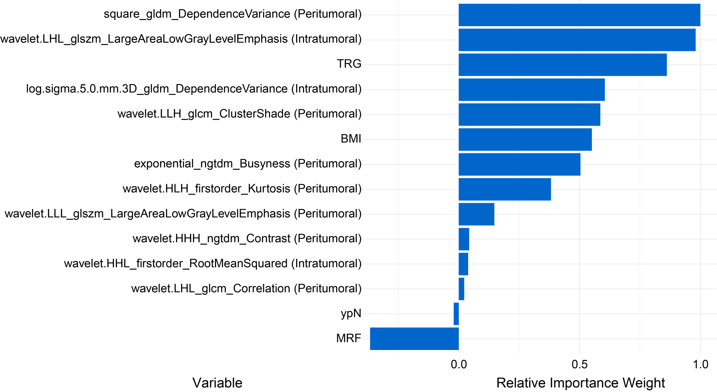
Permutation testing identifies the three most influencing features in the comprehensive model (Model 8 in this study) are, in order, a peritumoral feature, an intratumoral feature, and a clinicopathological feature. Abbreviations: TRG = tumor regression grade; BMI = body mass index; MRF = mesorectal fascia.