3636
Multi-Region MRI-Based Radiomics Predictive Model for METTL3 Expression in Hepatocellular Carcinoma1Hainan General Hospital (Hainan Affiliated Hospital of Hainan Medical University), Haikou, China, 2Philips Healthcare, Guangzhou, China
Synopsis
Keywords: Hepatobiliary, Radiomics
Motivation: To the best of our knowledge, our study is the first to non-invasively assess methyltransferase-like 3 (METTL3) expression in hepatocellular carcinoma (HCC) using multi-sequence MRI-based radiomics.
Goal(s): To develop a multi-region radiomics-based predictive model using multi-sequence MRI to assess METTL3 expression in HCC.
Approach: Three Models (Tumor, Tumor-Expand5, Tumor-Expand10) were constructed and evaluated.
Results: The Tumor-Expand5 model showed the highest efficacy with an AUC of 0.71 in the test set, outperforming both the Tumor and Tumor-Expand10 models. Multi-sequence MRI-based radiomics models hold the potential for preoperatively assessing METTL3 expression in HCC, aiding clinical decision-making.
Impact: This study introduces a novel multi-region radiomics-based model for predicting METTL3 expression in HCC using multi-sequence MRI. The results demonstrate the potential of radiomics, with an emphasis on the Tumor-Expand5 model, highlighting its promise for enhancing clinical decision-making in HCC.
Introduction
N6-methyladenosine (m6A) RNA methylation is the most prevalent modification of messenger RNAs (mRNAs) and is catalyzed by a multicomponent methyltransferase complex (MTC) [1,2]. methyltransferase-like 3 (METTL3) is one of the core components of MTC, which is localized in nuclear speckles and catalyzes the covalent transfer of a methyl group to adenine in a heterodimer form [3]. The unusual m6A modification resulting from differentially expressed METTL13, plays a critical role in the malignant progression of various cancers, such as bladder cancer, gastric cancer, and hepatocellular carcinoma (HCC) [4-6].Radiomics is a newly emerging form of medical image analysis that enables the extraction of high-throughput imaging features for quantitative analysis, significantly advancing precision medicine [7]. Radiomics-based models have been shown to accurately predict cancer diagnosis, therapeutic efficacy, and prognosis for clinical decision-making [8, 9].This study aims to develop a multi-region radiomics-based predictive model using multi-sequence MRI to assess METTL3 expression in hepatocellular carcinoma (HCC).
Methods
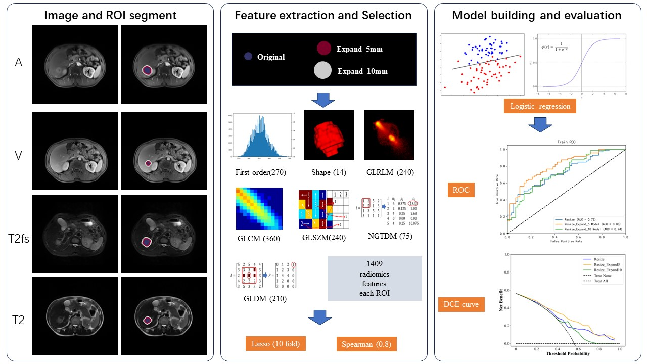
A total of 163 HCC patients, including 85 high METTL3 expression cases, diagnosed between January 2017 and August 2022, were enrolled, and randomly divided into training (n = 130, 80%) and testing (n = 33, 20%) sets. Four MRI sequences (T2-weighted imaging, T2WI with fat suppression, arterial phase, and venous phase) were employed. Tumor volumes of interest (VOIs) were manually delineated, with additional automatic 5mm and 10mm VOI expansions. A total of 1409 radiomic features were extracted from each VOI. Feature selection was performed using the least absolute shrinkage and selection operator (LASSO) and Spearman correlation analysis. Subsequently, three logistic regression models (Tumor, Tumor-Expand5, and Tumor-Expand10) were constructed based on radiomic features (see Figure 1). Model performance was assessed using receiver operating characteristic (ROC) analysis, net reclassification improvement (NRI), and integrated discrimination improvement (IDI). Furthermore, decision curve analysis (DCA) was utilized to evaluate the clinical value of these models.Results
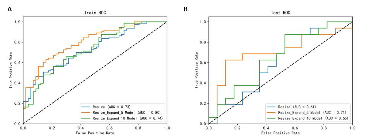
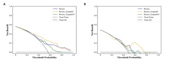
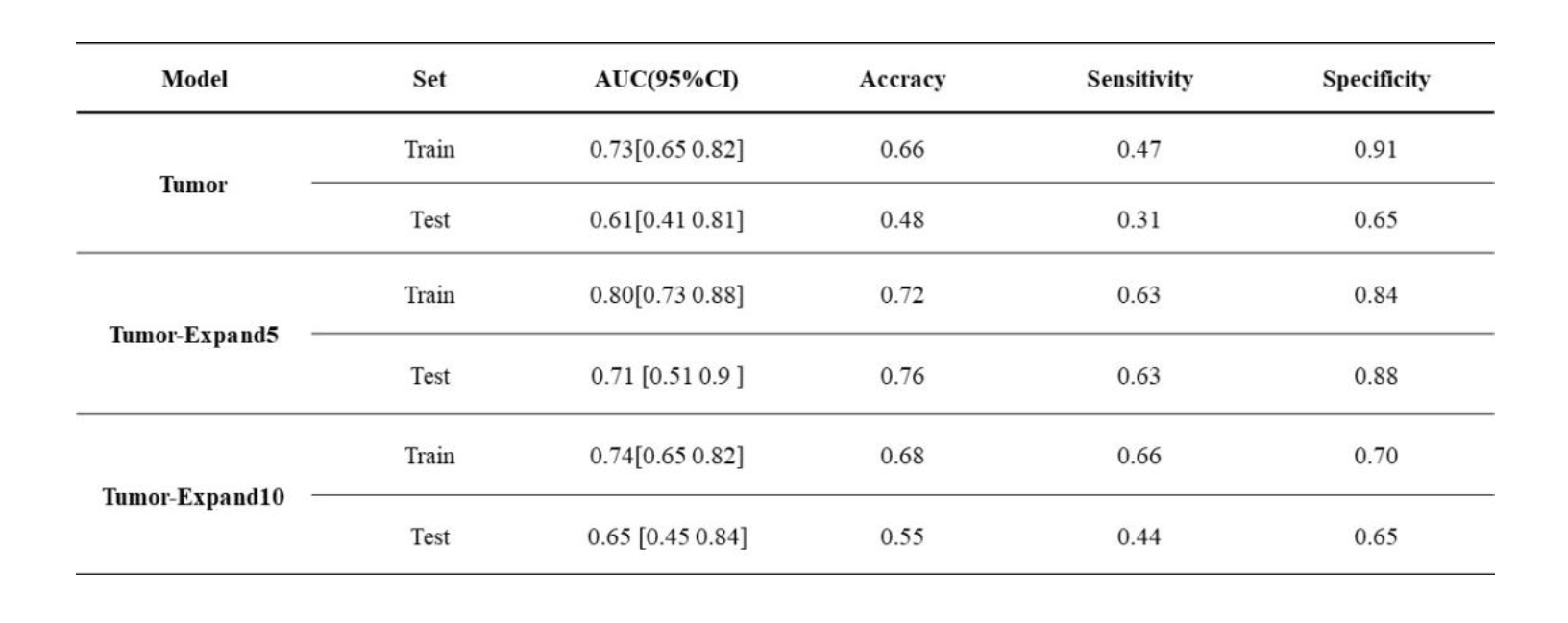
The study included 141 male and 22 female patients, with ages ranging from 26 to 84 years (mean age ± standard deviation: 51 ± 13 years). In the training set, the area under the curve (AUC) and 95% confidence intervals for the Tumor, Tumor-Expand5, and Tumor-Expand10 models were 0.73 (0.65-0.82), 0.80 (0.73-0.88), and 0.74 (0.65-0.82), respectively. In the test set, these models achieved AUCs of 0.61 (0.51-0.81), 0.71 (0.51-0.90), and 0.65 (0.45-0.84), respectively (Figure 2 and Table 1). Notably, the Tumor-Expand5 model outperformed both the Tumor and Tumor-Expand10 models, exhibiting an IDI of 0.11 and 0.14, as well as an NRI of 0.09 and 0.11, respectively. The DCA curve showed the advantages of the Tumor-Expand5 model over the Tumor and Tumor-Expand models in most threshold probability (Figure 3).Discussion
Radiomics has been widely applied at various stages of tumor diagnosis and treatment across different anatomical sites [8,9]. However, prior to this study, no research has explored the use of radiomics to predict METTL3 expression. Additionally, it’s important to consider the liver tumor microenvironment and its interactions with the tumor itself. This study is based on radiomic features from both the liver tumors and their surrounding tissues to predict METTL3 expression and investigate the influence of different sizes of peritumoral tissues on the prediction of METTL3 expression.The findings indicate that different radiomic models achieved good predictive performance in the training set, with the Tumor-Expand5 model showing the highest efficacy, a result that was consistently confirmed in the test set. This underscores the utility of radiomics in predicting METTL3 expression in hepatocellular carcinoma, with the 5mm expansion radiomics delivering the best predictive performance based on the radiomic features from the primary lesion.
However, this study has some limitations. First, it is a single-center study with a relatively small sample size, and the model's efficacy would benefit from validation in larger datasets encompassing multiple centers. Second, this study exclusively relies on radiomic features, and combining deep learning methods to further explore rich high-order features in images is expected to improve predictive performance.
Conclusion
The radiomics-based predictive model, utilizing features from both the tumor and peritumoral regions in multi-sequence MRI, holds promise as a preoperative tool for assessing METTL3 expression in HCC patients, with the prospect of augmenting clinical decision-making processes.Acknowledgements
No acknowledgement found.References
[1] Desrosiers R, Friderici K, Rottman F. Identification of methylated nucleosides in messenger RNA from Novikoff hepatoma cells. Proc Natl Acad Sci U S A. 1974;71(10):3971-3975. doi:10.1073/pnas.71.10.3971
[2] Liu J, Yue Y, Han D, et al. A METTL3-METTL14 complex mediates mammalian nuclear RNA N6-adenosine methylation. Nat Chem Biol. 2014;10(2):93-95. doi:10.1038/nchembio.1432
[3] Wang P, Doxtader KA, Nam Y. Structural Basis for Cooperative Function of Mettl3 and Mettl14 Methyltransferases. Mol Cell. 2016;63(2):306-317. doi:10.1016/j.molcel.2016.05.041
[4] Han J, Wang JZ, Yang X, et al. METTL3 promote tumor proliferation of bladder cancer by accelerating pri-miR221/222 maturation in m6A-dependent manner. Mol Cancer. 2019;18(1):110. Published 2019 Jun 22. doi:10.1186/s12943-019-1036-9
[5] Wang Q, Chen C, Ding Q, et al. METTL3-mediated m6A modification of HDGF mRNA promotes gastric cancer progression and has prognostic significance. Gut. 2020;69(7):1193-1205. doi:10.1136/gutjnl-2019-319639
[6] Liu X, Qin J, Gao T, et al. Analysis of METTL3 and METTL14 in hepatocellular carcinoma. Aging (Albany NY). 2020;12(21):21638-21659. doi:10.18632/aging.103959
[7] Lambin P, Leijenaar RTH, Deist TM, et al. Radiomics: the bridge between medical imaging and personalized medicine. Nat Rev Clin Oncol. 2017;14(12):749-762. doi:10.1038/nrclinonc.2017.141
[8] Gillies RJ, Kinahan PE, Hricak H. Radiomics: Images Are More than Pictures, They Are Data. Radiology. 2016;278(2):563-577. doi:10.1148/radiol.2015151169
[9] Bera K, Braman N, Gupta A, Velcheti V, Madabhushi A. Predicting cancer outcomes with radiomics and artificial intelligence in radiology. Nat Rev Clin Oncol. 2022;19(2):132-146. doi:10.1038/s41571-021-00560-7
[10] Yoon JH, Kim H. CT Radiomics in Oncology: Insights into the Tumor Microenvironment of Hepatocellular Carcinoma. Radiology. 2023;307(1):e222988. doi:10.1148/radiol.222988
Figures