3635
FetalSurfer: Automated Fetal Cortical Surface Reconstruction1Department of Biomedical Engineering, Tsinghua University, Beijing, China, 2Tanwei College, Tsinghua University, Beijing, China
Synopsis
Keywords: Fetal, Data Processing
Motivation: The cerebral cortex of the fetus is undergoing intricate development. Abnormal cortical development may potentially alter brain function. However the methods for processing fetal MRI images, especially cortical reconstruction, are still far behind those used for adults.
Goal(s): To develop an accurate cortical surface reconstruction method and perform morphological calculations for fetal MRI.
Approach: Trustworthy AI segmentation and refined Freesurfer were used to process T2-weighted fetal brain image.
Results: The proposed mothod (FetalSurfer) allows for trustworthy reconstruction of the cortical surfaces of the fetal brain and calculation of indicators to measure the morphology of fetal development.
Impact: FetalSurfer implements a novel fetal cerebral cortex reconstruction method without manual refinement by professional doctors, filling the gap of fetal image processing methods. Calculated indicators as curvature, thickness and sulcal depth can be used to perform morphological analysis.
Introduction
Fetuses develop rapidly during the second-to-third trimesters of pregnancy1, 2. The morphological properties of cerebral cortex, such as the thickness and folding are important biomarkers for investigating the healthy and diseased fetal brains in neuroscientific and clinical applications. Unfortunately, tools for automated cortical surface reconstruction and cortical morphometry are lacking, in contrast to several such software packages for adults and newborns and adults, such as FreeSurfer9,10 and its infant version3. This is partially because high isotropic resolution images are challenging to acquire on continuously moving fetuses and the commonly used T1-weighted contrast for differentiating gray and white matter in adult brains is poor in fetal brains which are still undergoing myelination3.Recent efforts performed fetal cortical reconstruction and analysis using atlas-based tissue segmentation results, which might be sub-optimal. Wu et al.4 used ANTs software package for the co-registration. Xu et al.5 used the refined structural MRI processing pipeline for Developmental Human Connectome Project (herein referred to as dhcp-structure-pipeline) originally designed for infants to perform segmentation and cortical analysis.
To address this challenge, we developed a new pipeline entitled “FetalSurfer” (Fig. 1) for automatically and more accurately reconstructing fetal cortical surfaces. FetalSurfer leveraged accurate and efficient tissue segmentation based on deep-learning models to replace time-consuming registration-based segmentation. FetalSurfer pipeline consists of slice-to-volume motion correction and high-resolution image volume reconstruction (SVR), brain tissue segmentation, cortices parcellation, and cortical surface reconstruction by refining FreeSurfer’s procedures. Compared to dhcp-structural-pipeline, FetalSurfer demonstrated superior accuracy and robustness in segmentation and cortical reconstruction.
Method and Materials
Data Acquisition. Fourteen pregnant women (22-36 weeks gestation) with normal fetal brains were scanned with written informed consent forms and IRB approval on a 1.5-T MRI scanner (PHILIPS Achieva Nova Dual hp) with spine and 16-channel body coils to obtain 2D T2-weighted turbo spin echo images, along axial, coronal, and sagittal directions, with 4 mm slice thickness and 0.8×0.8 mm2 in-plane resolution.High-resolution reconstruction. NiftyMIC6 (https://github.com/gift-surg/NiftyMIC) was employed to perform slice-to-volume motion correction and combine stacks of thick-slice images acquired along three directions for super-resolving 0.8 mm isotropic high-resolution T2-weighted image volume for each subject.
AI and Atlas segmentation. The trustworthy AI segmentation framework7 was used to segment each fetal brain volume into eight structures: extra-axial CSF, cortical gray matter, white matter, intra-axial CSF, deep gray matter, cerebellum, brainstem and corpus callosum. This approach offers a practical system to enhance the backbone AI system, leveraging the Dempster-Shafer theory. nnUnet8 model was chosen as the primary AI segmentation model. Specifically, when the primary model encounters challenges or is unable to produce reliable results, the fallback system can intervene to provide more dependable outcomes.
FBA is the structural fetal brain atlases4 for Chinese population ranging from 23w to 35w. The brain region labels including those for cortices were propagated from the atlas to each individual brain using a non-linear co-registration tool NiftyReg (https://github.com/KCL-BMEIS/niftyreg).
Surface reconstruction. Surface reconstruction followed Freesurfer9, 10 (https://surfer.nmr.mgh.harvard.edu/) which was designed for adult T1-weighted data. Briefly, AI model was used to replace Freesurfer's segmentation process for reconstructing the gray-white surface, which was subsequently expanded to find the boundary with the greatest contrast between gray matter and CSF. Steps such as white matter filling, hemispherical smoothing, and surface inflation are as standard. Cortical properties including the surface area, convexity, curvature, regional volume, sulcal depth, and thickness were quantified.
Results
Fig. 2 show successful super-resolution reconstruction, AI segmentation, atlas-based parcellation for 14 fetuses aging between 22 to 36 weeks.Fig. 3 shows cortical surfaces reconstructed from each individual brain volume. The cortex becomes more folded as fetuses age. The central sulcus appears around 28 weeks.
Fig. 4 compares gray-white and pial surfaces reconstructed from an exemplar brain volumeusing FetalSurfer and the dhcp-structural-pipeline. The dhcp-structural-pipeline misclassified the germinal matrix as gray matter (Fig. 4i), presumably due to atlas-based segmentation, and produced erroneous cortical folding (Fig. 4ii, iii). FetalSurfer improved upon these results by leveraging more accurate AI segmentation. All results were visually inspected by expert radiologists.
Fig. 5 demonstrates the quantified gray-CSF surface curvature and sulcal depth values, as well as the cortical thickness.
Discussion and Conclusion
An automated, accurate and efficient fetal cortical reconstruction pipeline FetalSurfer is proposed, which leverages state-of-the-art AI based brain segmentation. This tool has a high potential to advance fetal brain morphometry, aiding in the early diagnosis of neurological and developmental brain disorders and investigating fetal development assessment. Future work will systematically validate FetalSurfer and quantitatively compare it with other methods.Acknowledgements
Funding was provided by the Tsinghua University Startup Fund.References
1. Quezada, S., Castillo-Melendez, M., Walker, D.W. & Tolcos, M. Development of the cerebral cortex and the effect of the intrauterine environment. J Physiol-London 596, 5665-5674 (2018).
2. Garcia, K.E., Kroenke, C.D. & Bayly, P.V. Mechanics of cortical folding: stress, growth and stability. Philos Trans R Soc Lond B Biol Sci 373 (2018).
3. Zollei, L., Iglesias, J.E., Ou, Y., Grant, P.E. & Fischl, B. Infant FreeSurfer: An automated segmentation and surface extraction pipeline for T1-weighted neuroimaging data of infants 0-2 years. Neuroimage 218, 116946 (2020).
4. Wu, J. et al. Age-specific structural fetal brain atlases construction and cortical development quantification for chinese population. Neuroimage 241, 118412 (2021).
5. Xu, X. et al. Spatiotemporal atlas of the fetal brain depicts cortical developmental
gradient in Chinese population. (2022).
6. Ebner, M. et al. An automated framework for localization, segmentation and super-resolution reconstruction of fetal brain MRI. Neuroimage 206 (2020).
7. A Dempster-Shafer approach to trustworthy AI with application to fetal brain MRI segmentation.
8. Isensee, F., Jaeger, P.F., Kohl, S.A.A., Petersen, J. & Maier-Hein, K.H. nnU-Net: a self-configuring method for deep learning-based biomedical image segmentation. Nat Methods 18, 203-211 (2021).
9. Collins, D.L., Neelin, P., Peters, T.M. & Evans, A.C. Automatic 3D intersubject registration of MR volumetric data in standardized Talairach space. J Comput Assist Tomogr 18, 192-205 (1994).
10. Fischl, B., Liu, A. & Dale, A.M. Automated manifold surgery: constructing geometrically accurate and topologically correct models of the human cerebral cortex. IEEE Trans Med Imaging 20, 70-80 (2001).
Figures
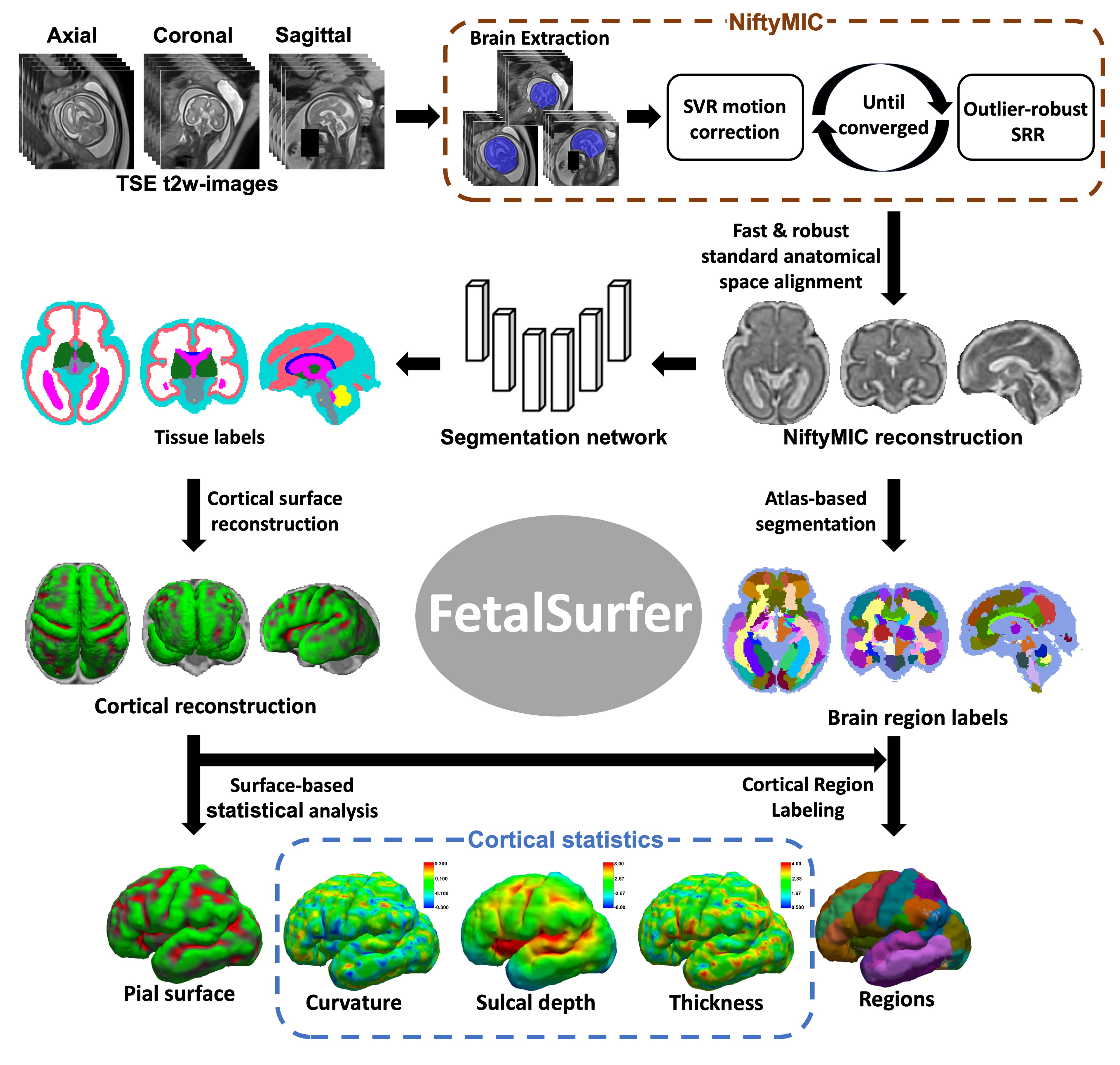
Figure 1. FetalSurfer Pipeline. High isotropic resolution image volumes are first reconstruction from stacks of thick-slice T2-weighted images using NiftyMIC. A trustworthy nnUnet model is then utilized for brain tissue segmentation. Cortical surfaces are reconstructed by refining Freesurfer procedures to extract cortical features as curvature, sulcal depth, thickness. Brain region labels are also propagated from the FBA atlas to each individual brain volume using non-linear co-registration for cortical parcellation.
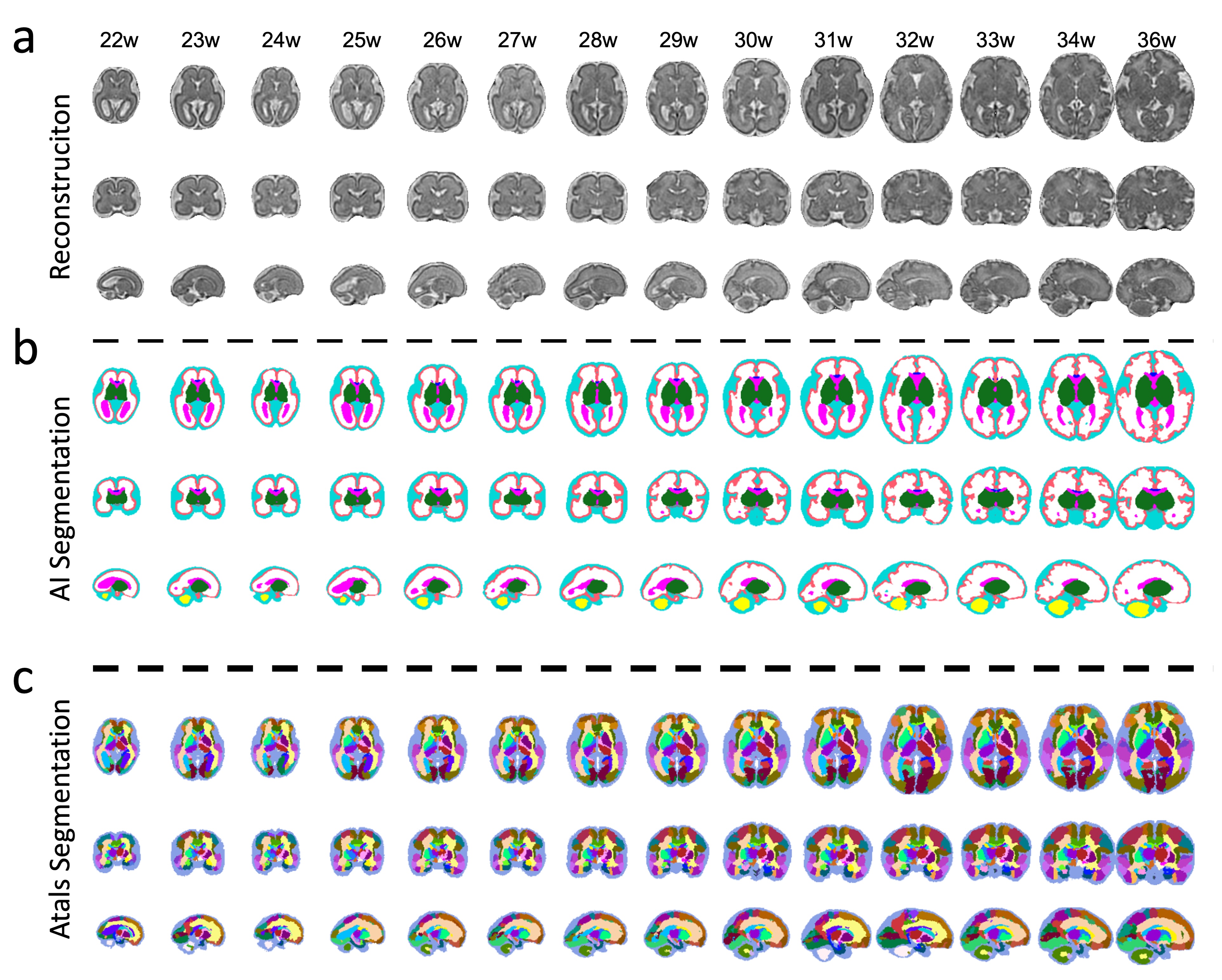
Figure 2. Reconstruction and segmentation. (a) High isotropic resolution T2-weighted image volume reconstruction results of NiftyMIC, (b) brain tissue segmentation results using the trustworthy nnUnet model, and (c) atlas based segmentation results using NiftyReg non-linear co-registration for fetuses aging between 22 to 36 weeks are displayed.
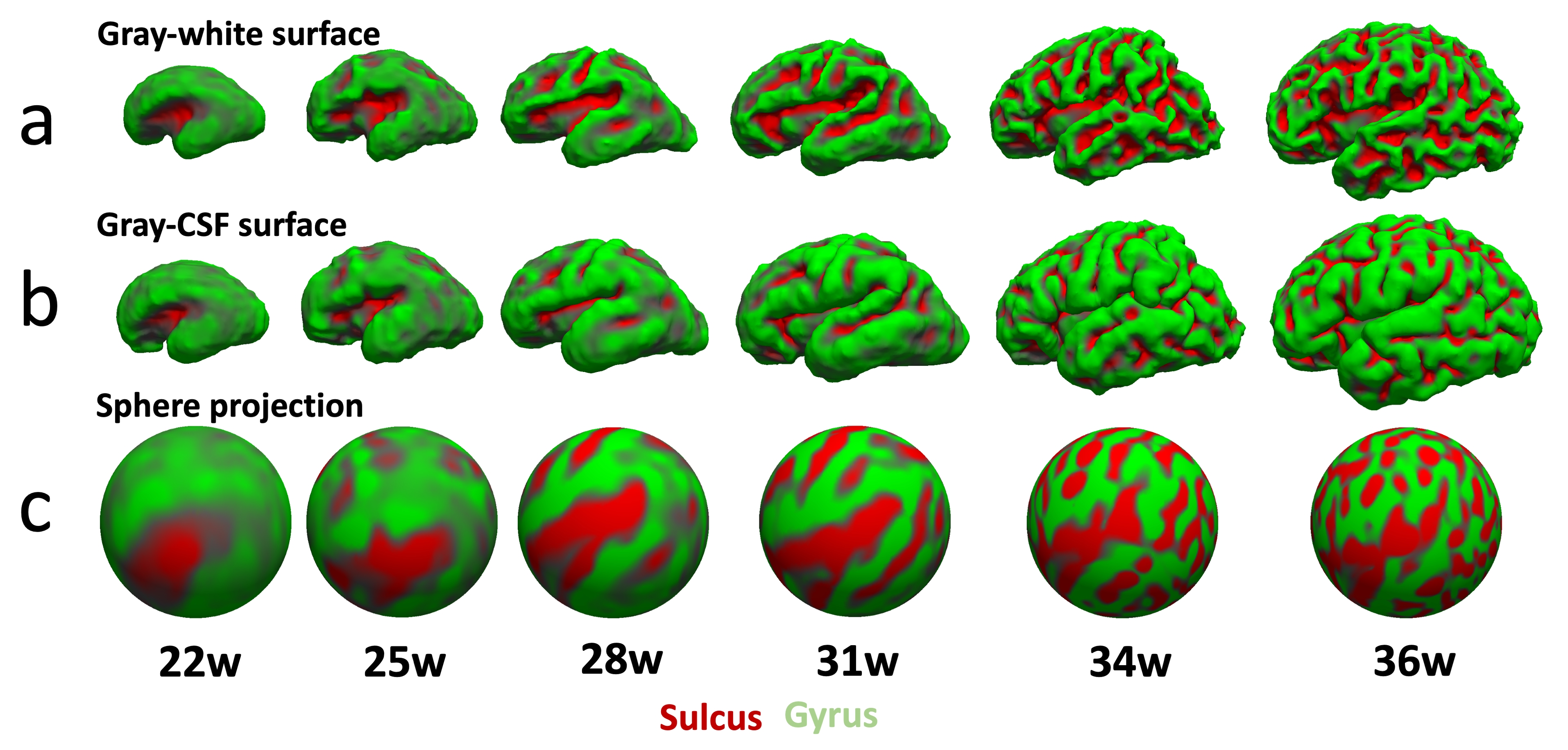
Figure 3. Cortical surfaces. (a) Gray–white interface surfaces, (b) gray–cerebrospinal fluid (CSF) interface surfaces, and (c) spherical projection reconstructed from high-resolution fetal brain image volumes for fetuses aging between 22 to 36 weeks are displayed.
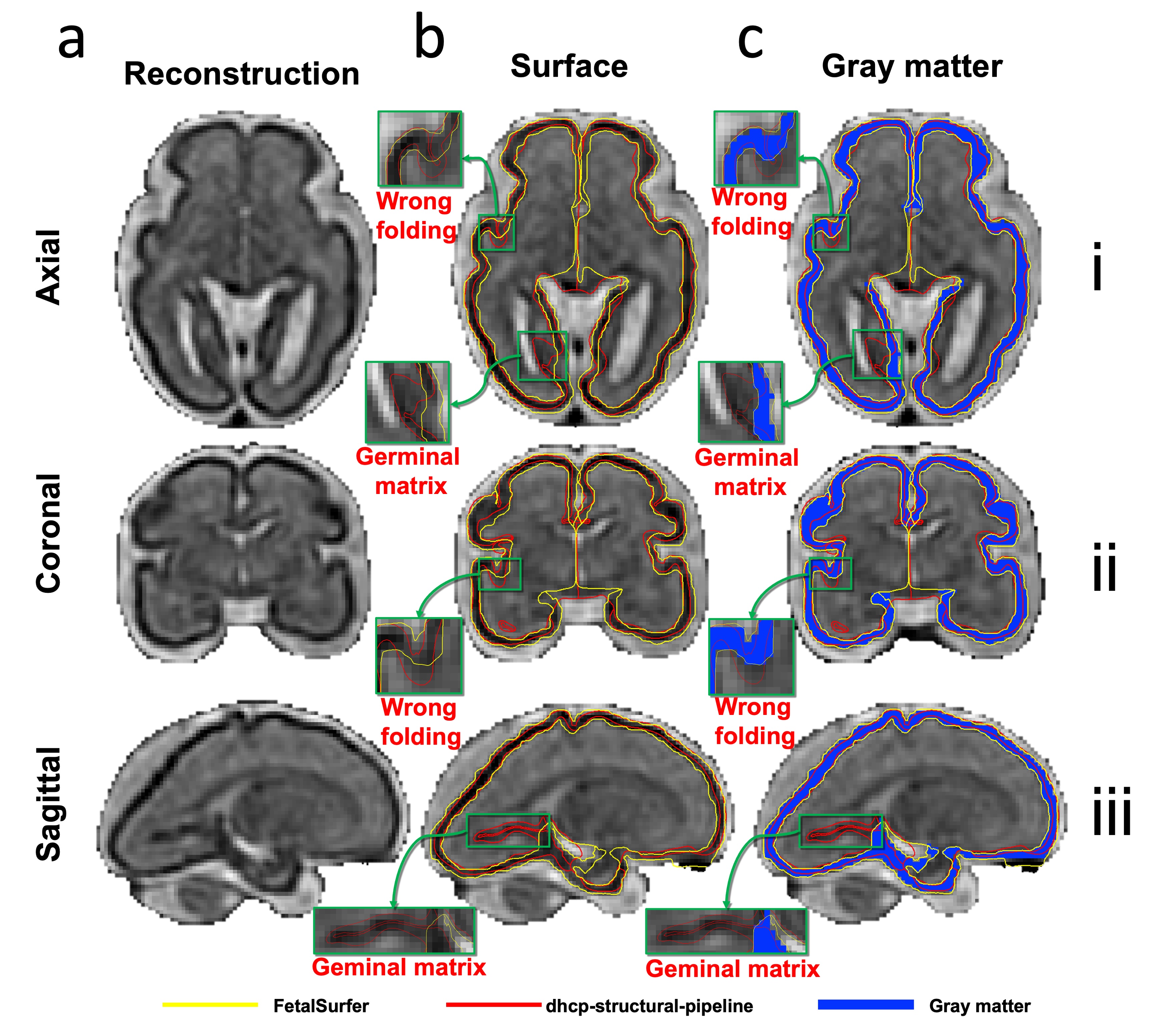
Figure 4. FetalSurfer vs. dhcp-structural-pipeline comparison. Gray–white and gray–cerebrospinal fluid (CSF) surfaces reconstructed from an exemplar fetal brain image volume using FetalSurfer and dhcp-structural-pipeline are displayed for comparison.
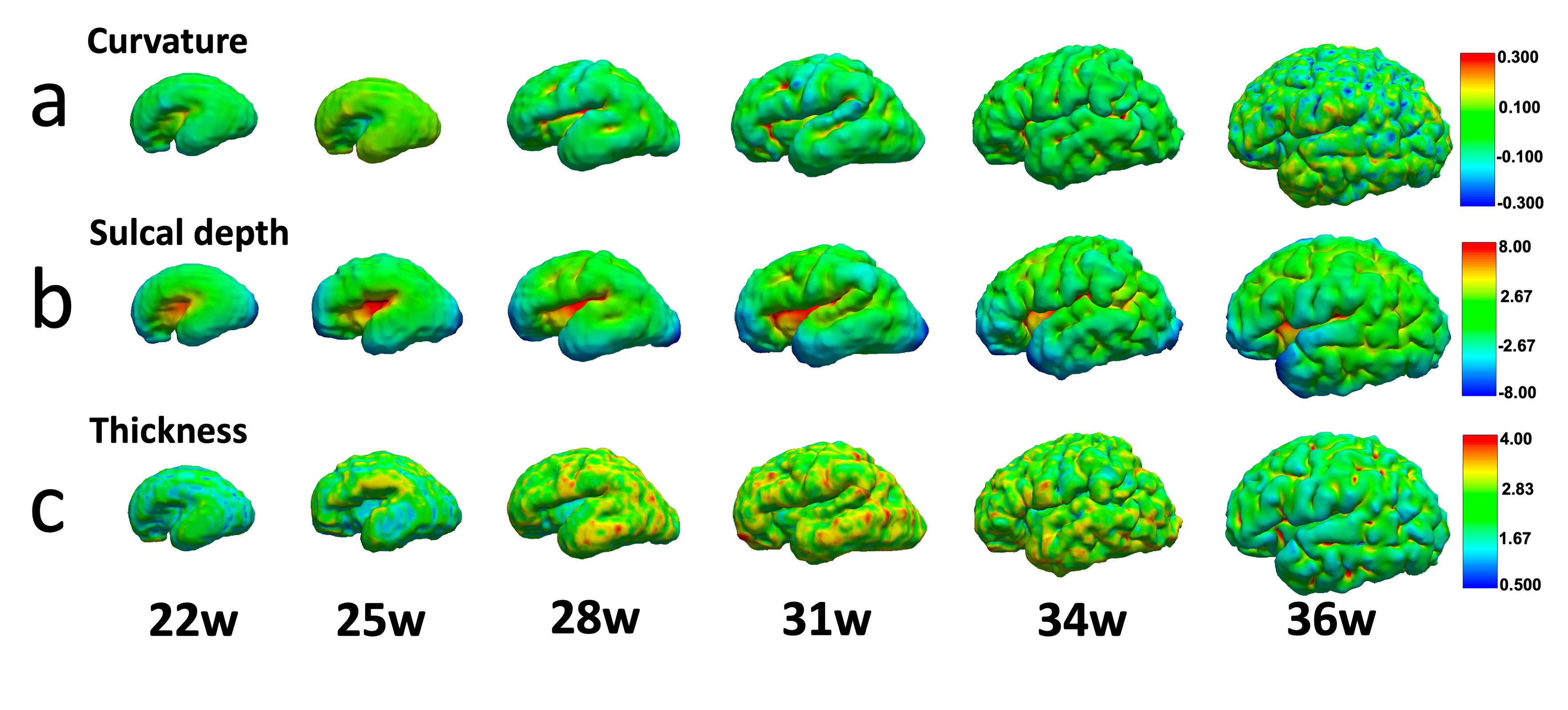
Figure 5. Quantification of morphological properties. Gray–cerebrospinal fluid (CSF) surface curvature and sulcal depth values, as well as the cortical thickness values of fetuses ranging between 22 to 36 weeks are displayed.