3634
MRI Radiomics Enhances Radiologists' Ability for Characterizing Intestinal Fibrosis in Patients with Crohn's Disease1Radiology, The First Affiliated Hospital, Sun Yat-Sen University, Guangzhou, China, 2Shenzhen University, Shenzhen, China, 3Pathology, The First Affiliated Hospital, Sun Yat-Sen University, Guangzhou, China, 4MR Research Collaboration Team, Siemens Healthineers, Guangzhou, China, 5Gastroenterology, The First Affiliated Hospital, Sun Yat-Sen University, Guangzhou, China, 6Gastrointestinal Surgery, The First Affiliated Hospital, Sun Yat-Sen University, Guangzhou, China
Synopsis
Keywords: Digestive, Radiomics, Crohn’s disease; Fibrosis; MR Enterography
Motivation: Fibrostenosis is a severe complication of Crohn's disease that significantly impacts patients' quality of life. Currently, there are no effective medical interventions for severe intestinal fibrosis
Approach: Radiomic models based on different MR sequence combinations were developed and validated in an independent test cohort.
Results: The radiologists’ interpretation aided by MRI-radiomics outperformed visual interpretation in characterizing intestinal fibrosis in Crohn's disease (AUC=0.86-0.93 vs. AUC=0.63-0.77).
Impact: The utilization of MRI-based radiomic models significantly enhances the diagnostic accuracy of radiologists in characterizing intestinal fibrosis.
Introduction
Fibrostenosis is a severe complication of Crohn's disease (CD) that significantly impacts patients' quality of life. Currently, there are no effective medical interventions for severe intestinal fibrosis, which necessitates endoscopic or surgical treatment1-4. Accurate detection and grading of intestinal fibrosis severity are crucial for selecting appropriate treatments and may facilitate the development of novel antifibrotic therapies.In recent years, radiomics analysis has increasingly been utilized in medical images to enhance the assessment of CD5-7. Radiomics extracts large amounts of high-dimensional features from images to uncover disease characteristics that may not be discernible through visual inspection or single parameter analysis8,9. In previous study10, a novel computed tomography enterography (CTE) radiomic model (RM) was developed and validated allowing for accurate characterisation of intestinal fibrosis in CD. However, because CTE has the potential risk of radiation, magnetic resonance enterography (MRE) is the preferred follow-up examination for patients with CD. Moreover, multi-sequence MRE performs better than CTE in the assessment of intestinal fibrosis11-13.
This study aims to develop and verify MRE-based RMs for the diagnosis of intestinal fibrosis, with the objective of improving radiologists' diagnostic accuracy and efficiency in characterizing this condition, as well as facilitating timely and appropriate management for patients with CD across various institutions.
Methods
This retrospective study included patients with refractory CD who underwent MR before surgery from November 2013 to September 2021. MRE was performed on two 3-T MR system (MAGNETOM Trio or Prisma; Siemens Healthcare, Erlangen, Germany) equipped with 12- (Trio) or 18-channel phased-array body coils (Prisma), including (1) T2-weighted imaging; (2) diffusion-weighted magnetic resonance imaging (DWI, b values of 800 s/mm2; the apparent diffusion coefficient map was reconstructed from the original DWI data); (3) T1-weighted imaging before and at 30s, 60s, 90s, 2 min, 3.5 min, and 7 min after an intravenous injection of 0.2 mL/kg gadopentetate dimeglumine (Gd-DTPA) at a rate of 2 mL/s (Trio) or 0.1 mL/kg gadobutrol at a rate of 1.5 mL/s (Prisma); and (4) magnetisation transfer imaging (MTI) was acquired by using two gradient-echo data sets with and without the application of an off-resonance pre-pulse (frequency offset, 1.2 kHz; duration, 9984 μsec; effective flip angle, 500°; bandwidth, 192 Hz).We have chosen T2WI, contrast-enhanced T1WI in venous phase, diffusion-weighted imaging (DWI), apparent diffusion coefficient (ADC), and MTI for radiomics analysis.RMs based on different MR sequence combinations (RM1: T2WI and enhanced-T1WI; RM2: T2WI, enhanced-T1WI, DWI, ADC); RM3: T2WI, enhanced-T1WI, DWI, ADC and MTI), were developed and validated in an independent test cohort. The RMs' diagnostic performance was compared to that of radiologists using identical sequences and a clinical model.
Results
The final population included 123 patients (81 men, 42 women; mean age: 30.26±7.98 years; training cohort, n=93; test cohort, n=30). The area under the receiver operating characteristic curve (AUC) of RM1, RM2, and RM3 was 0.86 (P=0.001), 0.88 (P=0.001), and 0.93 (P=0.02), respectively. The decision curve analysis confirmed a progressive improvement in diagnostic performance of three RMs with addition of more specific sequences. All RMs outperformed the corresponding radiologists’ interpretation (radiologist 1, AUC=0.65, P=0.56; radiologist 2, AUC=0.63, P=0.04; radiologist 3, AUC=0.77, P=0.002) and the clinical model compositing of C-reactive protein and erythrocyte sedimentation rate (AUC=0.60, P=0.13).Discussion
To address the lack of non-invasive and non-radiation tools for precisely detecting transmural fibrosis, we developed three multi-sequence MRE-based RMs, which showed satisfactory efficacy in distinguishing moderate-severe from none-mild intestinal fibrosis in CD. RM1, constructed using basic MRI sequences, already had moderate diagnostic accuracy. With more specific MRI sequences (DWI or MTI) added, the diagnostic performance of RM1, RM2, and RM3 improved gradually from moderate to excellent. Moreover, these three RMs ran steadily in an independent test cohort and in different subgroups, indicating their high reliability and stability. Additionally, all RMs outperformed the radiologist-performed visual interpretation and the clinical model in diagnosing bowel fibrosis.Conclusion
In conclusion, multi-sequence MRE-based RMs are a valuable tool for accurately characterizing intestinal fibrosis in CD and can significantly enhance radiologists' diagnostic ability. Our three RMs, constructed using different combinations of conventional, DWI and MTI sequences, meet diverse technical feasibility requirements for implementation in various institutions for patients with CD.Acknowledgements
This work was supported by the National Natural Science Foundation of China (82070680, 82270693, 81870451), Guangdong Basic and Applied Basic Research Foundation of China (2023B1515020070, 2020A1515010571), and Shenzhen-Hong Kong Institute of Brain Science-Shenzhen Fundamental Research Institutions of China (2021SHIBS0003).References
1. Rieder F, Fiocchi C. Mechanisms of tissue remodeling in inflammatory bowel disease. Dig Dis 2013;31(2):186-193.
2. Rieder F, de Bruyn JR, Pham BT et al. Results of the 4th scientific workshop of the ECCO (Group II): markers of intestinal fibrosis in inflammatory bowel disease. J Crohns Colitis 2014;8(10):1166-1178.
3. Latella G, Di Gregorio J, Flati V, Rieder F, Lawrance IC. Mechanisms of initiation and progression of intestinal fibrosis in IBD. Scand J Gastroenterol 2015;50(1):53-65.
4. Bouhnik Y, Carbonnel F, Laharie D, et al. Efficacy of adalimumab in patients with Crohn's disease and symptomatic small bowel stricture: a multicentre, prospective, observational cohort (CREOLE) study. Gut 2018;67(1):53-60.
5. Tabari A, Kilcoyne A, Jeck WR, Mino-Kenudson M, Gee MS. Texture Analysis of Magnetic Resonance Enterography Contrast Enhancement Can Detect Fibrosis in Crohn Disease Strictures. J Pediatr Gastroenterol Nutr 2019;69(5):533-538.
6. De Kock I, Bos S, Delrue L, et al. MRI texture analysis of T2-weighted images is preferred over magnetization transfer imaging for readily longitudinal quantification of gut fibrosis Eur Radiol 2023;33(9):5943-5952.
7. Makanyanga J, Ganeshan B, Rodriguez-Justo M, et al. MRI texture analysis (MRTA) of T2-weighted images in Crohn's disease may provide information on histological and MRI disease activity in patients undergoing ileal resection. Eur Radiol 2017;27(2):589-597.
8. Gillies RJ, Kinahan PE, Hricak H. Radiomics: images are more than pictures, they are data. Radiology 2016;278:563-577.
9.Lambin P, Leijenaar R, Deist TM, et al. Radiomics: the bridge between medical imaging and personalized medicine. Nat Rev Clin Oncol 2017;14:749-762.
10. Li X, Liang D, Meng J et al. Development and Validation of a Novel Computed-Tomography Enterography Radiomic Approach for Characterization of Intestinal Fibrosis in Crohn's Disease. Gastroenterology 2021;160(7):2303-2316.
11. Bruining DH, Zimmermann EM, Loftus EJ et al. Consensus Recommendations for Evaluation, Interpretation, and Utilization of Computed Tomography and Magnetic Resonance Enterography in Patients with Small Bowel Crohn's Disease. Gastroenterology 2018;154(4):1172-1194.
12. Sturm A, Maaser C, Calabrese E et al. ECCO-ESGAR Guideline for Diagnostic Assessment in IBD Part 2: IBD scores and general principles and technical aspects. J Crohns Colitis 2019;13(3):273-284.
13. Li XH, Mao R, Huang SY et al. Characterization of Degree of Intestinal Fibrosis in Patients with Crohn Disease by Using Magnetization Transfer MR Imaging. Radiology 2018;287(2):494-503
Figures
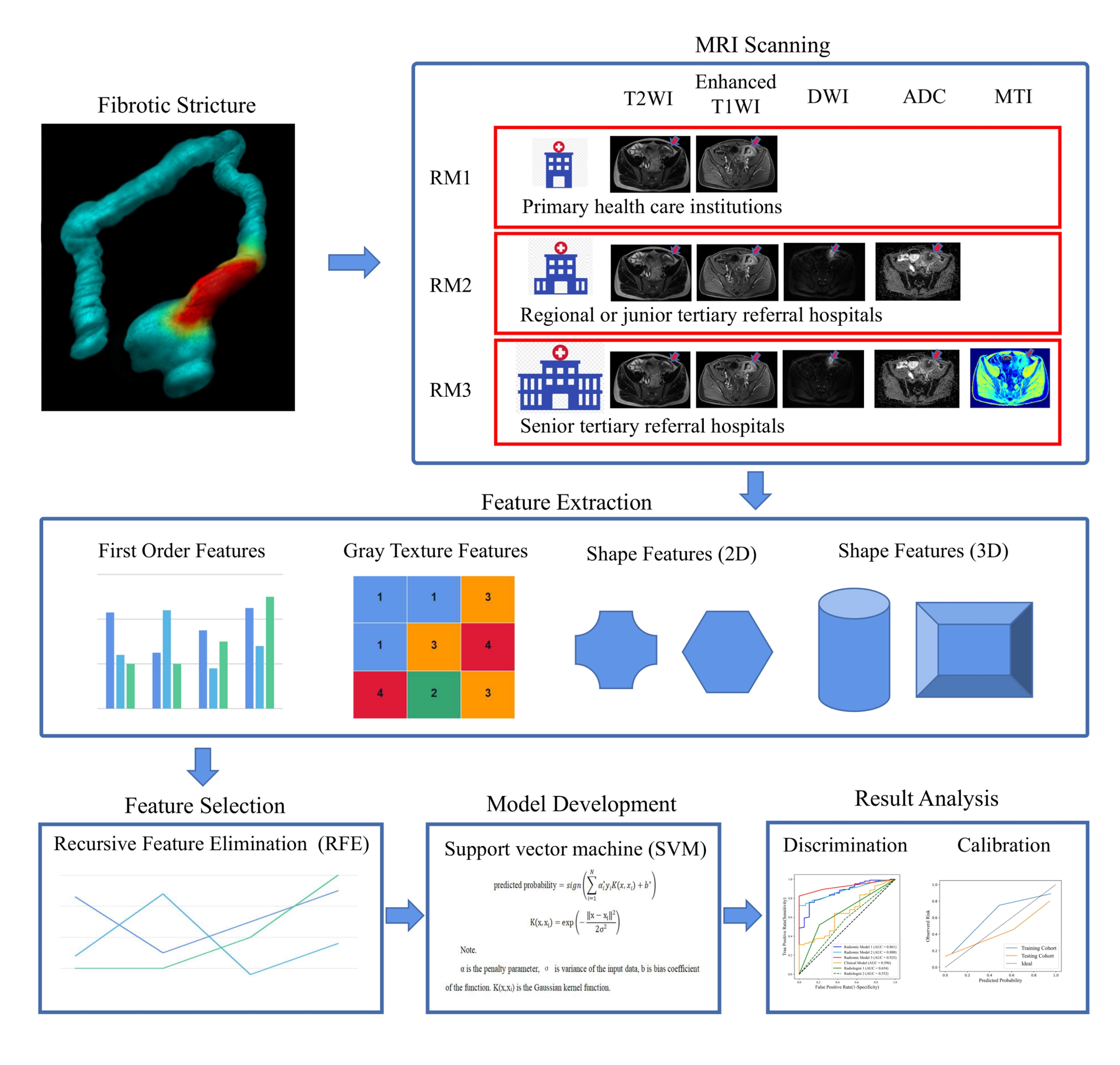
Figure 1. The radiomics analysis workflow: RM1, RM2, and RM3 established by different sequence combinations aid in the evaluation of Crohn’s disease fibrosis in health-care institutions at different levels.
RM, radiomic model
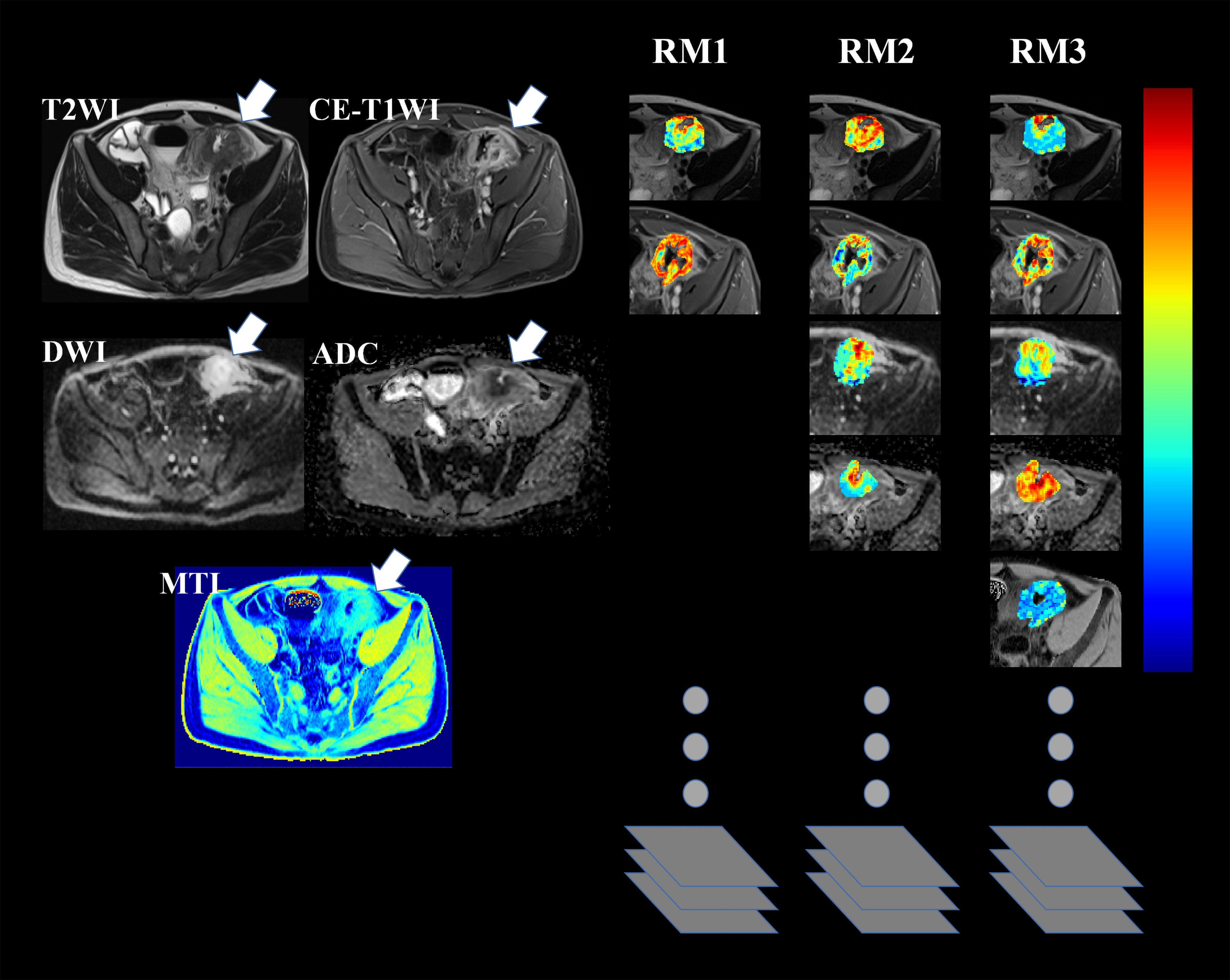
Figure 2. A 41-year-old man with Crohn’s disease with moderate-severe fibrotic stricture in descending colon. Radiomics features extracted from identical combinations of T2WI, contrast-enhanced T1WI, DWI, ADC, and MTI are used to constructed RM1-RM3; To generate feature maps, the features are calculated for each voxel in the segmented VOIs with a kernel radius of 3, and assigned to the center pixel.
ADC, apparent diffusion coefficient; DWI, diffusion-weighted imaging; MTI, magnetisation transfer MRI; RM, radiomic model
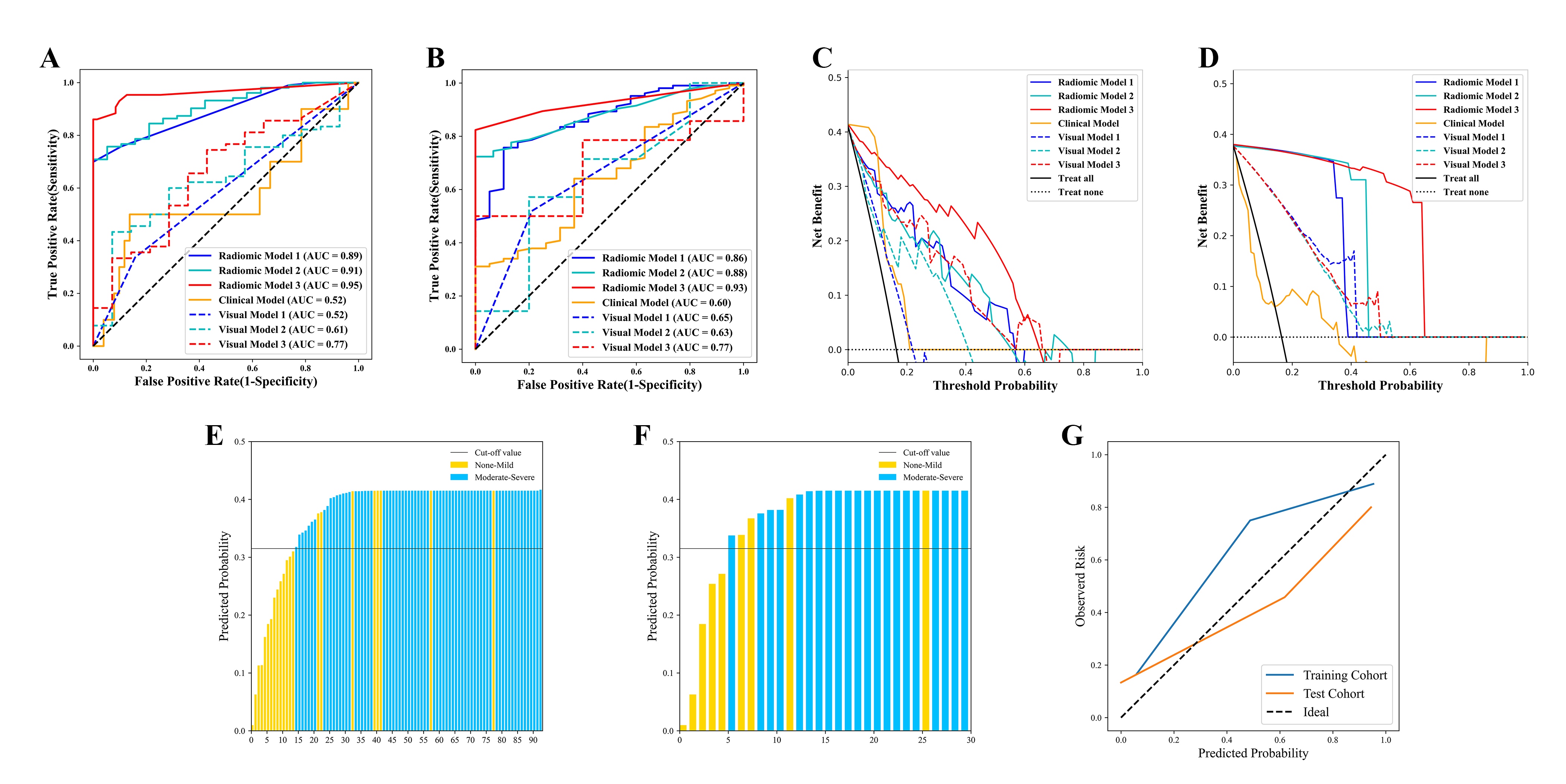
Figure 3. The diagnostic performance of the RMs is better than that of both the radiologists’ visual interpretations and clinical model (all P < 0.001) in the training (A) and test (B) cohorts. RMs have higher net benefit than the two radiologists’ visual interpretation and clinical model in the training (C) and test (D) cohorts. For the RM3, plots show predicted probabilities with a cut-off value of 0.360 (indicated by the black solid line) in the training (E) and test (F) cohorts, and the calibration curves (G) in both cohorts.