3633
AutoML Radiomics-Based Classification of Patients with Renal Cell Carcinoma Using Non-Contrast Enhanced Magnetic Resonance Imaging1Department of Radiology, Taichung Veterans General Hospital, Taiwan, Taichung, Taiwan, Taiwan, 2Ph.D. Program of Electrical and Communications Engineering, Feng Chia University, Taichung, Taiwan, taichung, Taiwan, 3Department of Automatic Control Engineering, Feng Chia University, Taichung, Taiwan, taichung, Taiwan
Synopsis
Keywords: Kidney, Radiomics
Motivation: Kidney cancer is often diagnosed as either clear cell renal carcinoma (ccRCC) or non-clear cell renal carcinoma (non-ccRCC) to determine treatment recommendations. Additionally, many patients with kidney cancer cannot receive contrast medium due to renal function disorders.
Goal(s): for the distinction of ccRCC from other types of RCC without contrast medium administration
Approach: A model using automated machine learning (AutoML) based on radiomics features
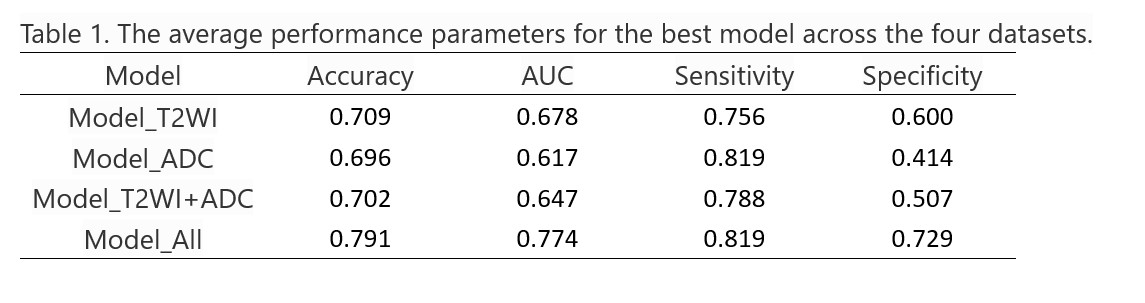
Results: Our results indicate that the best model from the AutoML process demonstrated a mean sensitivity of 0.819 and a mean specificity of 0.729 in distinguishing between ccRCC and non-ccRCC.
Impact: To demonstrated that the TPOP-radiomics-based classification model can effectively discriminate between ccRCC and non-ccRCC using MRI without the need for contrast medium.
A better understanding of the biology of renal cell carcinoma (RCC) has led to a significant shift in the treatment approach for this disease. The two most common subtypes of malignant RCC are clear cell RCC (ccRCC) and non-clear cell renal carcinoma(non-ccRCC) [1]. None-ccRCC subtypes have shown relatively better survival rates compared to ccRCC [2]. The discovery of the VHL gene in 1993 [3] marked a pivotal moment in the quest to develop effective therapies for ccRCC. Therefore, the ability to distinguish between ccRCC and non-ccRCC is crucial for the treatment of RCC. Multiple MRI parameters can provide insights into the intratumoral contents, including T2-weighted (T2WI), T1-weighted (T1WI), chemical shift imaging, and diffusion-weighted imaging (DWI) with apparent diffusion coefficient (ADC) values. These imaging features can be valuable for distinguishing RCC from benign lesions and for classifying RCC subtypes and histological grades [4]. Radiomics and machine learning (ML) have opened new horizons for accurate detection, diagnosis, prediction, and prognosis in various diseases. However, there is a growing need for machine learning solutions that can automatically handle feature selection, algorithm choice, and model optimization. To address this demand, automated machine learning (AutoML) researchers have developed systems to automate the design and optimization of ML pipelines. One such AutoML algorithm is the Tree-based Pipeline Optimization Tool (TPOT), which automatically designs and optimizes ML pipelines for specific problem domains, without any need for human intervention [5]. This study aimed to develop a TPOP-radiomics classification model for discriminating between ccRCC and non-ccRCC using multiple MR images.
Methods
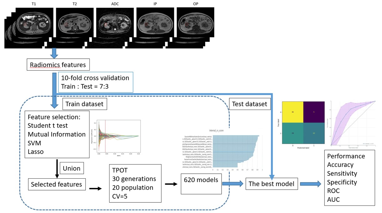
Image Data: We enrolled 53 patients with ccRCC and 23 patients with non-ccRCC, as identified by pathology. We collected five different MRI sequences from each patient, including T1WI, T2WI, in-phase (IP), out of phase (OP), and ADC. Two experienced research assistants selected the region of interest (ROI) by examining each level of the kidney tumor and then reviewed with slice-by-slice by the senior radiologist. Figure 1 illustrates the TPOP-radiomics model workflow. Feature Extraction: Pyradiomics (version 1.3.0) was used to automatically extract radiomic features. A total of 3102 radiomic features were extracted from the original image and filtered image for each of the T1WI, T2WI, IP, OP, and ADC. Feature selection: First, Radiomics features with significant differences were retained using Student's t-test (p < 0.05). Second, the features were selected by combining the results of four optimal feature screening methods, including ANOVA, L1 regularized LASSO regression analysis model, Mutual Information, and linear Support Vector Machines (SVM). Third, the final selected features were normalized for feature scaling. We employed TPOT to search for the best model using its default configuration, which includes all data operators and machine learning classification models. The TPOT search was set to run for 30 generations and 20 population size sets with five-fold cross-validation. 620 TPOT experiments were conducted for each dataset. Additionally, we performed 10 random repetitions for the dataset in which the training and test data were split at a ratio of 70% for training and 30% for testing. Four datasets were used to compare the performance of AutoML-radiomics model in discriminating between ccRCC and non-ccRCC. These datasets were generated based on MRI sequences and included the following: a model based on sole T2WI (Model_T2WI), a model based on sole ADC (Model_ADC), a model based on both T2WI and ADC (Model_T2WI+ADC), and a model based on all five MRI sequences (Model_All). Performance evaluation: In comparing all the models, various performance evaluations were employed, including accuracy, sensitivity, specificity, and the area under the curve (AUC).
Results
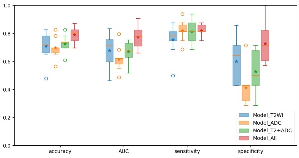
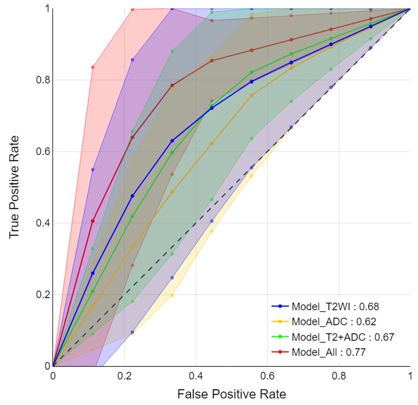
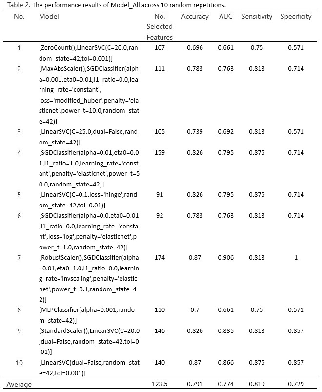
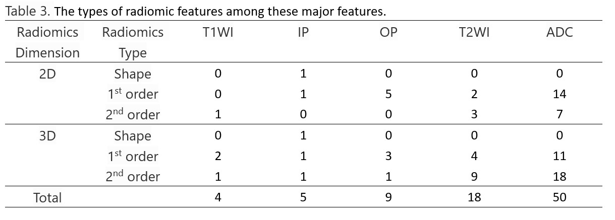
Table 1 displays the average performance parameters for the best model across the four datasets. Figure 2 presents box plots for the accuracy, sensitivity, specificity, and AUC of the four models across 10 random repetitions. Figure 3 displays the receiver operating characteristic (ROC) curves for these models. Among all the models studied, Model_All demonstrated the best performance with mean sensitivity of 0.819 and mean specificity of 0.729 in distinguishing between ccRCC and non-ccRCC. Table 2 displays the performance results of Model_All across 10 random repetitions. Major features of Model_All, which are effective for discriminating between ccRCC and non-ccRCC, were identified as features selected more than 3 times in the 10 random repetitions, are presented in Table 3.
Conclusion
In conclusion, our study demonstrated that the TPOP-radiomics-based classification model can effectively discriminate between ccRCC and non-ccRCC using MRI without the need for contrast medium.
Acknowledgements
Supported by Taichung Veterans General Hospital (TCVGH-FCU1128205 Joint Research Program and TCVGH-1125504A)References
1. Sun MR, Ngo L, Genega EM, Atkins MB, Finn ME, Rofsky NM, Pedrosa I. Renal cell carcinoma: dynamic contrast-enhanced MR imaging for differentiation of tumor subtypes--correlation with pathologic findings. Radiology. 2009 Mar;250(3):793-802. doi: 10.1148/radiol.2503080995. PMID: 19244046.
2. Cheville JC, Lohse CM, Zincke H, Weaver AL, Blute ML. Comparisons of outcome and prognostic features among histologic subtypes of renal cell carcinoma. Am J Surg Pathol. 2003 May;27(5):612-24. doi: 10.1097/00000478-200305000-00005. PMID: 12717246.
3. Latif F, Tory K, Gnarra J, Yao M, Duh FM, Orcutt ML, Stackhouse T, Kuzmin I, Modi W, Geil L, et al. Identification of the von Hippel-Lindau disease tumor suppressor gene. Science. 1993 May 28;260(5112):1317-20. doi: 10.1126/science.8493574. PMID: 8493574.
4. Lopes Vendrami C, Parada Villavicencio C, DeJulio TJ, Chatterjee A, Casalino DD, Horowitz JM, Oberlin DT, Yang GY, Nikolaidis P, Miller FH. Differentiation of Solid Renal Tumors with Multiparametric MR Imaging. Radiographics. 2017 Nov-Dec;37(7):2026-2042. doi: 10.1148/rg.2017170039. PMID: 29131770.
5. Olson, R.S., Urbanowicz, R.J., Andrews, P.C., Lavender, N.A., Kidd, L.C., Moore, J.H. (2016). Automating Biomedical Data Science Through Tree-Based Pipeline Optimization. In: Squillero, G., Burelli, P. (eds) Applications of Evolutionary Computation. EvoApplications 2016. Lecture Notes in Computer Science(), vol 9597. Springer, Cham. https://doi.org/10.1007/978-3-319-31204-0_9
6. Cheng D, Abudikeranmu Y, Tuerdi B. Differentiation of Clear Cell and Non-clear-cell Renal Cell Carcinoma through CT-based Radiomics Models and Nomogram. Curr Med Imaging. 2023;19(9):1005-1017. doi: 10.2174/1573405619666221121164235. PMID: 36411581; PMCID: PMC10556396.
Figures