3627
FOCUS DWI and FOCUS DWI with deep learning-based reconstruction in breast MRI: A comparison with conventional DWI1Department of Radiology, West China Hospital of Sichuan University, Chengdu, China, 2GE HealthCare MR Research, Beijing, China
Synopsis
Keywords: Breast, Image Reconstruction
Motivation: DWI MRI is widely used in diagnosis and treatment evaluation of breast cancer but is prone to artifacts due to the breast's superficial location and large field of view (FOV).
Goal(s): To investigate the feasibility and performance of reduced-FOV FOCUS DWI and FOCUS DWI with deep learning-based reconstruction (DLR) for breast MRI in Asian patients with small breast volumes.
Approach: Both subjective and objective methods were used to compare the image quality of FOCUS DWI, FOCUS-DLR DWI and conventional DWI for breast cancer imaging.
Results: Our results demonstrated that FOCUS-DLR DWI showed improved image quality and higher image scores compared to conventional DWI.
Impact: FOCUS-DLR DWI enhances the visibility of lesion details, offering a novel approach to optimize breast MRI. This technique also holds promise for improving diffusion imaging in other regions of the human body, particularly small organs with surrounding tissue.
Introduction
DWI plays a significant role in the diagnosis and prognosis evaluation of breast diseases. However, conventional DWI images are prone to artifacts and distortion1,2. By shortening the readout echo-train length, reduced-FOV FOCUS DWI is less prone to these issues3,4. Several studies have shown that FOCUS DWI can improve the image quality of pancreatic, prostate, and rectal cancers5-7. Since Asian women typically have smaller mammary glands, FOCUS DWI can effectively display all glandular tissue and axillary lymph nodes during imaging. Although with reduced artifacts and distortion, FOCUS DWI images always show relatively low signal-to-noise ratio (SNR) due to the use of a smaller excitation FOV8. Therefore, this study aimed to employ deep-learning based reconstruction (DLR) to improve the SNR of FOCUS DWI for breast imaging in Asian patients, and compare the image quality of FOCUS DWI, FOCUS DWI with DLR (FOCUS-DLR), and conventional DWI.Methods
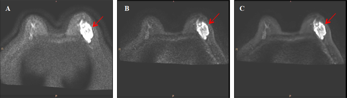
Patients: The IRB-approved MR examinations were performed on a 3.0T MR scanner (SIGNATM Premier, GE Healthcare, Milwaukee, WI) equipped with a dedicated 8-channel bilateral breast coil. Between June 2023 and September 2023, 15 Asian women patients (age range, 40-70 years) with suspected breast cancer were enrolled in the study.Imaging parameters: All patients were positioned in the feet-first prone position and underwent bilateral breast MRI. The clinical breast MRI protocol was extended to include both conventional single-shot DWI sequence and FOCUS DWI with b-values of 1000 s/mm2, as shown in Figure 1. The detailed imaging parameters of conventional DWI were as follows: FOV=34×34mm2, TR/TE=4525/53.4ms, slices=32, matrix=128×168, NEX=3.00 (b1000), acquired voxel size=2.7×2.0×4.0mm3, bandwidth=3906.25Hz/Px, acquisition time=127s. The imaging parameters of FOCUS DWI were: FOV=34×17 mm2, TR/TE=10079/51.0ms, slices=32, matrix=128×84, NEX=4.00 (b1000), acquired voxel size=2.7×2.0×4.0 mm3, bandwidth =3906.25Hz/Px, acquisition time=143s.
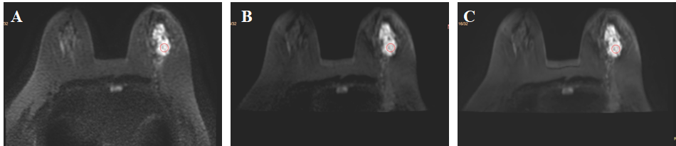
Data processing: A prototype version of DLR (AIR Recon DL) was applied to reconstruct the k-space data of FOCUS DWI to obtain images of FOCUS DWI with DLR (FOCUS-DLR). Subjective image quality was assessed by one radiologist using a 5-point Likert scale (1=poor, 5=excellent) in terms of the overall image quality, lesion conspicuity, artifacts, and geometric distortion3,9,10. For quantitative objective assessment, SNR11 of lesion, contrast-to-noise ratio (CNR)4,12 between lesion and surrounding tissue, and apparent diffusion coefficient (ADC) of lesion were also measured for comparison. The ROI of lesion was carefully drawn on the slice (b1000) with the largest cross section of the lesion, as shown in Figure 2. The ROI of surrounding tissue was positioned on homogeneous glandular tissue around the lesion. The ROI of background was drawn in air region on the same slice.
Statistical analysis: The Likert scales and quantitative parameters were compared using Friedman's test and one-way ANOVA, and Dunn-Bonferroni post hoc tests were used to adjust for all significant pairwise comparisons. P<0.05 was considered statistically significant.
Results
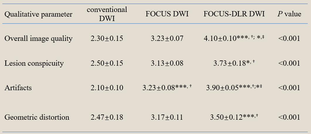
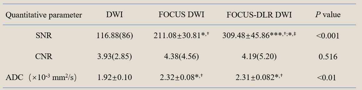
As shown in Table 1, in the qualitative evaluation, there were significant differences between conventional DWI, FOCUS DWI, and FOCUS-DLR DWI in terms of image quality, lesion conspicuity, artifacts, and distortion. The overall image quality of FOCUS-DLR DWI was better than that of conventional DWI and FOCUS DWI (P<0.001, P<0.05). FOCUS-DLR DWI had higher scores in terms of conspicuity, artifacts and distortion than conventional DWI (P<0.05, P<0.001, P<0.001). FOCUS DWI had higher score in terms of artifacts than conventional DWI (P<0.001). As shown in Table 2, the SNR of FOCUS DWI and FOCUS-DLR DWI was higher than that of conventional DWI (P<0.05, P<0.001), SNR of FOCUS-DLR DWI was higher than that of FOCUS DWI (P<0.05). The CNR values between lesion and surrounding tissue were not statistically significant among three sequences. The mean ADC values of lesion in FOCUS-DLR and FOCUS DWI were higher than that of conventional DWI (P<0.05, P<0.05).Discussion
This study showed that FOCUS-DLR and FOCUS DWI was superior to conventional DWI in terms of overall image quality, lesion clarity, artifacts, and distortion, which aligns with previous results on prostate cancer and optic nerve MRI3,5. Images of FOCUS-DLR has the highest SNR, but CNR was not significantly different from conventional DWI. FOCUS-DLR and FOCUS DWI had higher ADC than conventional DWI, which is consistent with a previous result5. In this study, the utilization of FOCUS-DLR DWI in breast MR imaging yielded improved image quality, enabling clearer visualization of breast glandular tissues and lesions, which offers valuable information for the diagnosis and treatment of breast diseases.Conclusion
Our findings indicate that FOCUS DWI with deep learning-based reconstruction produces superior images than conventional DWI, enhancing the applicability of this technique in clinical practice. Deep learning-based reconstruction provides a new direction for optimizing DWI imaging techniques in Asian breast MRI with small breast volumes.Acknowledgements
No acknowledgement found.References
1.MCDONALD E S, HAMMERSLEY J A, CHOU S-H S, et al. Performance of DWI as a Rapid Unenhanced Technique for Detecting Mammographically Occult Breast Cancer in Elevated-Risk Women With Dense Breasts [J]. American Journal of Roentgenology, 2016, 207(1): 205-16.
2.DONG H, LI Y, YU K, et al. Comparison of image quality and application values on different field-of-view diffusion-weighted imaging of breast cancer [J]. Acta Radiologica, 2016, 57(1): 19-24.
3.TIAN Y, WANG J, LI M, et al. Comparison of field-of-view optimized and constrained undistorted single-shot diffusion-weighted imaging and conventional diffusion-weighted imaging of optic nerve and chiasma at 3T [J]. Neuroradiology, 2018, 60(9): 903-12.
4.PARK J Y, SHIN H J, SHIN K C, et al. Comparison of Readout Segmented Echo Planar Imaging (EPI) and EPI With Reduced Field-of-View Diffusion-Weighted Imaging at 3T in Patients With Breast Cancer [J]. Journal of Magnetic Resonance Imaging, 2015, 42(6): 1679-88.
5.FENG Z, MIN X, SAH V K, et al. Comparison of field-of-view (FOV) optimized and constrained undistorted single shot (FOCUS) with conventional DWI for the evaluation of prostate cancer [J]. Clinical Imaging, 2015, 39(5): 851-5.
6.BAI Y, PEI Y, LIU W V, et al. MRI: Evaluating the Application of FOCUS-MUSE Diffusion-Weighted Imaging in the Pancreas in Comparison With FOCUS, MUSE, and Single-Shot DWIs [J]. Journal of Magnetic Resonance Imaging, 2023, 57(4): 1156-71.
7.CHENG Y, JIANG H, WANG H, et al. Application of Field-of-View Optimized and Constrained Undistorted Single Shot (FOCUS) with Intravoxel Incoherent Motion (IVIM) in 3T in Locally Advanced Rectal Cancer [J]. Disease Markers, 2021, 2021: 5565902.
8.WANG Y F, REN Y, ZHU C F, et al. Optimising diffusion-weighted imaging of the thyroid gland using dedicated surface coil [J]. Clinical Radiology, 2022, 77(11): e791-e8. 9.WARNDAHL B A, BORISCH E A, KAWASHIMA A, et al. Conventional vs. reduced field of view diffusion weighted imaging of the prostate: Comparison of image quality, correlation with histology, and interreader agreement [J]. Magnetic Resonance Imaging, 2018, 47: 67-76.
10.EL HOMSI M, BATES D D B, MAZAHERI Y, et al. Multiplexed sensitivity-encoding diffusion-weighted imaging (MUSE) in diffusion-weighted imaging for rectal MRI: a quantitative and qualitative analysis at multiple b-values [J]. Abdominal Radiology, 2022, 48(2): 448-57.
11.CHEN M, FENG C, WANG Q, et al. Comparison of reduced field-of-view diffusion-weighted imaging (DWI) and conventional DWI techniques in the assessment of Cervical carcinoma at 3.0T: Image quality and FIGO staging [J]. European Journal of Radiology, 2021, 137.
12.LEE E J, CHANG Y-W, SUNG J K, et al. Feasibility of deep learning k-space-to-image reconstruction for diffusion weighted imaging in patients with breast cancers: Focus on image quality and reduced scan time [J]. European Journal of Radiology, 2022, 157: 110608.
Figures