3622
Few-shot Learning for Differentiation of Malignant and Benign Breast Cancer Lesions Using Dynamic Contrast-enhanced MRI1Lunenfeld-Tanenbaum Research Institute, Sinai Health System, University of Toronto, Toronto, ON, Canada, 2Joint Department of Medical Imaging, University Health Network, Sinai Health System and Women’s College Hospital University of Toronto, Toronto, ON, Canada, 3Radiology Department, Instituto Nacional do Cancer (INCa), Rio de Janeiro, Brazil, Rio de Janiro, Brazil
Synopsis
Keywords: Diagnosis/Prediction, Breast
Motivation: Breast cancer (BrCa) is the most prevalent malignancy among women. MRI is a useful tool for BrCa early detection and characterization. However, high false-positive rates can lead to unnecessary biopsies and patient distress. To enhance diagnostic accuracy, deep learning presents a promising avenue, but training deep neural networks (DNN) requires a large, annotated dataset.
Goal(s): Introduce a novel method for BrCa classification, utilizing a minimally labeled dataset.
Approach: We employ a few-shot learning (FSL) approach to differentiate between benign and malignant breast tumors.
Results: Our FSL-based model significantly surpasses the diagnostic performance of trained radiologists in breast cancer classification (p < 0.0001).
Impact: Our FSL model streamlines machine learning by reducing data labeling needs outperforms radiologists in detecting breast cancer, and could reduce unnecessary biopsies, sparing patients from potential harm.
Introduction
Breast MRI is a highly sensitive imaging modality for women at higher risk 1. Dynamic contrast-enhanced (DCE) MRI is notable for its high temporal resolution, enabling detailed analysis of the kinetics of contrast media uptake, which is crucial for assessing breast cancer (BrCa) 2-4. Despite its sensitivity, differentiating between benign and malignant tumors remains challenging due to the variations in tumor presentations on DCE images. Deep learning (DL) has the potential to detect these subtle signs of cancer, possibly reducing errors that can occur with human interpretation.Truhn et al. assessed the efficacy of a deep neural network (DNN) model against a radiomic approach in classifying breast lesions. They employed a dataset comprising multiparametric (mp)MRI scans with 1294 identified enhancing lesions. On a 10% test set, the DNN model exhibited sensitivity and an area under the receiver operating characteristic curve (AUROC) of 78% and 0.88, respectively 5. Zheng et al. implemented a dense convolutional long short-term memory to classify lesions from a DCE MRI dataset of 72 patients. By integrating data manually extracted from diagnostic and pathological reports, they improved the model's ability to generalize, achieving an accuracy of 84.7% 6. In a different study, a neural network trained on both image features from DCE MRI, and analytical features (geometric and contrast kinetics) demonstrated an 87.7% accuracy in classifying breast lesions, using a test dataset representing 20% of 130 MRI patient scans 7.
Training state-of-the-art DNN for cancer-type prediction requires a large sample size. When such data are scarce, integrating existing clinical information, like radiology or pathology reports, becomes crucial to guide the training process. However, this integration can be a labor-intensive task and such information may not always be available in hospital records. FSL has emerged as a promising solution for image classification tasks in such data-constrained scenarios. This technique allows a model to learn from a small subset of examples and generalize to new, unseen data. We introduced a novel FSL-based approach that employs a Siamese network architecture to differentiate BrCa types using co-registered wash-in and wash-out phases of DCE MRIs (BrCa Few-shot Siamese Network, BrCa-FSN). This approach was evaluated against the diagnostic performance of radiologists, showing its potential for accurate BrCa classification with limited labeled data.
Method
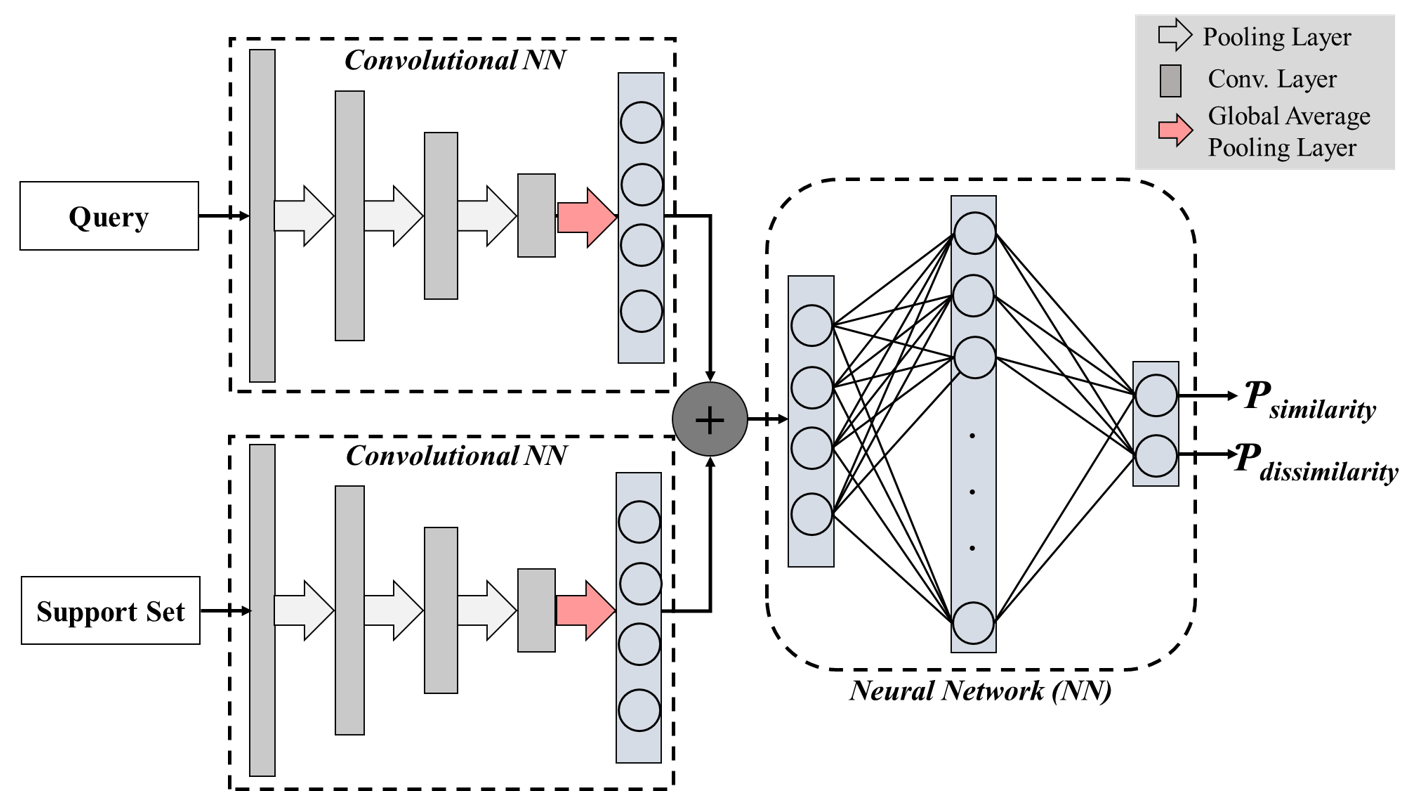
This study comprised 414 consecutive women with 520 biopsy-confirmed lesions who received an exemption informed consent from our local institutional review board. A dedicated radiologist with 10 years of experience (R.P) contoured all suspicious lesions on mpMRI. The imaging data were acquired at the 1.5-T or 3.0-T system. The DCE protocol consisted of a pre-contrast scan followed by four post-contrast scans, where wash-in and wash-out parameters were calculated at 1 minute and 4 minutes post-contrast administration.The images were divided into two sets: 400 for training and 120 for testing, respectively. From these, was generated 160,000 training pairs categorized as similar or dissimilar based on the DCE MRI characteristics of the benign and malignant lesions. The Siamese network was trained to predict the similarity/dissimilarity between image representations learned through two independent DNNs. The BrCa-FSN takes a pair of the query (i.e., test image) and support samples (i.e., labeled image) in the test phase to predict the similarity/dissimilarity between image representations. To enrich the image representation, a two-channel image was created from co-registered wash-in and wash-out DCE, and latent attributes of the lesions were concatenated for final prediction. Moreover, a support set consisted of 10 benign and 10 malignant cases to be compared with the test image. The majority vote system was applied for the final prediction. Figure 1 shows an overview of the proposed method.
The accuracy of the BrCa-FSN was compared to the radiologist's accuracy. To estimate radiologist accuracy, lesions were first labeled as benign or malignant using a BI-RAD score determined by the radiologist (1 to 5), such that lesions with BI-RADS > 3 were labeled malignant. These assessments were then cross-referenced with pathological findings for accuracy validation.
Results
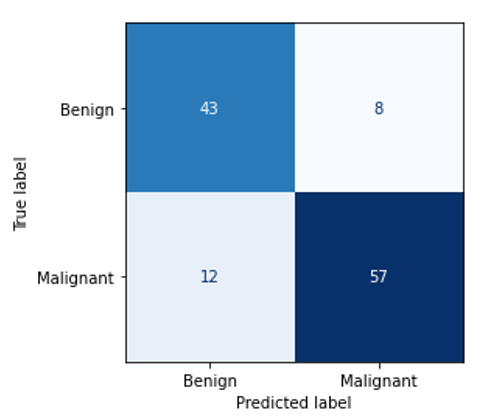
The proposed method was assessed using 120 lesions (51 benign and 69 malignant). Our evaluation reported an accuracy of 83%, AUROC of 0.835, and a positive predictive value of 88%. Figure 2 shows the confusion matrix. Notably, BrCa-FSN significantly outperformed radiologists’ performance (83% vs 54%; p-value < 0.0001).Conclusion and Discussion
We introduced a novel FSL-based algorithm to differentiate benign from malignant lesions using DCE-MRI. The results demonstrated that employing FSL facilitates training a DNN with limited labeled data, achieving results that are comparable with those reported in the current literature derived from much larger datasets or data enriched with radiology-pathology information.Acknowledgements
The Ontario Institute of Cancer Research, Sinai Hospital Foundations, and University Medical Imaging Toronto, Ontario, Canada, supported this work.References
[1] Stephan P. Mammography and Breast MRIs. In: Hayat MA, editor. Lung and breast carcinomas. Elsevier. 2010; http://breastcancer.about.com/od/mammograms/a/mammo_vs_mri_2.htm. 2010. Accessed October 16, 2023.
[2] Helbich TH, Roberts TP, Gossmann A, et al. Quantitative gadopentetate-enhanced MRI of breast tumors: testing of different analytic methods. Magn Reson Med. 2000;44:915–924.
[3] Huang W, Tudorica LA, Li X, et al. Discrimination of benign and malignant breast lesions by using shutter-speed dynamic contrast-enhanced MR imaging. Radiology. 2011;261:394–403.
[4] Li L, Wang K, Sun X, et al. Parameters of dynamic contrast-enhanced MRI as imaging markers for angiogenesis and proliferation in human breast cancer. Med Sci Monit. 2015;21:376–382.
[5] Truhn D, Schrading S, Haarburger C, et al. Radiomic versus Convolutional Neural Networks Analysis for Classification of Contrast-enhancing Lesions at Multiparametric Breast MRI. Radiology. 2019;290(2):290-297.
[6] Nagarajan MB, Huber MB, Schlossbauer T, et al. Small Lesion Classification in Dynamic Contrast Enhancement MRI for Breast Cancer Early Detection. Medical Image Computing and Computer Assisted Intervention – MICCAI 2018: 21st International Conference, Granada, Spain, September 16-20, 2018, Proceedings, Part IISep 2018Pages 876–884https://doi.org/10.1007/978-3-030-00934-2_97.
[7] Wu Y, Wu J, Dou Y, et al. A deep learning fusion model with evidence-based confidence level analysis for differentiation of malignant and benign breast tumors using dynamic contrast enhanced MRI. Biomedical Signal Processing and Control. 2022;72:103319.
Figures