3619
Diagnostic value of combining radiomics and clinical features in placenta accreta spectrum1Radiology, The First Affiliated Hospital of Guangxi Medical University, Nanning, China, 2Philips Healthcare, Guangzhou, China, Guangzhou, China
Synopsis
Keywords: Diagnosis/Prediction, Radiomics, Placenta
Motivation: Placenta accrete spectrum disorder (PAS) is a dangerous pregnancy complication that posed a threat to the safety of pregnant women, and its incidence is still on the rise.
Goal(s): To develop a machine learning model for effective diagnosis of PAS.
Approach: We developed machine learning models based on T2WI radiomics, clinical features, and clinical-radiomics features in the diagnosis of PAS.
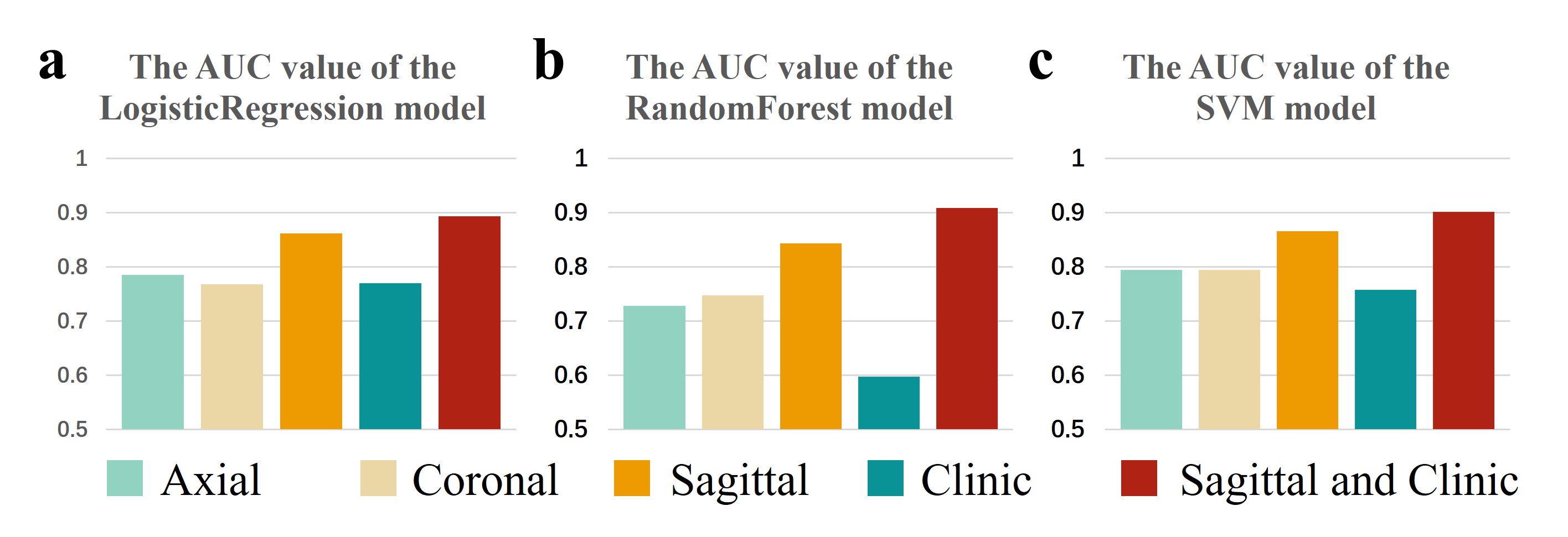
Results: Radiomics models have a great diagnostic performance for PAS, with sagittal-based model shows better performance. The clinical-radiomics model exhibits the highest diagnostic performance in this study.
Impact: Machine learning models that combined with radiomics and clinical features can improve the diagnosis of PAS, and benefit PAS patients. Furthermore, our results provide new insights for future research.
Introduction
Placenta accrete spectrum disorder (PAS) is a group of disorders in which the placental villi abnormally invade beyond the decidua basalis1. PAS can lead to preterm birth and significant bleeding, and even endanger the lives of pregnant women. Previous studies have confirmed that a history of uterine surgery and placenta praevia are the two most important risk factors for PAS2. Magnetic Resonance Imaging (MRI) with its high soft tissue contrast is considered as the most valuable non-invasive imaging examination and has played a crucial role in diagnosing PAS. However, the morphology of the placenta varies greatly among individuals and is largely dependent on the ability of the radiologist. This can result in different conclusions being reached by different radiologists. Therefore, the development of more objective and efficient methods for diagnosis of PAS is urgently needed. Radiomics can reveal features that are difficult to recognize with the naked eye and have found widespread applications in various diseases3. This study aims to investigate the diagnostic value of machine learning models based on radiomics and clinical features for PAS.Methods
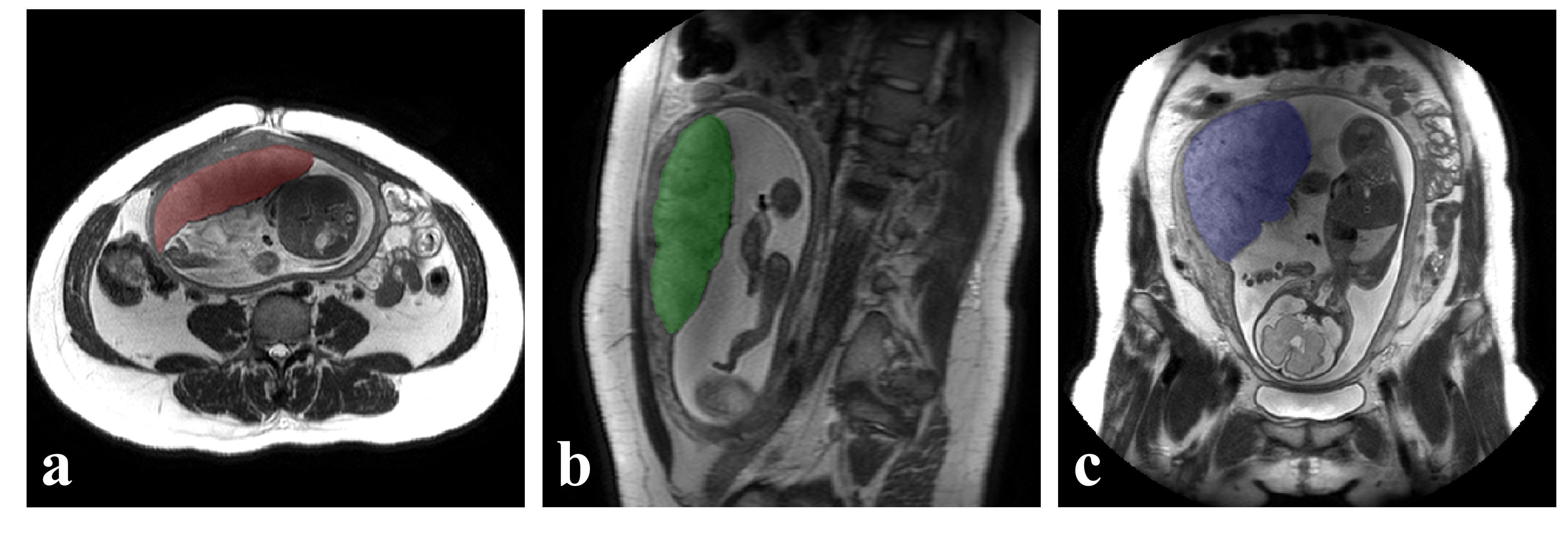
We retrospectively reviewed patients who underwent MRI (Achieva 3.0T; Philips Healthcare, Best, the Netherlands) examination at our hospital from 2017 to 2022. Patients with inadequate image quality and those with incomplete clinical data were excluded. The placental images were manually segmented by one radiologist using ITK-SNAP (v3.8.0). Segmentation was performed for the axial, coronal, and sagittal T2WI images. The results were then assessed by another radiologist, and in cases of disagreement, a third radiologist (with over 10 years of experience in abdominal imaging diagnosis) participated in discussion to make a final decision. Clinical features include the history of uterine surgery and placenta praevia. The data was divided into a training set and a testing set with a 7:3 ratio. The least absolute shrinkage and selection operator (LASSO) was used for radiomics features selection. Logistic Regression (LR), Random Forest (RF), and Support Vector Machine (SVM) models were established separately for radiomics features based on axial, coronal, and sagittal images, as well as clinical features. The optimal radiomics model was combined with clinical features to establish a clinical-radiomics model. Receiver Operating Characteristic (ROC) curve analysis was conducted, and the diagnostic performance of different models was assessed using the Area Under the ROC Curve (AUC).Results
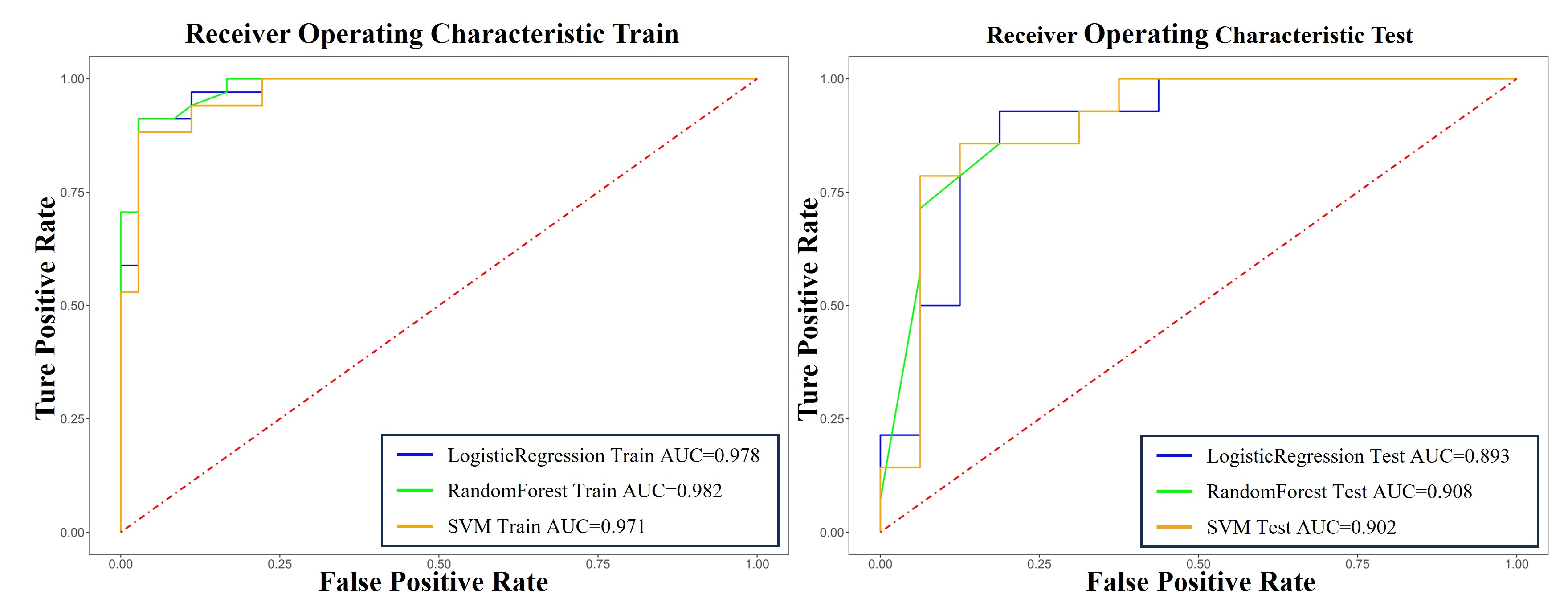
We collected a total of 100 patients, including 48 cases of PAS and 52 cases without PAS. In the radiomics models, the SVM showed superior performance compared to LR and RF, and the best-performance model among radiomics SVM models is the sagittal-radiomics SVM model (AUC=0.8661). The clinical models (AUC=0.7698) showed slightly lower diagnostic performance compared to the radiomics models. The combined model, which combined with sagittal radiomics features and clinical features, achieved higher diagnostic performance than radiomics models and clinical model: LR model (training set AUC=0.9779, test set AUC=0.8929), RF model (training set AUC=0.9824, test set AUC=0.9085), and SVM model (training set AUC=0.9706, test set AUC=0.9018) .Discussion
In this study, we developed radiomics models, clinical models, and clinical-radiomics models based on T2WI radiomics features and clinical features to diagnose PAS. The results showed that the sagittal-based radiomics models exhibited better diagnostic performance, which can be attributed to the site of PAS: the site of PAS is often the anterior and posterior wall of the uterus, and the sagittal image can be regarded as favorable4, so the radiomics features extracted from the sagittal image are better suited to reflect and predict the PAS. Clinical features are important diagnostic criteria for diseases, and the history of uterine surgery and placenta previa have been widely recognized as significant risk factors for PAS. However, the clinical information is relatively limited. Therefore, using clinical features alone for the diagnosis of PAS may not achieve the desired level of performance. Relying solely on clinical features or radiomics features for prediction still has certain limitations. Combining different types of features can overcome the shortcomings of poor predictive performance and weak generalization ability5. In this study, the combination of the clinical feature and radiomics features to establish the clinical-radiomics models can make full use of their respective characteristics, thus further enhancing the diagnostic efficacy. In this situation, it is possible to minimize the severe complications of PAS as much as possible, which can greatly improve the prognosis of PAS patients.Conclusion
The T2WI radiomics model has demonstrated good diagnostic performance for PAS, and when combined with clinical features and radiomics features, it can achieve higher diagnostic performance. This study provides new insights for future research.Acknowledgements
Not applicable.References
1. Do QN, Lewis MA, Xi Y, et al. MRI of the Placenta Accreta Spectrum (PAS) Disorder: Radiomics Analysis Correlates With Surgical and Pathological Outcome. J Magn Reson Imaging. 2020;51(3):936-946. doi:10.1002/jmri.26883
2. Comstock CH, Bronsteen RA. The antenatal diagnosis of placenta accreta. BJOG. 2014;121(2):171-181; discussion 181-182. doi:10.1111/1471-0528.12557
3. Wu Q, Yao K, Liu Z, et al. Radiomics analysis of placenta on T2WI facilitates prediction of postpartum haemorrhage: A multicentre study. EBioMedicine. 2019;50:355-365. doi:10.1016/j.ebiom.2019.11.010
4. Zaghal AA, Hussain HK, Berjawi GA. MRI evaluation of the placenta from normal variants to abnormalities of implantation and malignancies. J Magn Reson Imaging. 2019;50(6):1702-1717. doi:10.1002/jmri.26764
5. Ye Z, Xuan R, Ouyang M, Wang Y, Xu J, Jin W. Prediction of placenta accreta spectrum by combining deep learning and radiomics using T2WI: a multicenter study. Abdom Radiol (NY). 2022;47(12):4205-4218. doi:10.1007/s00261-022-03673-4
Figures