3617
The Value of Magnetic Resonance Imaging Peritumoral Radiomics in Predicting 21-gene Recurrence Score of ER+/HER2- Breast Cancer1Department of Radiology, Fudan University Shanghai Cancer Center, Shanghai, China, 2Department of Oncology, Shanghai Medical College, Fudan University, Shanghai, China, 3GE Healthcare, MR Research, Beijing, China
Synopsis
Keywords: Diagnosis/Prediction, Radiomics, Breast neoplasms; Peritumoral; Neoplasm recurrence; Image interpretation
Motivation: 21-gene assay is recommend to guide decision on the use of adjuvant chemotherapy. However, this test is expensive and time-consuming, so it is not widely used in clinic.
Goal(s): To develop a radiomics model for predicting 21-gene recurrence score based on MRI intratumoral and peritumoral features.
Approach: Prediction models in tumor and different peritumoral regions (2 mm, 4 mm, 6 mm, 8 mm, 10 mm) were established using machine learning method. Feature-fusion and logistics-regression methods were used to fuse information.
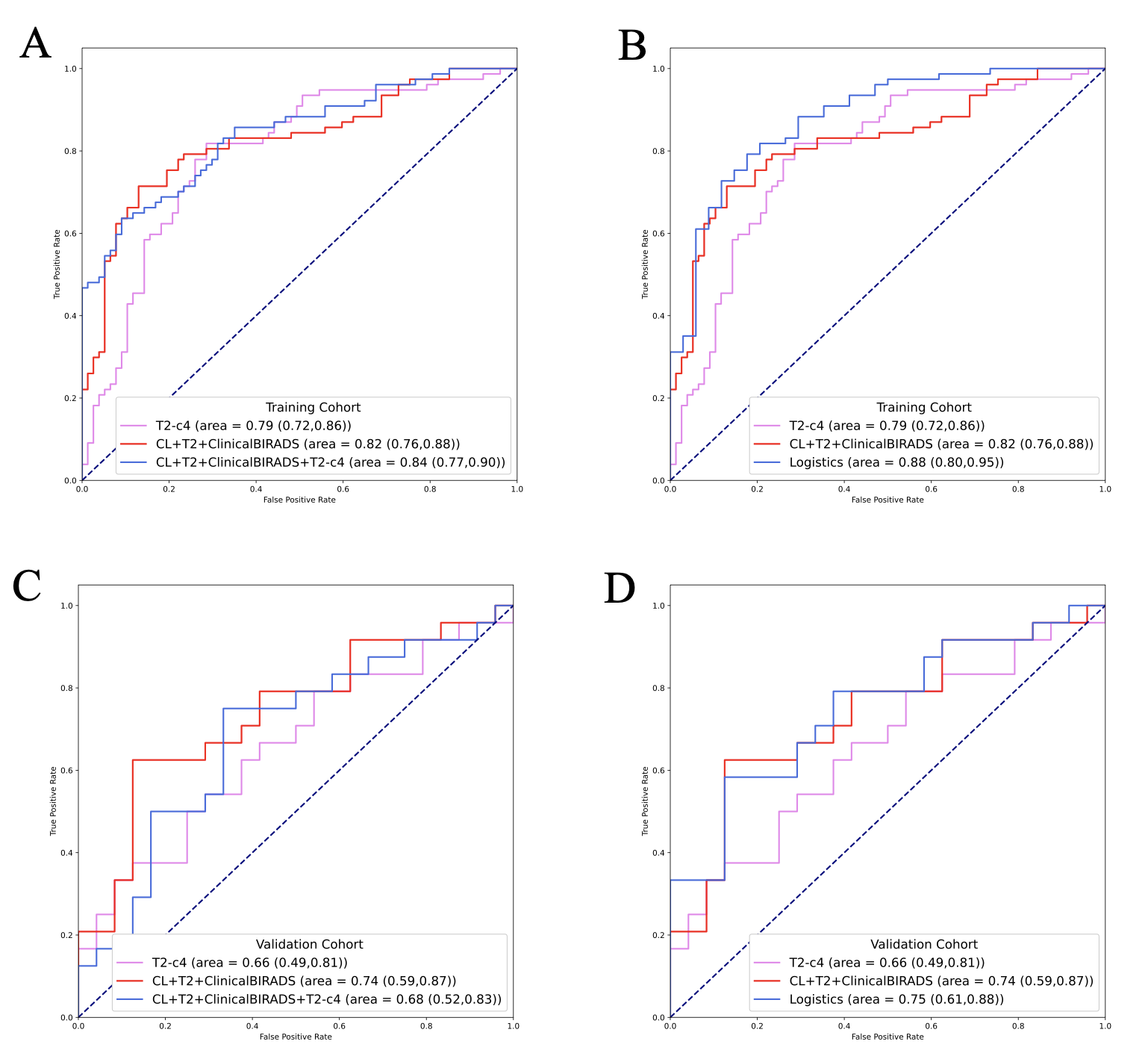
Results: Combining 4-mm peritumoral model on T2WI (AUC = 0.66) with intratumoral and clinical-imaging features, the fusion model performed best (AUC = 0.75).
Impact: To provide an alternative for patients who cannot afford Oncotype 21-gene assay and to reduce the medical costs for those who could afford it.
INTRODUCTION
The 21-gene assay has been recommended by the National Comprehensive Cancer Network (NCCN) guidelines to evaluate the distant recurrence and guide decision on the use of adjuvant systemic therapy in ER+/HER2- patients. However, the 21-gene test is expensive and the time-consuming, so it have not been widely used in clinic.Breast MRI is the most sensitive technology for diagnosis and treatment response evaluation in breast cancer patients[1-4]. The development of radiomics makes it possible to extract richer information from medical images. Tumor microenvironment has been shown to play an important role in development and progression of breast cancer[5, 6]. As a result, the region of interest in radiomics extends from the tumor to the peritumoral tissues. However, as far as we know, only one published study focused on the correlation between recurrence score (RS) and peritumoral environment[7], in which only 62 subjects and dynamic contrast-enhancement (DCE) sequence were included.
Therefore, the objective of this study was to develop a 21-gene RS prediction model based on DCE and T2WI peritumoral radiomics, as well as magnetic resonance imaging features and clinical information.
METHODS
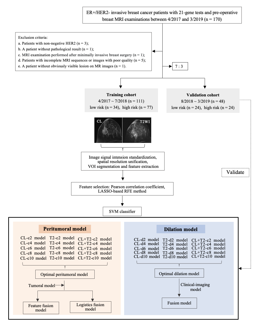
The data of ER+/HER2- breast cancer patients who came to our hospital from April 2017 to March 2019 were reviewed, and the patients who underwent 21-gene test and preoperative MRI were screened out. Patients were divided into training group and validation group in chronological order and 7:3 ratio. The patients were divided into low risk group (RS < 26) or high risk group (RS ≥ 26) according to RS threshold recommended by the 2022 version of the NCCN guidelines.MR images were evaluated and the results were recorded according to the 2013 version of Breast Imaging Reporting and Data System lexicon. Volume of interest (VOI) was manually delineated along the lesion contour on the last-enhanced phase of the DCE images (CL) and the VOI was then copied to T2W images. The VOI contour was automatically expanded outward by different ranges (2 mm, 4 mm, 6 mm, 8 mm and 10 mm) using Python 3.7. After VOI expansion, both the peritumoral VOIs (excluding the internal tumor area) and the dilation VOIs (tumor + peritumoral area) with different ranges were obtained.
For each VOI, radiomics features were extracted, and then, z-score standardized processing was performed. The Pearson correlation coefficient screening method and recursive feature elimination method were used for feature screening, and the synthetic minority oversampling technique was used for resampling patients in the training group. The recurrence risk prediction models were constructed using linear support vector machine (SVM) method. The fusion models were constructed by combining the best peritumoral model and dilatation model, with the intratumoral, clinical and imaging features (feature-fusion method and logistics-regression method). Prediction performance of the models was evaluated by the area under the receiver operating characteristic curve (AUC).
RESULTS
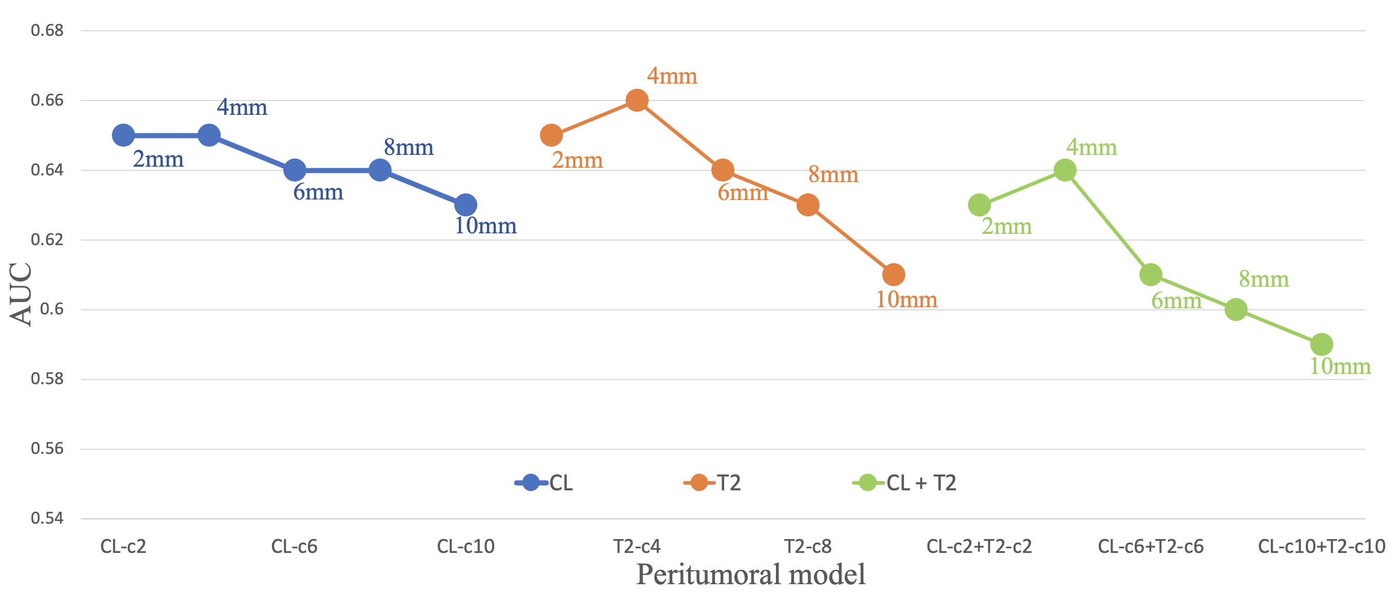
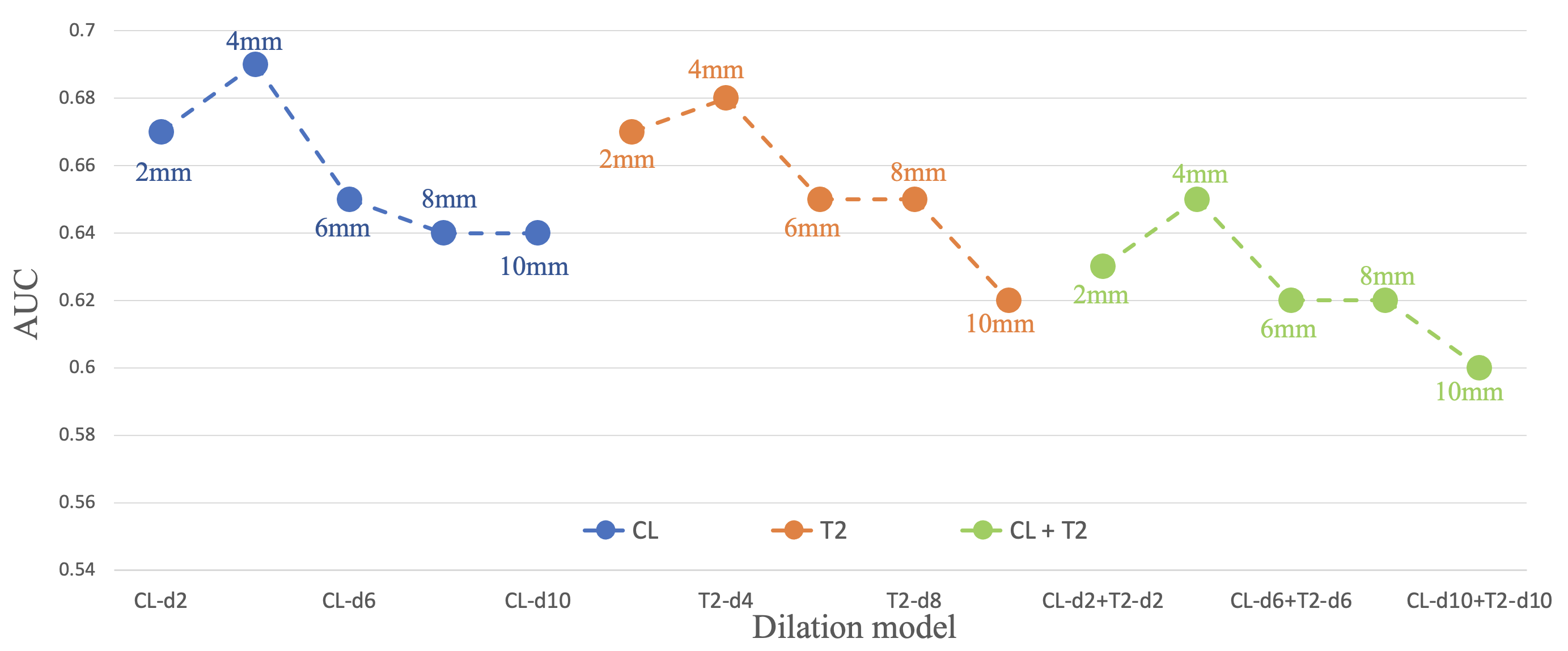
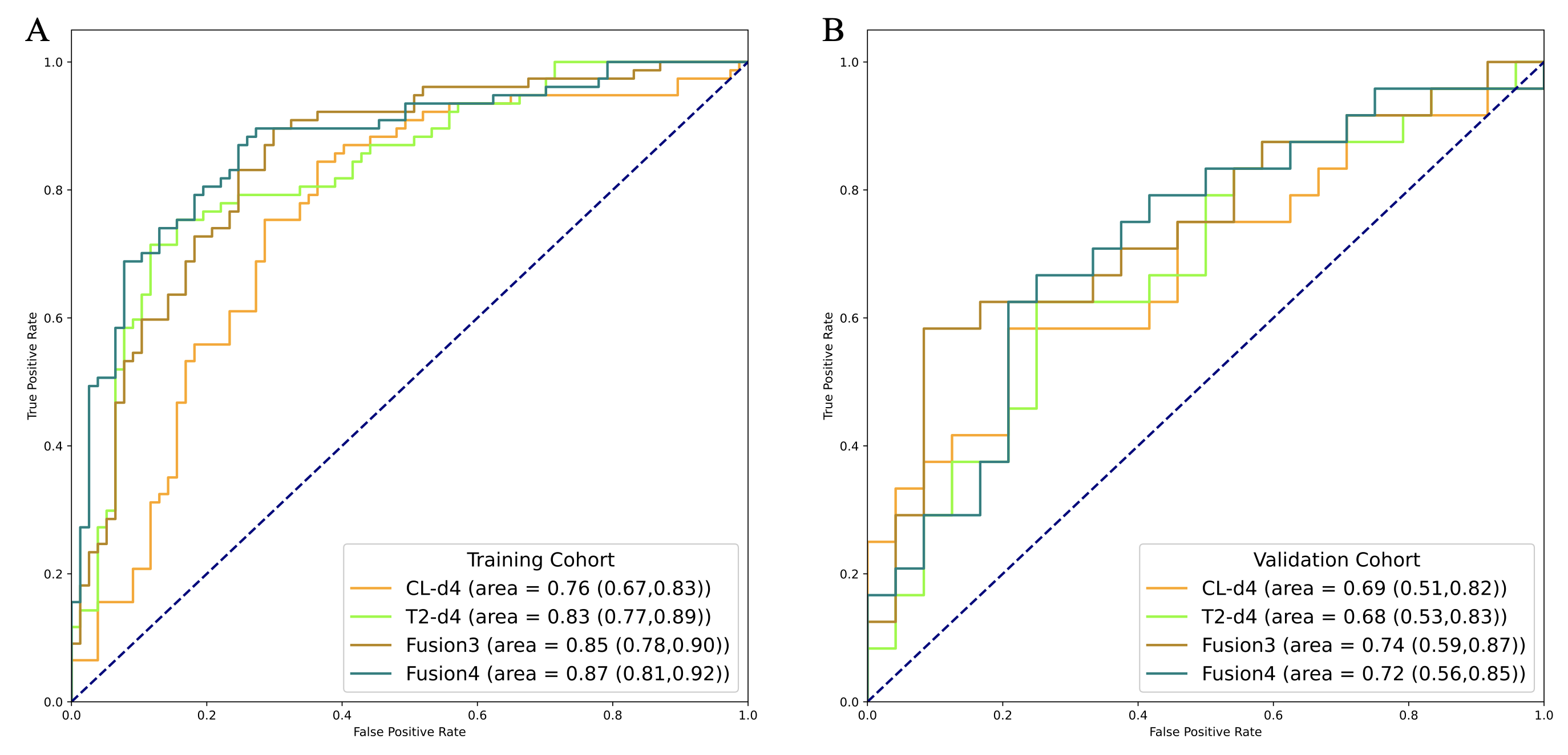
A total of 159 patients were enrolled, with 111 in the training group (34 in the low-risk group and 77 in the high-risk group) and 48 in the validation group (24 in the low-risk group and 24 in the high-risk group). With the increase of the peritumoral area, the variation trend of AUCs of peritumoral models was similar to that of dilation models: the AUCs of peritumoral 4-mm models were higher than or equal to the AUCs of peritumoral 2-mm models, and the AUCs gradually decreased with the range increasing from peritumoral 4 mm to peritumoral 10 mm.Among all peritumoral models, peritumoral 4-mm model on T2WI had the highest AUC (AUC = 0.66, 95% CI: 0.99-0.81). After combining with intratumoral, clinical and MR imaging features (feature-fusion method and logistics-regression method), the AUCs of the fusion models increased, and the AUC of logistics-regression fusion model was higher (AUC = 0.75, 95% CI: 0.61-0.88).
Among all the dilation models, 4-mm CL dilation model and 4-mm T2WI dilation model had higher AUCs, with AUCs of 0.69 (95% CI: 0.51-0.82) and 0.68 (95% CI:0.53-0.83), respectively. The AUCs of fusion models increased by adding clinical and MR imaging features (AUC = 0.74, 95% CI: 0.59-0.87; AUC = 0.72, 95% CI: 0.56-0.85).
CONCLUSION
Magnetic resonance imaging peritumoral radiomics may have potential in assessing recurrence risk of ER+/HER2- breast cancers, and the predictive performance could be improved by combining intratumoral, peritumoral, clinical, and MR imaging features.Acknowledgements
No acknowledgement found.References
- Mann RM, Balleyguier C, Baltzer PA et al (2015) Breast MRI: EUSOBI recommendations for women's information. Eur Radiol 25:3669-3678
- Marino MA, Helbich T, Baltzer P, Pinker-Domenig K (2018) Multiparametric MRI of the breast: A review. J Magn Reson Imaging 47:301-315
- Mann RM, Cho N, Moy L (2019) Breast MRI: State of the Art. Radiology 292:520-536
- Menezes GL, Knuttel FM, Stehouwer BL, Pijnappel RM, van den Bosch MA (2014) Magnetic resonance imaging in breast cancer: A literature review and future perspectives. World J Clin Oncol 5:61-70
- Braman N, Prasanna P, Whitney J et al (2019) Association of Peritumoral Radiomics With Tumor Biology and Pathologic Response to Preoperative Targeted Therapy for HER2 (ERBB2)-Positive Breast Cancer. JAMA Netw Open 2:e192561
- Soysal SD, Tzankov A, Muenst SE (2015) Role of the Tumor Microenvironment in Breast Cancer. Pathobiology 82:142-152
- Chiacchiaretta P, Mastrodicasa D, Chiarelli AM et al (2023) MRI-Based Radiomics Approach Predicts Tumor Recurrence in ER + /HER2 - Early Breast Cancer Patients. J Digit Imaging. 10.1007/s10278-023-00781-5
Figures