3610
Assessment of Functional Liver Reserve Based on Gd-EOB-DTPA Enhanced MRI Radiomics and Delta Radiomics1Shanxi Medical University, Taiyuan, China, 2Department of Radiology, First Hospital of Shanxi Medical University, Taiyuan, China
Synopsis
Keywords: Diagnosis/Prediction, Radiomics, Functional liver reserve; Delta radiomics; Gd-EOB-DTPA;
Motivation: The current indocyanine green clearance test is not yet widespread. Gd-EOB-DTPA enhanced MRI imaging can assess the overall liver function, but it is challenging to quantitatively and accurately evaluate it.
Goal(s): Our goal is to assess the ability of Gd-EOB-DTPA enhanced MRI radiomics and Delta radiomics for predicting functional liver reserve.
Approach: We obtained MRI images from 117 patients, trained models on 70% of them, and tested models on the remaining patients.
Results: The prediction models based on Gd-EOB-DTPA enhanced MRI radiomics and Delta radiomics are effective diagnostic tools for the quantitative assessment of functional liver reserve.
Impact: Gd-EOB-DTPA enhanced MRI radiomics and Delta radiomics models can quantitatively assess functional liver reserve, aiding clinicians in selecting the right treatment, monitoring treatment progress, and predicting patient survival.
Introduction
Indocyanine green retention rate at 15 minutes (ICG-R15) is a widely used method to quantitatively assess functional liver reserve in cirrhosis patients1. However, it relies on costly equipment and reagents, making it impractical in some healthcare facilities. Nonetheless, the excretion patterns of gadolinium-ethoxybenzyl diethylenetriamine pentaacetic acid (Gd-EOB-DTPA) and indocyanine green (ICG) are similar1-3. To address this limitation, we aimed to develop Hepatobiliary phase (HBP) radiomics and Delta radiomics models to predict ICG-R15 quantitatively and classify patients' liver function. This approach provides clinicians with integrated anatomical and functional information, reducing unnecessary tests for patients, and providing a more convenient option for assessing functional liver reserve in medical facilities without ICG clearance testing capabilities.Methods
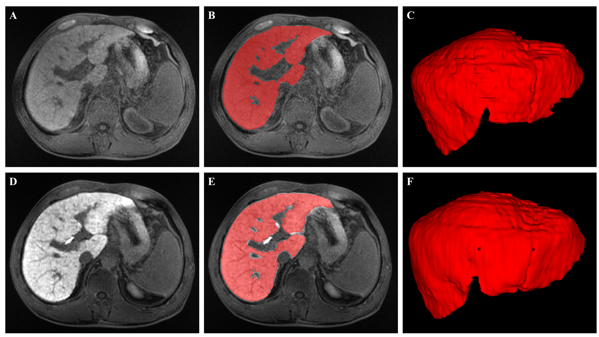
This study retrospectively collected data from 117 patients with clinically confirmed liver cirrhosis who underwent Gd-EOB-DTPA-enhanced MRI at our hospital between October 2018 and April 2023. According to the ratio of 7:3, they were randomly divided into a training set and a validation set. We collected patients' liver MRI images and clinical information. The MRI images (MASK and HBP scans) were pre-processed by 3D-Slicer, and the liver region of interest was manually delineated layer by layer (Figure 1). The radiomics features were extracted by FAE software. Features with high reliability were selected through intraclass correlation coefficient calculations. We also created Delta radiomics features by subtracting the MASK features value from the same HBP features. In the training group, Z-score was used to normalize the data, and Lasso and ten-fold cross-validation were used to identify the radiomics features that most correlated with the true value of ICG-R15 from the HBP and Delta images. Subsequently, we constructed prediction models and evaluated their effectiveness through correlation analysis, correlation coefficients, and root mean square error. At the same time, we also divided the patients into 4 data sets according to ICG-R15 thresholds (10%, ≤20%, ≤30%, and ≤40%). Pearson correlation analysis, Lasso, ten-fold cross validation and backward stepwise regression were used to identify the relevant HBP and Delta radiomics features in each dataset. The HBP and Delta radiomics classification models were constructed. The receiver operating characteristic (ROC) curve was drawn and the area under curve (AUC), sensitivity and specificity of the model were calculated. Delong test was used to compare the AUC values between different models, and the clinical decision curve was drawn to analyze the clinical diagnostic value of the model.Results
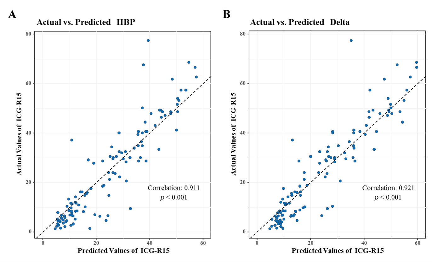
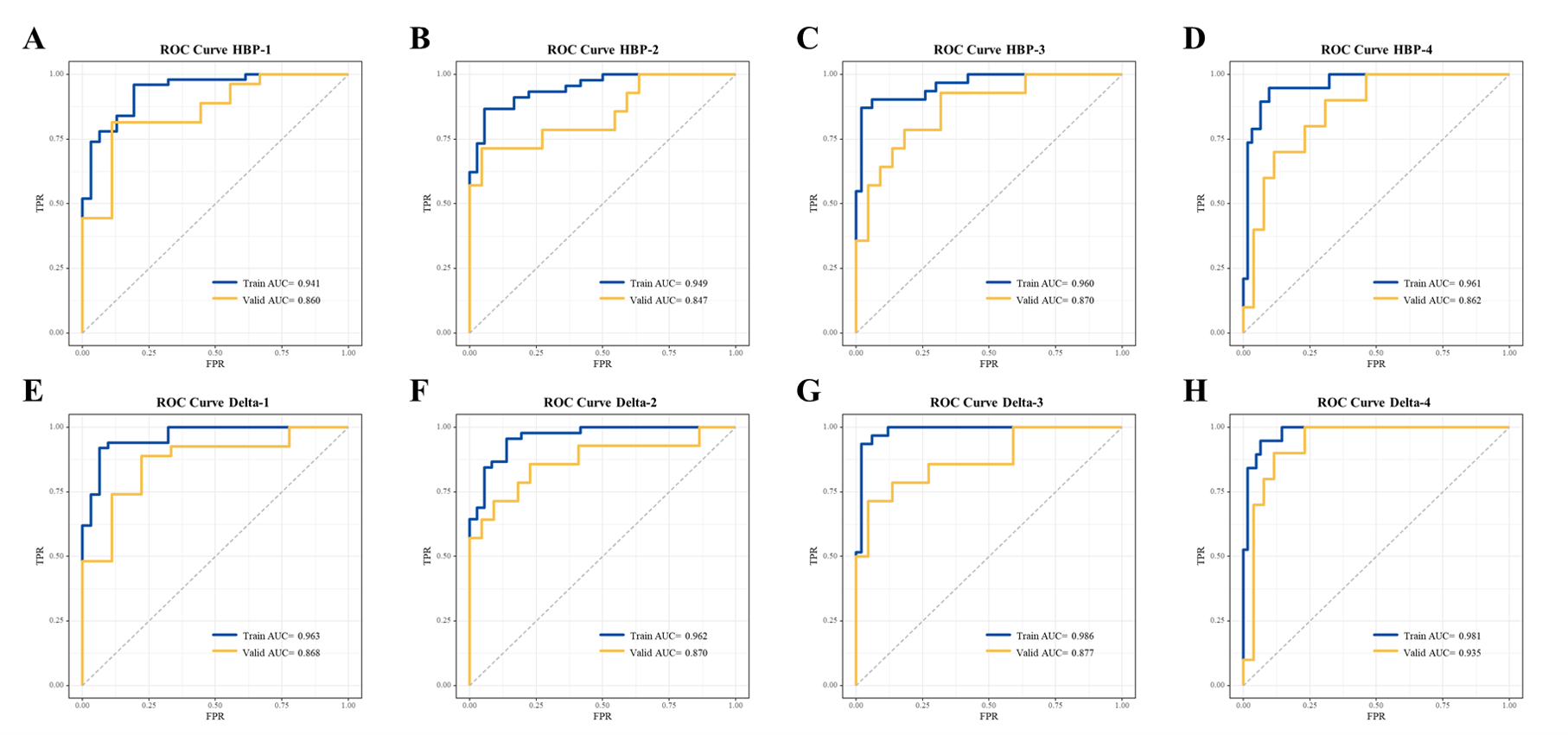
In this study, 1316 radiomics features were selected from the MASK and HBP images, and 1125 Delta radiomics features were calculated. The 9 HBP radiomics features and 7 Delta radiomics features that highly correlated with ICG-R15 were selected to construct prediction model. Correlation analysis revealed significant relationships between the predicted values of ICG-R15 and the true values (R HBP=0.911; R Delta=0.921) (Figure 2). For datasets 1 to 4, we selected 7, 4, 6, and 5 HBP radiomics features related to ICG-R15 to build classification models, respectively. The validation set showed AUC values of 0.860, 0.847, 0.870, and 0.862, respectively. We also selected 8, 7, 9, and 5 Delta radiomics features for ICG-R15 classification models. The AUC values of validation set were 0.868, 0.870, 0.877, and 0.935, respectively (Figure 3). The results of the DeLong test indicated that there was no statistically significant difference in performance between the HBP and Delta radiomics models in each dataset (p > 0.05). Calibration and clinical decision curves confirmed the accuracy and clinical applicability of all models.Discussion
Prior research first established a significant correlation between Gd-EOB-DTPA-enhanced MRI radiomic features and ICG-R15 values in liver cancer patients4. Additionally, radiomic models based on Gd-EOB-DTPA-enhanced MRI and CT have shown promise in assessing functional liver reserve4,5. Our study delved into the value of Delta radiomics in assessing functional liver reserve for the first time. We found a strong correlation between ICG-R15 predictions from both HBP and Delta radiomic models and actual values. And the Delta radiomic model demonstrated a slightly stronger correlation. This indicates that Gd-EOB-DTPA-enhanced MRI radiomic features and Delta radiomic features can be used to predict functional liver reserve, with the Delta model showing potential advantages. Moreover, both HBP and Delta radiomic models proved effective in categorizing different ICG-R15 categories, consistent with prior research4,5. However, differences in ICG-R15 threshold settings between our study and previous research may stem from variations in standards for maximum safe liver resection between countries6.Conclusion
The prediction models based on Gd-EOB-DTPA enhanced MRI radiomics and Delta radiomics are effective diagnostic tools for the quantitative assessment of functional liver reserve, with the potential to provide references for clinical evaluation of functional liver reserve.Acknowledgements
This study was supported by grants from the National Natural Science Foundation of China (No. 82071893 and 82371941).References
1. Luerken L, Dollinger M, Goetz A, et al. Diagnostic Accuracy of Indocyanine Green Clearance Test for Different Stages of Liver Fibrosis and Cirrhosis. Diagnostics (Basel). 2023;13(16). doi:10.3390/diagnostics131626632. Sakka SG. Assessing liver function. Curr Opin Crit Care. 2007;13(2):207-14. doi:10.1097/MCC.0b013e328012b268
3. Goodwin MD, Dobson JE, Sirlin CB, Lim BG, Stella DL. Diagnostic challenges and pitfalls in MR imaging with hepatocyte-specific contrast agents. Radiographics. 2011;31(6):1547-68. doi:10.1148/rg.316115528
4. Shi Z, Cai W, Feng X, et al. Radiomics Analysis of Gd-EOB-DTPA Enhanced Hepatic MRI for Assessment of Functional Liver Reserve. Acad Radiol. 2022;29(2):213-218. doi:10.1016/j.acra.2021.04.019
5. Zhu L, Wang F, Chen X, et al. Machine learning-based radiomics analysis of preoperative functional liver reserve with MRI and CT image. BMC Med Imaging. 2023;23(1):94. doi:10.1186/s12880-023-01050-1
6. Clavien PA, Petrowsky H, DeOliveira ML, Graf R. Strategies for safer liver surgery and partial liver transplantation. N Engl J Med. 2007;356(15):1545-59. doi:10.1056/NEJMra065156
Figures