3608
Using Machine Learning to Predict the Efficacy of Neoadjuvant Chemoradiotherapy for Local Advanced Rectal Cancer Based on Texture Features of MRI1Department of Radiology, The First Affiliated Hospital of USTC(Anhui Provincial Cancer Hospital), HeFei, China, 2The First Affiliated Hospital of USTC, Anhui Provincial Cancer Hospital, HeFei, China
Synopsis
Keywords: Diagnosis/Prediction, Machine Learning/Artificial Intelligence, Rectal Cancer
Motivation: To explore and validate the association between magnetic resonance image texture features and the efficacy of Neoadjuvant Chemoradiotherapy for rectal cancer.
Goal(s): To predict the efficacy of neoadjuvant chemoradiotherapy for locally advanced rectal cancer using machine learning methods.
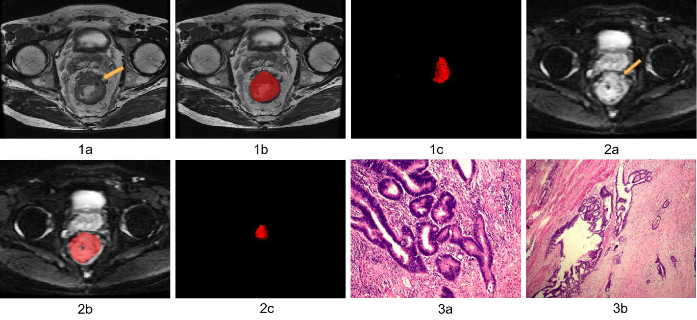
Approach: The wavelet texture parameters of all lesions in patients' MRI images were extracted, and feature selection was performed using random forest classifier model, and then classification learning was performed using the XGBoost classifier.
Results: The model based on wavelet texture feature analysis of MRI can effectively predict the effect of neoadjuvant radiochemotherapy for rectal cancer patients.
Impact: Through this study, we explored and validated the relationship between MRI texture features and the efficacy of Neoadjuvant Chemoradiotherapy for rectal cancer, providing new guidance and decision support for individualized treatment strategies.
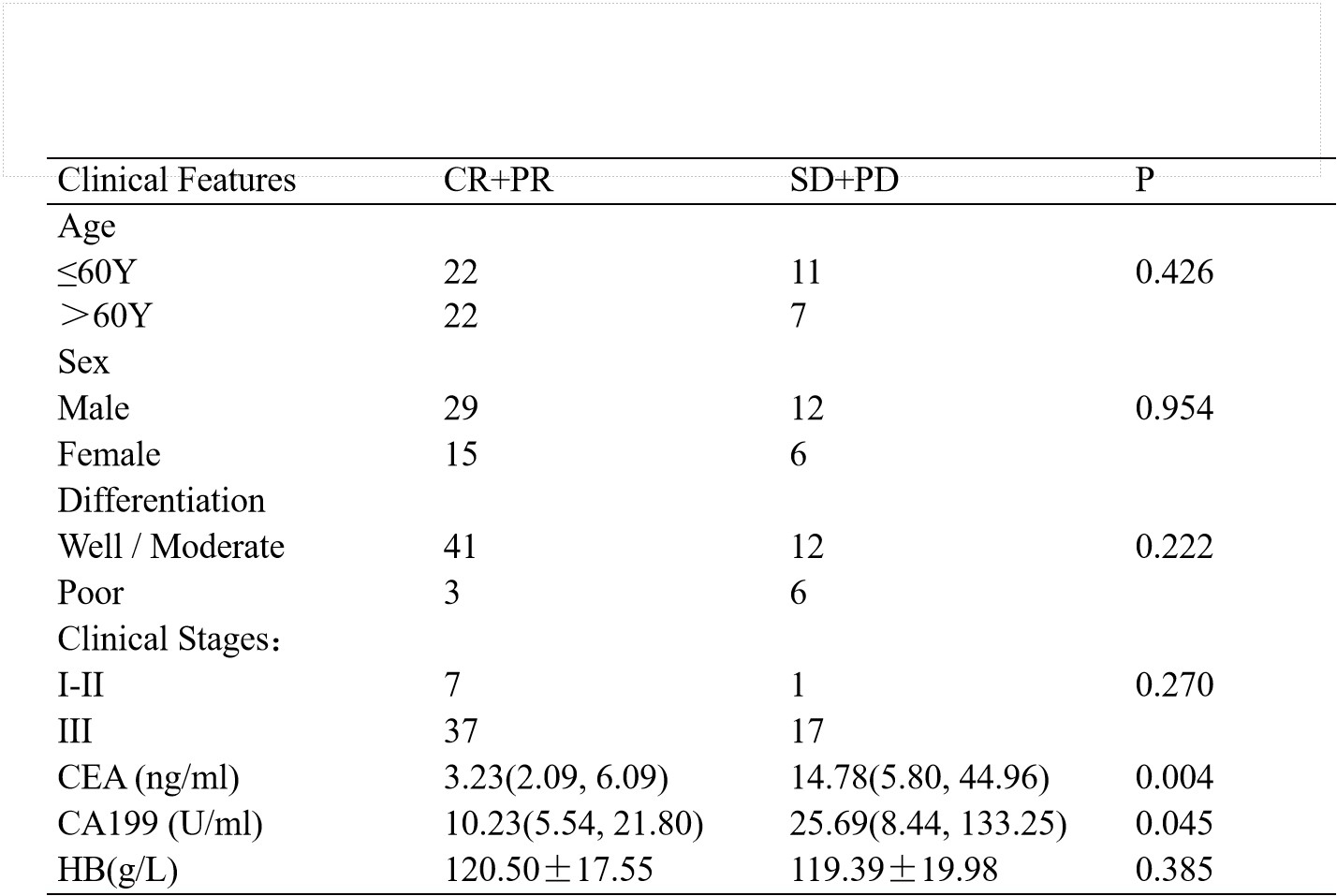
Methods:A retrospective collection was conducted involving 62 patients with locally advanced rectal cancer between June 2016 and December 2020. Clinical characteristics of the patients were collected. Efficacy assessment was conducted according to the revised RECIST (1.1 version) criteria.MRI examinations were performed on a 3.0-T system (Signa HDXT, General Electric Healthcare, Waukesha, WI, USA). For the T1WI sequence, the following parameters were used: repetition time (TR) of 500 ms, echo time (TE) of 7.2 ms, number of excitations (NEX) of 1, field of view (FOV) of 32 cm × 32 cm, slice thickness of 6 mm, and no interslice gap. The T2WI sequence was acquired with parameters including a TR of 3500 ms, TE of 109.1 ms, NEX of 4, FOV of 24 cm × 24 cm, slice thickness of 3 mm, and no interslice gap. IVIM-DWI was performed using 10 different b-values (0, 10, 20, 50, 100, 200, 400, 800, 1200, 2000 s/mm²), with a TR of 4000 ms, TE of 65 ms, and NEX of 6. Wavelet texture feature parameters of all lesions in patients' MRI images were extracted. The Random Forest Classifier model was employed for feature selection, followed by classification learning using the XGBoost classifier.
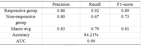
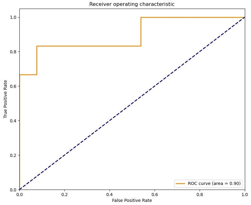
Results :A total of 62 patients with 1656 magnetic resonance wavelet texture feature parameters from all lesions were obtained. Feature selection using the Random Forest Classifier identified the most important four wavelet texture features, which included DWI wavelet-HHH glcm Maximum Probability, DWI wavelet-LLL firstorder Minimum, T2 wavelet-LHH firstorder Kurtosis, and T2 wavelet-HLL glcm MCC. Utilizing the XGBoost classifier for predictive classification yielded a final accuracy of 84.21% and an AUC value of 0.90. The precision, recall, and F1 score for the responsive treatment group were 0.86, 0.92, and 0.89, respectively. For the non-responsive treatment group, the precision, recall, and F1 score were 0.80, 0.67, and 0.73, respectively. The macro-averaged values of precision, recall, and F1 score across all labels were 0.83, 0.79, and 0.81, respectively.
Discussion:Machine learning can automatically extract key features from extensive data, construct prediction models, provide fast and accurate prediction results, alleviate clinicians' burdens, and offer more reliable references for clinical decision-making.In this study, a Random Forest model was employed to extract key features from a vast array of wavelet texture features. The Random Forest model is capable of handling high-dimensional datasets and a multitude of features, exhibiting excellent scalability[7]. It is adept at managing non-linear relationships and interaction effects. It performs well when dealing with mixed data containing discrete and continuous features, aiding in the identification of features of higher importance to the target variable.Random Forest can estimate the importance of features, measuring the contribution of features to the model. By selecting and analyzing texture features from different regions of MRI images, we achieved a predictive accuracy of 84.21% and an AUC value of 0.90 for nCRT efficacy prediction of localized advanced rectal cancer in this experiment. Therefore, this study demonstrates that on our dataset, the XGB Classifier model exhibits excellent predictive performance.
Conclusion:The model established based on wavelet texture feature analysis of magnetic resonance images can effectively predict the efficacy of neoadjuvant radiochemotherapy for rectal cancer patients. The XGB Classifier model demonstrates excellent performance in handling small, imbalanced datasets, thus showing promising clinical application value.
Acknowledgements
No acknowledgement found.References
[1] Sung H, Ferlay J, Siegel RL, Laversanne M, Soerjomataram I, Jemal A, Bray F. Global Cancer Statistics 2020: GLOBOCAN Estimates of Incidence and Mortality Worldwide for 36 Cancers in 185 Countries. CA Cancer J Clin. 2021 May;71(3):209-249.
[2] Benson AB, Venook AP, Al-Hawary MM, Azad N, Chen YJ, Ciombor KK, Cohen S, Cooper HS, Deming D, Garrido-Laguna I, Grem JL, Gunn A, Hecht JR, Hoffe S, Hubbard J, Hunt S, Jeck W, Johung KL, Kirilcuk N, Krishnamurthi S, Maratt JK, Messersmith WA, Meyerhardt J, Miller ED, Mulcahy MF, Nurkin S, Overman MJ, Parikh A, Patel H, Pedersen K, Saltz L, Schneider C, Shibata D, Skibber JM, Sofocleous CT, Stotsky-Himelfarb E, Tavakkoli A, Willett CG, Gregory K, Gurski L. Rectal Cancer, Version 2.2022, NCCN Clinical Practice Guidelines in Oncology. J Natl Compr Canc Netw. 2022 Oct;20(10):1139-1167.[3] Huang Q, Qin H, Xiao J, He X, Xie M, He X, Yao Q, Lan P, Lian L. Association of tumor differentiation and prognosis in patients with rectal cancer undergoing neoadjuvant chemoradiation therapy. Gastroenterol Rep (Oxf). 2019 Aug;7(4):283-290.
[4] Xu W, Zhu Y, Shen W, Ding W, Wu T, Guo Y, Chen X, Zhou M, Chen Y, Cui L, Du P. Combination of CDX2 expression and T stage improves prognostic prediction of colorectal cancer. J Int Med Res. 2019 May;47(5):1829-1842.
[5] Hong J, Wu J, Huang O, He J, Zhu L, Chen W, Li Y, Chen X, Shen K. Early response and pathological complete remission in Breast Cancer with different molecular subtypes: a retrospective single center analysis. J Cancer. 2020 Oct 6;11(23):6916-6924.
[6] Sun Y, Wu X, Zhang Y, Lin H, Lu X, Huang Y, Chi P. Pathological complete response may underestimate distant metastasis in locally advanced rectal cancer following neoadjuvant chemoradiotherapy and radical surgery: Incidence, metastatic pattern, and risk factors. Eur J Surg Oncol. 2019 Jul;45(7):1225-1231.
[7] Sharma A, Yadav DP, Garg H, Kumar M, Sharma B, Koundal D. Bone Cancer Detection Using Feature Extraction Based Machine Learning Model. Comput Math Methods Med. 2021 Dec 20;2021:7433186.
Figures