3604
Multimodal MRI-Based Radiomics Combining 3D Deep Transfer Learning for Predicting Cervical Stromal Invasion in Endometrial Carcinoma1Medical school, Kunming University of Science and Technology, Kunming, China, 2The First People's Hospital of Yunnan Province, Kunming, China, 3MR Research Collaboration Team, Siemens Healthineers Ltd, Shanghai, China
Synopsis
Keywords: Diagnosis/Prediction, Radiomics, Deep learning
Motivation: Cervical stromal invasion (CSI) plays a critical role in distinguishing between stage I and II endometrial carcinoma (EC) and serves as a key prognostic indicator.
Goal(s): Assisting clinicians in achieving precise preoperative treatment and prognostic assessments.
Approach: This study constructed innovative machine learning models that merge radiomics and 3D deep transfer learning to preoperatively and non-invasively predict CSI.
Results: Novel machine learning model has significant superiority over radiologists for preoperative prediction of CSI.
Impact: Constructing a non-invasive preoperative prediction model to increase the diagnostic accuracy of CSI, makes up for the limitations of traditional imaging observation in the assessment of CSI and subsequently directs clinicians in preoperative precise treatment and prognostic evaluation.
Introduction
CSI significantly impacts the progression-free and overall survival rates of EC patients by increasing the risk of lymph node metastases1-4. Accurate preoperative prediction of CSI is crucial for tailoring treatment strategies and evaluating prognoses in EC patients. However, conventional MRI often exhibits low sensitivity in detecting CSI5,6, leading to a lack of non-invasive tools for its accurate prediction. This study aims to develop and compare various models, including radiomics, 3D deep learning transfer, and integrated models, using single-modality and multimodality MRI for preoperative CSI prediction.Methods
Data from 466 early-stage EC patients across three medical centers were collected and divided into training, external validation group 1, and external validation group 2. MR images were obtained from five 3.0 T MRI scanners (Siemens Prisma 3.0 T, GE Signa HDXt 3.0 T, Philips Ingenia 3.0 T, GE Pioneer 3.0 T, and GE Premier 3.0 T) and two 1.5 T MRI scanners (Siemens Aera 1.5 T). Radiomics models, employing five classifiers based on T2-weighted imaging (T2WI), diffusion-weighted imaging (DWI), apparent diffusion coefficient (ADC) maps, contrast-enhanced T1-weighted imaging (CE-T1WI), and four sequences with combined features, were constructed. Additionally, 3D deep transfer learning models (Densenet121 and Resnet50) were employed. The optimal radiomics model and the optimal deep learning model had the highest average area under the curve (AUC) in the external validation groups. Two integrated models were created using ensemble and stacking algorithms based on the optimal radiomics and deep learning models. Model performance and clinical benefits were assessed using various metrics, including AUC, decision curve analysis (DCA), net reclassification index (NRI), integrated discrimination index (IDI), and the Delong test for model comparisons.Results
The fused radiomics feature model exhibited the highest average AUC (0.872) and accuracy (0.857) in external validation groups 1 and 2 (AUC=0.987 and 0.757, respectively), making it the optimal radiomics model. Similarly, the fused deep learning feature model achieved the highest average AUC (0.856) and accuracy (0.827) in external validation groups 1 and 2 (AUC=0.893 and 0.819, respectively), establishing it as the optimal deep learning model. Both fusion models displayed excellent performance. The ensemble model in external validation groups 1 and 2 (AUC=0.961 and 0.851, respectively) with an average AUC of 0.906 and accuracy of 0.865 was superior. The stacking model recorded the highest average AUC (0.908) and accuracy (0.883) in external validation groups 1 and 2 (AUC=0.965 and 0.851, respectively) and emerged as the best predictive model for CSI. Additionally, all models significantly outperformed radiologist diagnosis (AUC=0.587 and 0.581, respectively) according to the Delong test (P<0.05). In terms of net benefits, the optimal radiomics model, optimal deep learning model, stacking model, and ensemble model all demonstrated favorable outcomes in DCA, NRI, and IDI, with the stacking model yielding the highest net benefit.Discussion
This study introduces pioneering techniques involving 3D deep transfer learning for EC staging prediction and leverages stacking and ensemble algorithms to create models that merge radiomics and deep learning for comprehensive evaluation.Conclusion
Multiple models based on multimodal MRI radiomics, combined with 3D deep transfer learning, effectively and non-invasively predict CSI in EC patients, outperforming radiologists in diagnostic performance. Among these models, the two fusion models have significant superiority and the stacking model stands out as the most robust predictive tool.Acknowledgements
No acknowledgement found.References
1 Bhatla, N. et al. Revised FIGO staging for carcinoma of the cervix uteri. Int J Gynaecol Obstet 145, 129-135, doi:10.1002/ijgo.12749 (2019).
2 Berek, J. S. et al. FIGO staging of endometrial cancer: 2023. Int J Gynaecol Obstet 162, 383-394, doi:10.1002/ijgo.14923 (2023).
3 Kim, S. I. et al. Prediction of lymphovascular space invasion in patients with endometrial cancer. Int J Med Sci 18, 2828-2834, doi:10.7150/ijms.60718 (2021).
4 Taşkın, S. et al. Cervical stromal involvement can predict survival in advanced endometrial carcinoma: a review of 67 patients. Int J Clin Oncol 18, 105-109, doi:10.1007/s10147-011-0351-y (2013).
5 Goel, G., Rajanbabu, A., Sandhya, C. J. & Nair, I. R. A Prospective Observational Study Evaluating the Accuracy of MRI in Predicting the Extent of Disease in Endometrial Cancer. Indian J Surg Oncol 10, 220-224, doi:10.1007/s13193-018-0832-9 (2019).
6 Soneji, N. D., Bharwani, N., Ferri, A., Stewart, V. & Rockall, A. Pre-operative MRI staging of endometrial cancer in a multicentre cancer network: can we match single centre study results? Eur Radiol 28, 4725-4734, doi:10.1007/s00330-018-5465-4 (2018).
Figures
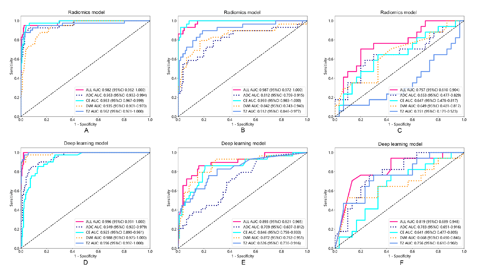
Figure 1. Receiver operator characteristic (ROC) curves of radiomics models(A-C) and deep learning models(D-E) in the training group (A, D), external validation group 1 (B, E), and external validation group 2 (C, F). The results show that the fused radiomics feature model (ALL) and the fused deep learning feature model (ALL) outperform other single-modality models.
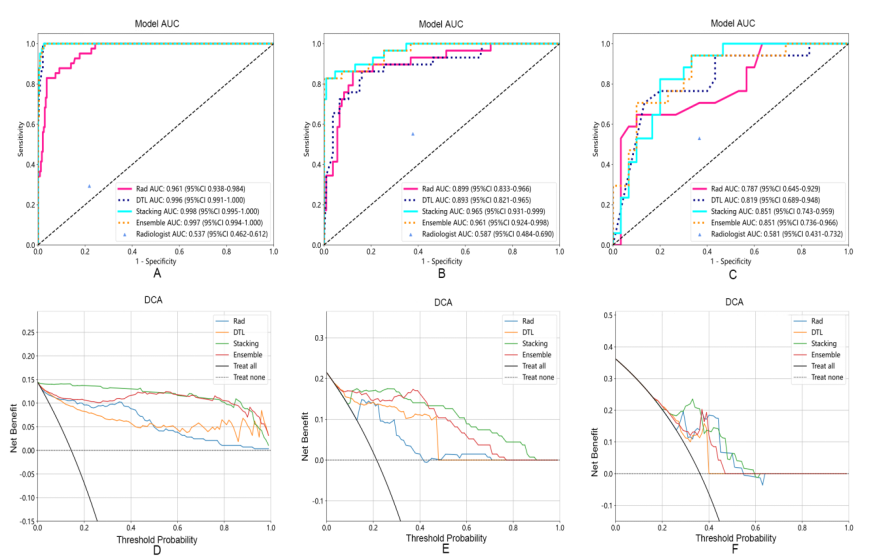
Figure 2. Receiver operator characteristic (ROC) curves (A-C) and decision curve analysis (DCA)(D-F) of different models in the training group (A, D), external validation group 1 (B, E), and external validation group 2 (C, F). The results show that the models compared to radiologists all have better net clinical gains, with the stacking model having the highest net gain.
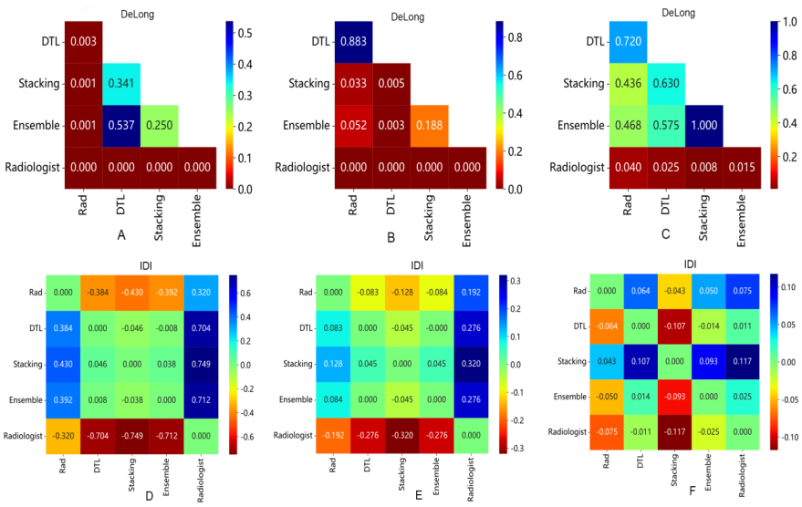
Figure 3. DeLong test (A-C) and integrated discrimination index (IDI)(D-F) of different models in the training group (A, D), external validation group 1 (B, E), and external validation group 2 (C, F). The results show that the optimal radiomics model, the optimal deep learning model, the stacking model, and the ensemble model all achieve good net benefits.
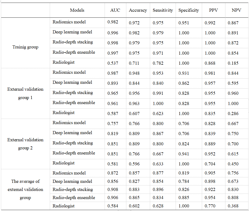
Table 1. The performance of various models. All models outperformed radiologists, with both fusion models outperforming the optimal radiomics and deep learning models, with the stacking model exhibiting the highest average AUC and accuracy in the external validation group.