3601
Prediction of Tumor-Stroma Ratio in Prostate Cancer using multiparametric MRI-Based Radiomics Mode1The Second Affiliated Hospital of Chongqing Medical University, Chongqin, China, 2Clinical Science, Philips Healthcare, Chengdu, China
Synopsis
Keywords: Diagnosis/Prediction, Prostate, magnetic resonance imaging, radiomics, tumor-stroma ratio, tumor microenvironment
Motivation: Tumor stroma is considered one of the key participants in prostate cancer development, progression, and even treatment resistance as an independent predictor, is associated with aggressiveness in a variety of malignancies.
Goal(s): We would like to apply the value of stroma cells in clinical practice for assessing the aggressiveness of PCa.
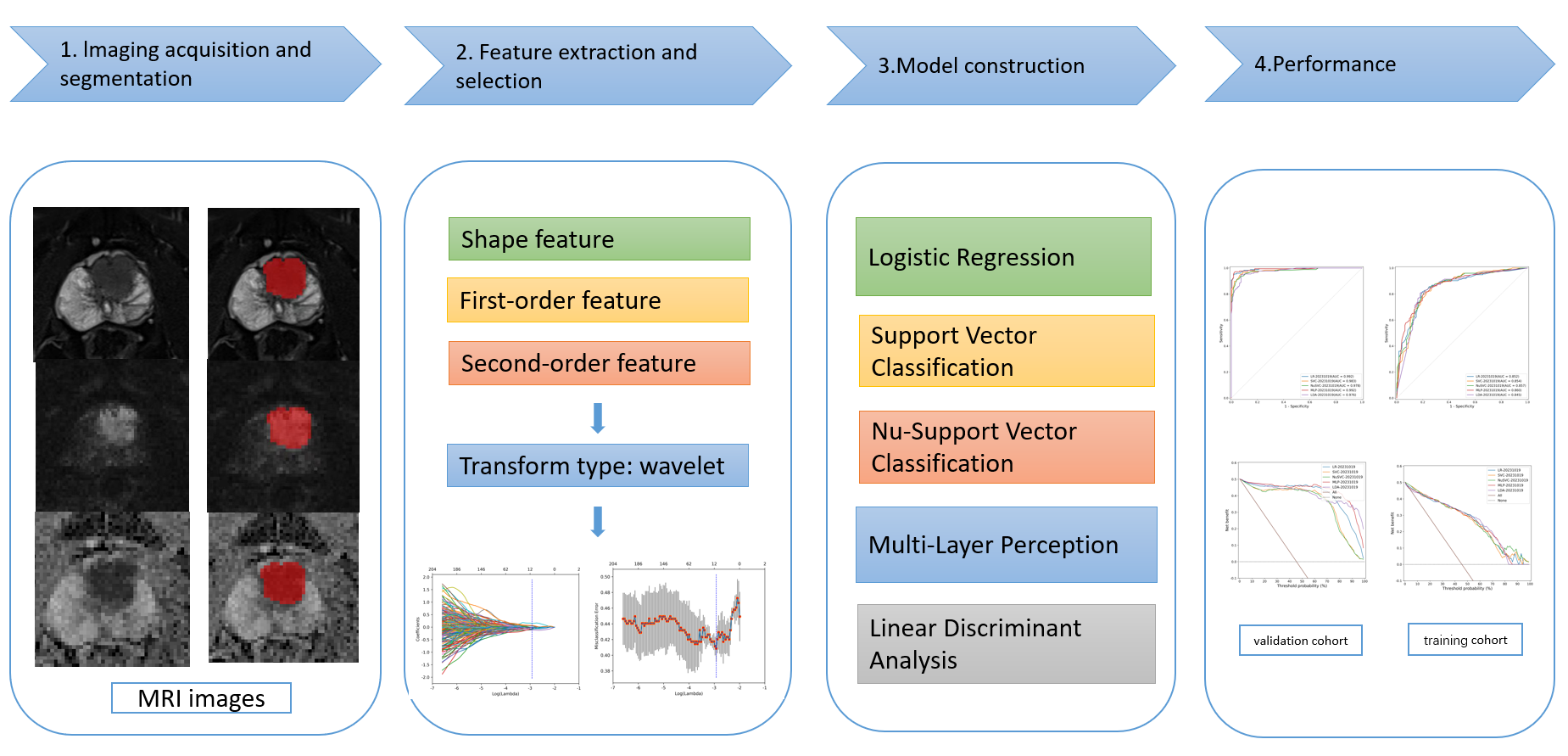
Approach: Five multiparametric magnetic resonance imaging (mp- MRI) radiomics feature-based machine learning models were developed and assessed to predict the tumor-stroma ratio (TSR) of PCa.
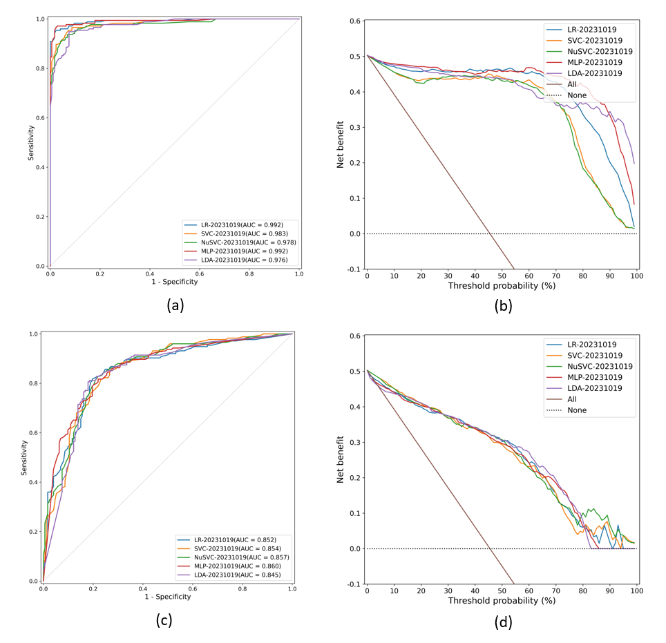
Results: The developed Multi-Layer Perception model showed excellent performance at predictive the TSR in prostate cancer with the area under the ROC curve (AUC) at 0.860.
Impact: This study constructed a mp-MRI-based radiomics model which is capable of accurately predicting the TSR of PCa and may serve as a complementary tool for assisting in risk stratification and guiding treatment decisions.
Active treatment of high-risk prostate cancer is an important measure to reduce prostate cancer (PCa) mortality. In previous studies, serum prostate-specific antigen level, Gleason score and tumor stage (T-stage) are important indicators of prostate cancer prognosis1–4. However, in terms of the current incidence and mortality of prostate cancer, these prognostic indicators are not enough, and more valuable prognostic indicators are urgently needed to optimize treatment decisions. Studies have shown that interactions between prostate stromal cells and prostate cancer cells can regulate tumor progression5. This study aims to develop a multiparametric MRI (mp-MRI) radiomics feature-based machine learning model to predict the tumor-stroma ratio (TSR) for assessing the aggressiveness of PCa, thus avoiding overtreatment of indolent PCa and undertreatment of aggressive PCa.
Methods
This retrospective study was approved by our Institutional Review Board. A total of 191 PCa patients confirmed through biopsy were recruited. Based on previous studies6,7, the TSR was defined as the percentage of stromal cells in tumor tissue and was retrospectively measured by one uropathology. TSR was evaluated within the tumor area with the highest Gleason score (GS) in the biopsy set. Based on the results, patients were categorized into either a high-TSR (stromal cell < 50%) or low-TSR (stromal cell > 50%) group. The MRI images were acquired using a 3.0 T MRI scanner (MAGNETOM Prisma; SIEMENS A Tim Dot System) equipped with an 8-channel phased-array software coil, which included the axial fat suppression (FS) T2-weighted imaging (T2WI), and diffusion-weighted imaging (DWI) sequences. Regions of interest (ROIs) for PCa were delineated on T2WI, DWI, and apparent diffusion coefficient (ADC) maps. Patients were divided into training and testing sets in a 7:3 ratio. Five radiomics models were constructed using the Least Absolute Shrinkage and Selection Operator (LASSO) algorithm for feature selections, including Logistic Regression (LR), Support Vector Classification (SVC), Nu-Support Vector Classification (NuSVC), Multi-Layer Perception (MLP), Linear Discriminant Analysis (LDA). Model performances were assessed using Receiver Operating Characteristic (ROC) curves, Decision Curve Analysis (DCA), and DeLong's test. The differences in clinical data between L-TSR and H-TSR patients were assessed using SPSS version 27.0.
Results
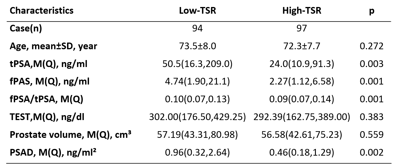
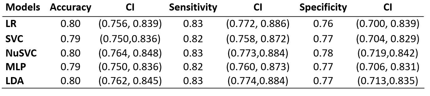
94 patients were classified as Low-TSR and 97 patients as High-TSR patients. The independent sample t-test and the Mann–Whitney U test showed that there were no significant differences in terms of age, testosterone (TEST) and prostate volume. Prostate-specific antigen(PSA), free-PSA(fPSA), fPSA/PSA and Prostate-specific antigen density(PSAD) showed statistically significant differences (all P < 0.010) between the two groups. Compared to other models, the MLP Model demonstrated superior performance in distinguishing between Low-TSR and High-TSR. In the validation and training cohorts, its area under the ROC curve (AUC) was 0.860[95% confidence interval (CI),0.820,0.900] and 0.992[95% CI,0.982,0.998], respectively. The model exhibited a classification accuracy of 0.79[95% CI,0.750,0.836], sensitivity of 0.82[95% CI,0.760,0.0.873], and specificity of 0.77[95% CI,0.706,0.831]. Decision Curve Analysis indicated that this radiomic model possesses strong clinical utility.
Discussion
For the progress and treatment of prostate cancer, people are more concerned about the tumor cells. However, as a solid tumor, prostate cancer occurs and develops under the continuous, dynamic and interaction between tumor cells and tumor microenvironments (TME). The TME includes stromal cells, endothelial cells, immune cells, and regulatory factors produced by these cells that support tumor growth8,9. As more and more mechanisms of TME determining tumor aggressiveness have been revealed, TSR, as a vital component of tumor microenvironments, has been widely confirmed as an independent prognostic factor for a variety of solid cancers5,10. In this study, we employed T2WI, DWI, and ADC maps using a high-throughput radiomics approach, and successfully constructed a MLP radiomics model to accurately predict the TSR of PCa. Radiomic methods can consistently extract high-dimensional image features that are highly correlated with intra-tumor heterogeneity, which are related to shape and texture. T2WI can clearly display the anatomic features of tumor lesions in prostate cancer patients, and the images contain more valuable texture features. DWI and ADC images objectively reflect the limited diffusion of water molecules in the tissue, indicating the malignancy of the tumor. At the same time, ADC images avoid the penetration effect of DWI caused by the excessive T2 decay time of the tissue. The combination of the three sequences can lead to more accurate and comprehensive tumor information. In our study, the radiomics model showed good performance in predicting L-TSR and H-TSR, with high AUC in both the training and validation groups.
Acknowledgements
The authors would like to thank InferScholar platform for the assistance in the performance of the construction of the radiomics model.References
1. YA N, JL G. What Goes Up Must Come Down: Identifying Truth from Global Prostate Cancer Epidemiology[J/OL]. European urology, 2020, 77(1)[2023-09-13]. https://pubmed.ncbi.nlm.nih.gov/31627967/. DOI:10.1016/j.eururo.2019.09.018.
2. PARKER C, CASTRO E, FIZAZI K, et al. Prostate cancer: ESMO Clinical Practice Guidelines for diagnosis, treatment and follow-up[J/OL]. Annals of Oncology, 2020, 31(9): 1119-1134. DOI:10.1016/j.annonc.2020.06.011.
3. PALLER C J, ANTONARAKIS E S. Management of biochemically recurrent prostate cancer after local therapy: evolving standards of care and new directions[J]. Clinical Advances in Hematology & Oncology: H&O, 2013, 11(1): 14-23.
4. SIEGEL R L, MILLER K D, WAGLE N S, et al. Cancer statistics, 2023[J/OL]. CA: A Cancer Journal for Clinicians, 2023, 73(1): 17-48. DOI:10.3322/caac.21763.
5. AYALA G, TUXHORN J A, WHEELER T M, et al. Reactive Stroma as a Predictor of Biochemical-Free Recurrence in Prostate Cancer[J].
6. SMIT M, VAN PELT G, ROODVOETS A, et al. Uniform Noting for International Application of the Tumor-Stroma Ratio as an Easy Diagnostic Tool: Protocol for a Multicenter Prospective Cohort Study[J/OL]. JMIR research protocols, 2019, 8(6): e13464. DOI:10.2196/13464.
7. SÆTER T, VLATKOVIC L, WAALER G, et al. The prognostic value of reactive stroma on prostate needle biopsy: A population‐based study[J/OL]. The Prostate, 2015, 75(6): 662-671. DOI:10.1002/pros.22957.
8. XIAO Y, YU D. Tumor microenvironment as a therapeutic target in cancer[J/OL]. Pharmacology & Therapeutics, 2021, 221: 107753. DOI:10.1016/j.pharmthera.2020.107753.
9. KWON J T W, BRYANT R J, PARKES E E. The tumor microenvironment and immune responses in prostate cancer patients[J/OL]. Endocrine-Related Cancer, 2021, 28(8): T95-T107. DOI:10.1530/ERC-21-0149.
10. PENG C, LIU J, YANG G, et al. The tumor-stromal ratio as a strong prognosticator for advanced gastric cancer patients: proposal of a new TSNM staging system[J/OL]. Journal of Gastroenterology, 2018, 53(5): 606-617. DOI:10.1007/s00535-017-1379-1.
Figures