3599
Utilizing XGBoost and LR to find the significant predictive factors of MR-guided high intensity focused ultrasound ablation in uterine fibroids1Radiology Department, The First Affiliated Hospital of Xi'an Jiaotong University, Xi'an, China
Synopsis
Keywords: Diagnosis/Prediction, Radiomics, MR-HIFU, Treatment of Uterine Fibroids, XGBoost, SHAP, LR, Coefficients
Motivation: MR-HIFU offers a new treatment option for women with uterine fibroids. However, there is currently a lack of quantitative models to predict the efficacy of MR-HIFU based on T2WI of fibroids for guiding preoperative clinical decisions.
Goal(s): We hope to identify the most important predictive factors of MR-HIFU treatment for uterine fibroids and predict the efficacy using radiomics data combine with clinical data.
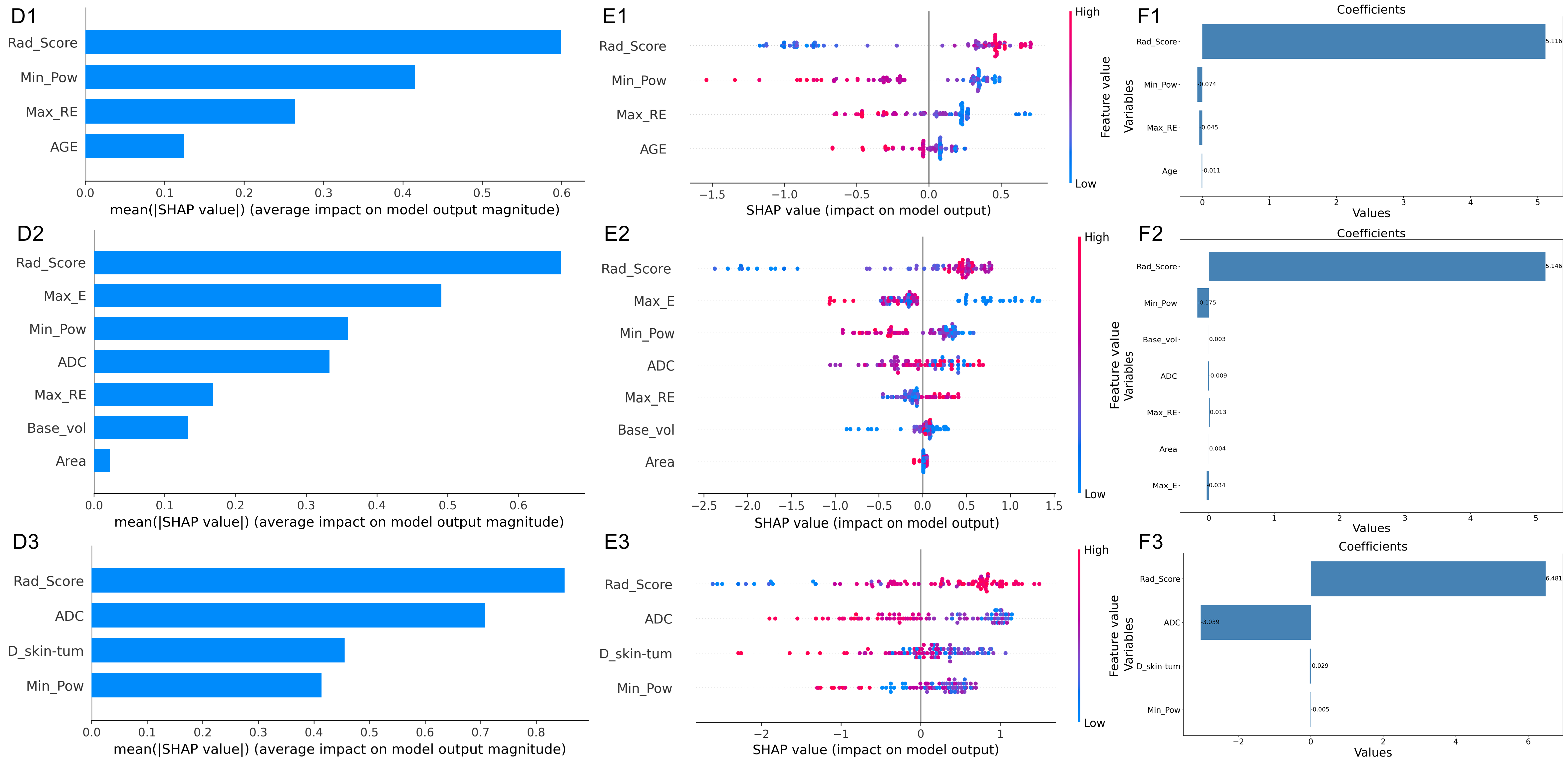
Approach: We employed XGBoost and logistic regression (LR) to build two prediction models. SHAP values of XGBoost and LR coefficients were used to pinpoint significant predictive factors.
Results: Both models achieved outstanding results and the significant predictive factors are consistent.
Impact: Our excellent model results have identified the optimal predictive factors for assessing the efficacy of MR-HIFU in the treatment of uterine fibroids. These factors aid physicians in preoperative guidance and clinical strategy formulation, clarifying which patients will achieve better outcomes.
Introduction
Research reveals that T2-weighted imaging (T2WI) signal intensity is vital for predicting the success of Magnetic Resonance guided High-Intensity Focused Ultrasound (MR-HIFU) ablation in treating uterine fibroids, yet quantitative methods for assessing T2WI signal intensity and its heterogeneity remain undeveloped[1,2,3,4,5,6]. Our study aims to achieve this by establishing predictive models for MR-HIFU treatment outcomes, including ablation effects, symptom improvement and fibroid shrinkage, using preoperative T2WI Radiomics scores (Radscore)[7]. By integrating clinical factors with Radiomics scores, we employed XGBoost and logistic regression (LR) models, whose significant prediction factors were assessed using explainable AI techniques, the SHapley Additive exPlanations (SHAP) values, and LR coefficients [8,9,10,11,12,13,14].Materials and Methods:
For the study, all participants underwent MR-HIFU treatment and were scanned using sagittal fat-suppressed T2WI TSE with parameters like TR/TE at 3800/120ms, NSA at 2, FOV at 250×250mm, Slice at 3.0mm, Gap at 0.3mm, Matrix at 256×256, Scan time at 148s, and Flip Angle at 90°. The region of interest (ROI) in T2WI data were manually segmented using ITK-SNAP (Version 3.6) by two radiologists, and Dice Similarity Coefficient (DSC) was performed to evaluate the consistency[15]. Feature extraction was employed by PyRadiomics tool[16]. Statistical evaluations were done using the uAI Research Portal system (v.1.6) and MedCalc software (MedCal 20.0). Select K Best, Least Absolute Shrinkage and Selection Operator (LASSO) method was employed for filtering radiomics features[17,18].For three outcomes, three main evaluation metrics are: Non-perfused volume ratio (NPVR), NPVR (%) = (Non-perfused volume / Fibroid volume) × 100%; Volume shrinkage ratio (VSR), VSR (%) = (Baseline volume - Volume at m months) / Baseline volume × 100%; Symptom severity score reduction ratio (SSSRR), SSSRR (%) = (Baseline SSS - SSS at m months) / Baseline SSS × 100%.To interpret results, SHAP values were used for XGBoost, offering insights into feature impacts on predictions. In contrast, LR used coefficients, indicating the change in outcome odds for predictor variations.Results:
Patients:In this study, a total of 252 patients with uterine fibroids were screened, and ultimately 109 (43.3%) were included. MRI screening failures accounted for 94 cases (37.3%), which were the primary reason for participants not being enrolled in the study, and 49 individuals (19.4%) withdrew from the study after signing the informed consent forms. The number of fibroids included was 113, with a ratio of 7:3 for training (N=79) and testing cohort (N=34).
Prediction Factor Analysis:
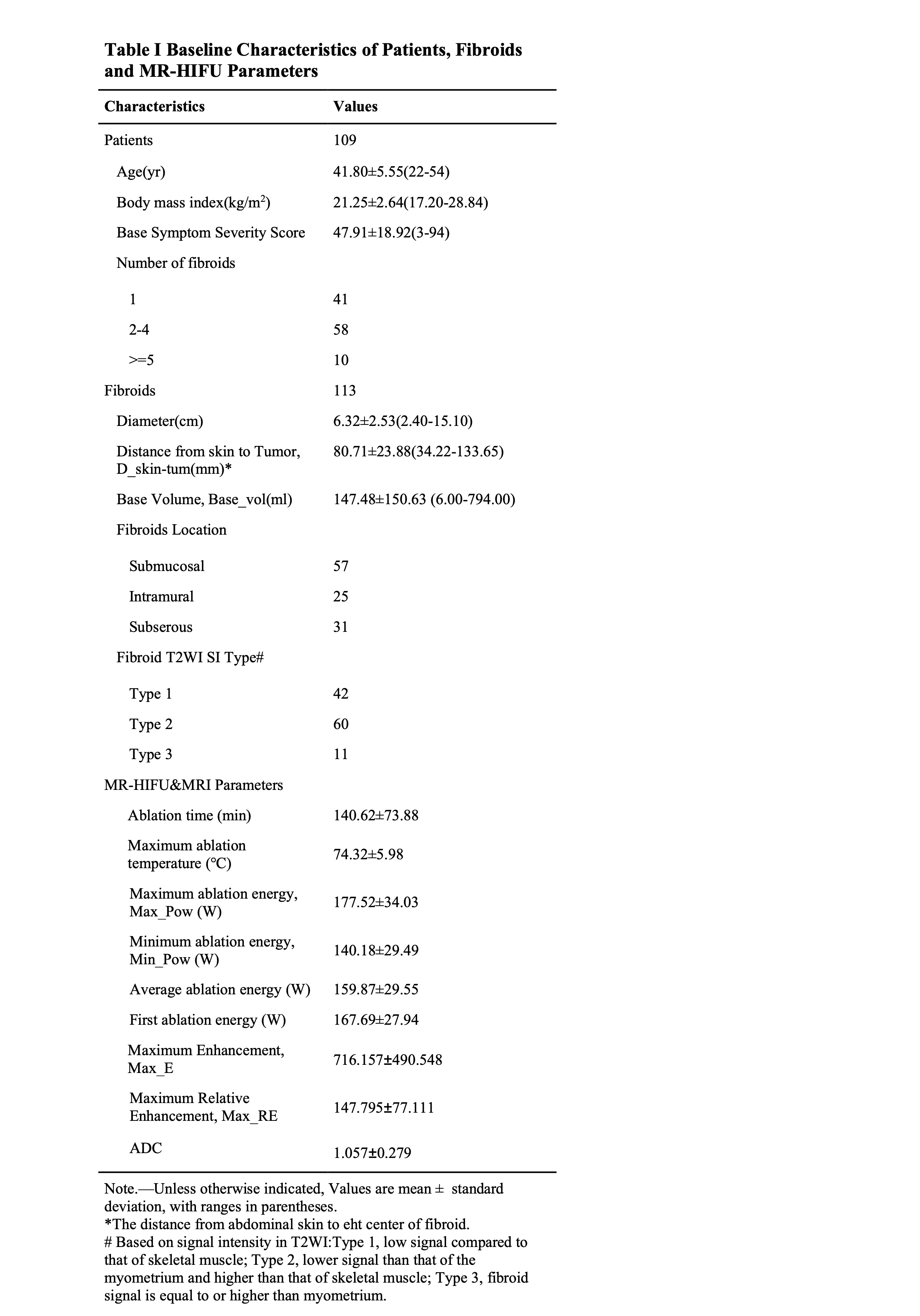
The collected patient information includes: age, body mass index, base symptom severity score, number of fibroids, fibroid general characteristics, and MR-HIFU ablation parameters such as Maximum Enhancement, (Max_E) and Maximum Relative Enhancement, (Max_RE). Details and distribution can be found in Figure I.
For the Radscore:
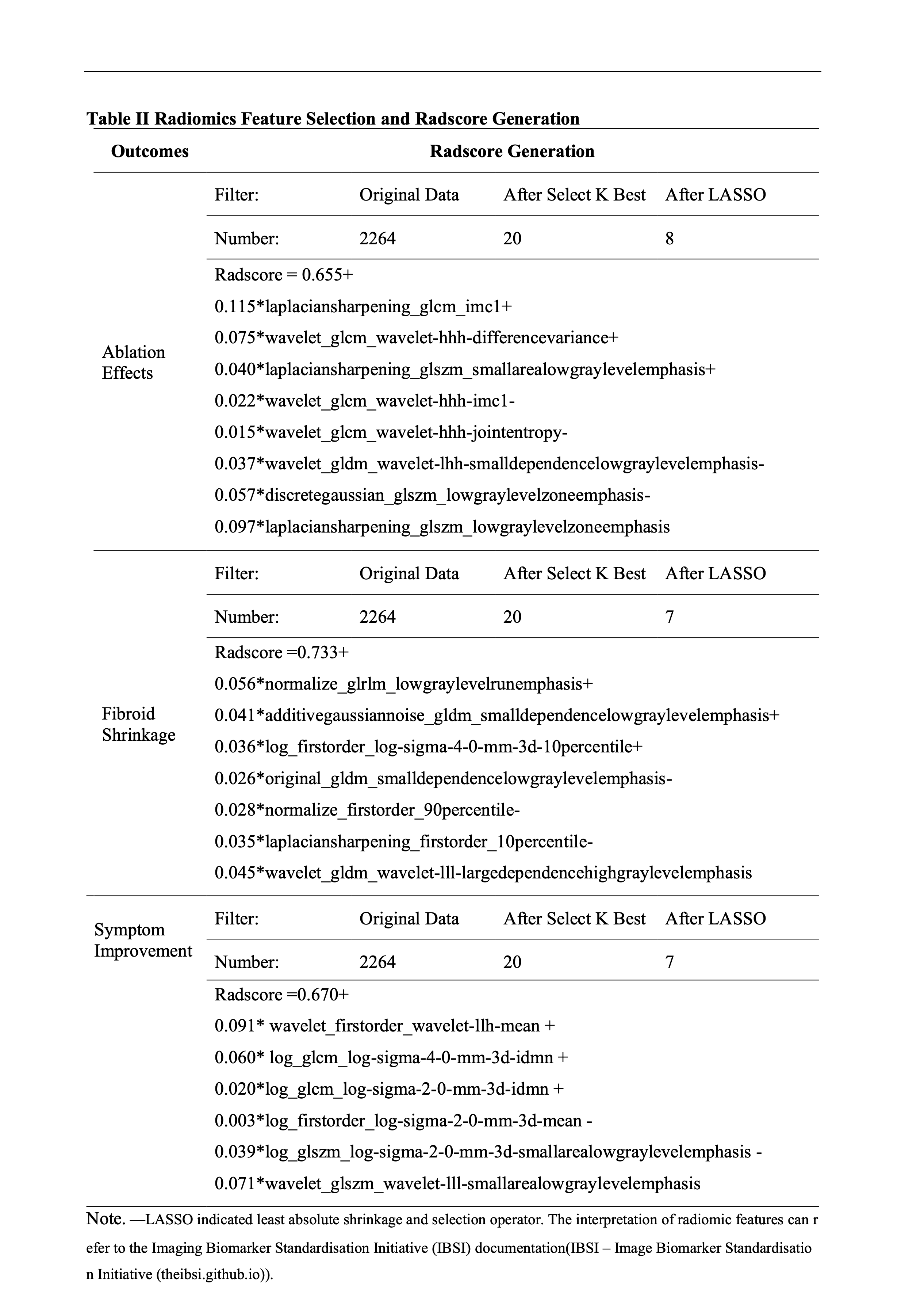
Using NPVR≥60%, VSR≥20%, and SSSRR≥30% as the three outcome grouping criteria, the Select K Best method was employed to filter the top 20 factors by weight. Subsequently, LASSO regression was used to generate the Radscore. The number of factors retained to build radscore for the three outcomes were 8, 7, and 6, respectively. For detailed information and the Radscore, please refer to Figure II.
Models Results:
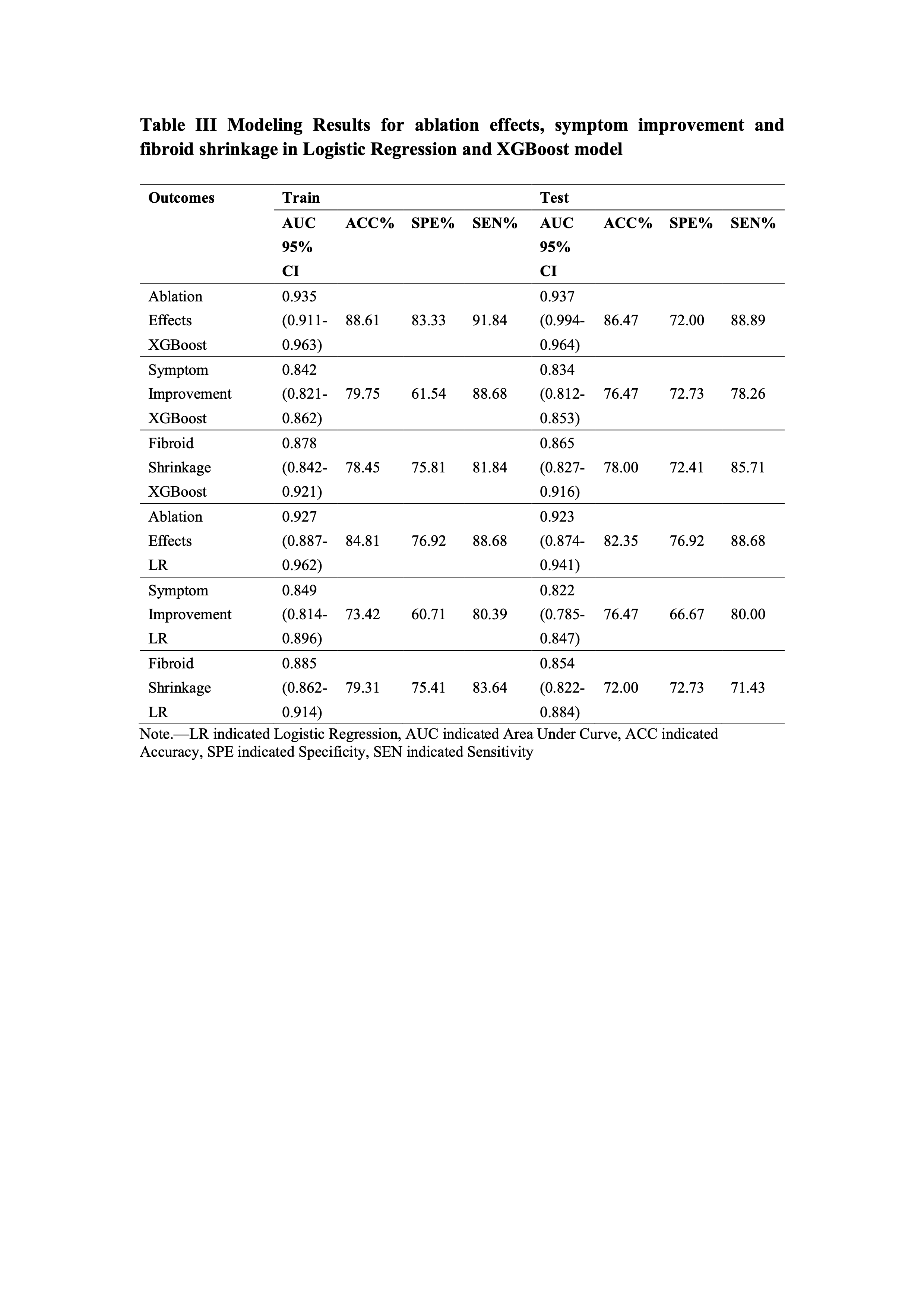
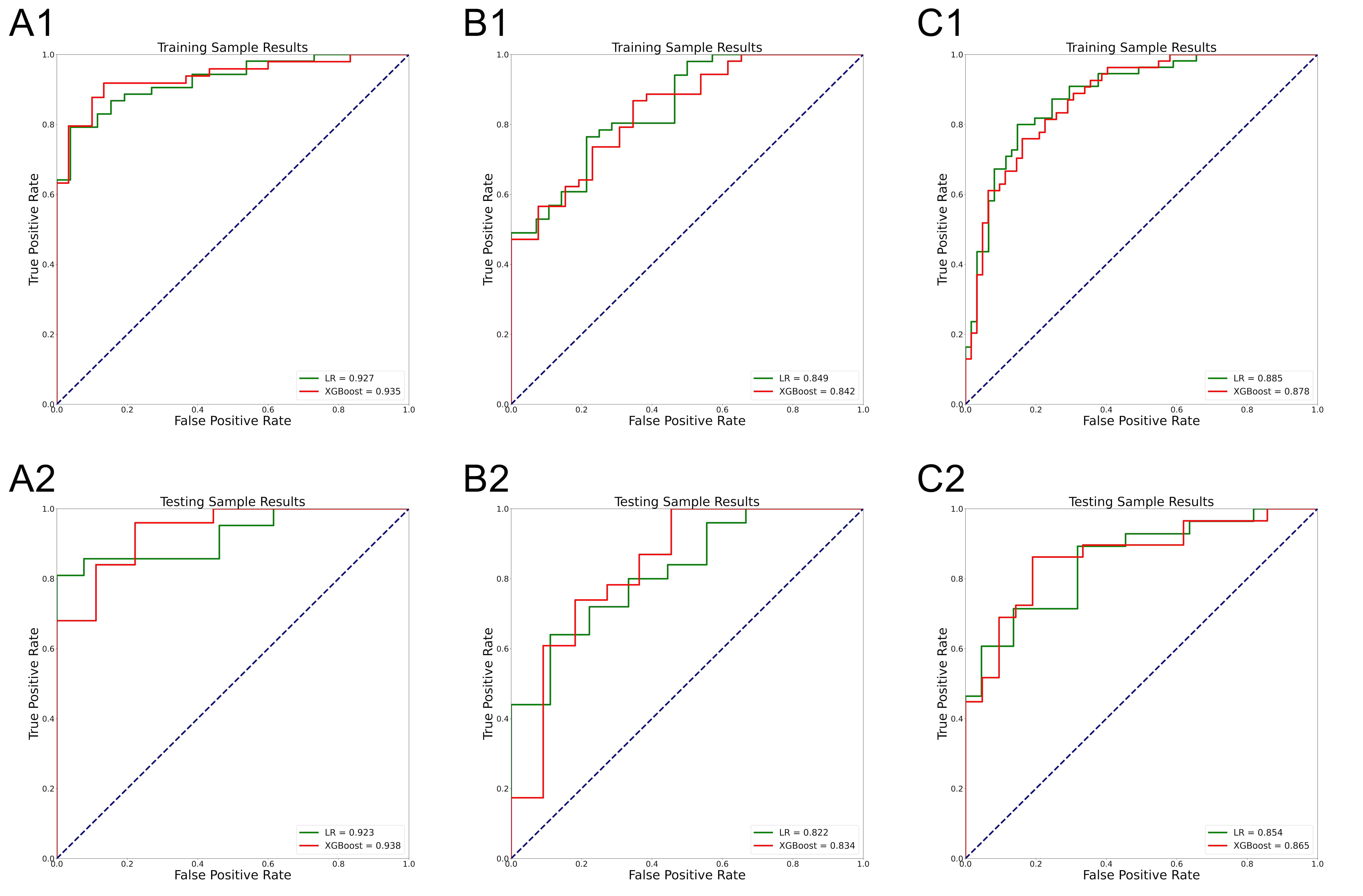
For three outcomes, the parameters included in the LR model and the XGBoost model are respectively: Radscore, AGE, Min_Pow, Max_RE; Radscore, Min_Pow, Base_vol, ADC, Max_RE, Area, Max_E; Radscore, D_skin-tum, Min_Pow, ADC. Upon evaluation, the XGBoost model showcased results with Area Under the Curve (AUC) values of 0.935/0.937, 0.842/0.834, and 0.878/0.865 for training and testing cohorts respectively. Similarly, the LR model displayed nearly AUC values of 0.927/0.923, 0.849/0.822, and 0.885/0.854. Both models demonstrated excellent classification performance and detailed results can be shown in Figure III and Figure IV. The top three coefficients in LR model and the top three average SHAP values in XGBoost model were consistent, which could be seen in Figure V.
Disscussion
Both classification models have demonstrated exceptional predictive abilities in MR-HIFU prognosis. Notably, a comparison between the two modeling techniques reveals that they both pinpoint the same top three features for each of the outcome indicators, it suggests a consistency in the influence of the features across both models. This convergence suggests that the identified parameters are robust predictors across different model architectures, thus reinforcing their significance in clinical decision-making for MR-HIFU treatments. Moreover, the Radiomics score (Radscore) showing the highest logistic regression coefficients and average SHAP values across the prediction models indicates that radiomic features play the most significant role in predicting the efficacy of MR-HIFU treatment for uterine fibroids.Acknowledgements
This work was supported by the Shaanxi Provincial KeyResearch and Development Program (2022SF-105).References
[1] Funaki, Kaoru, et al. "Magnetic resonance-guided focused ultrasound surgery for uterine fibroids: relationship between the therapeutic effects and signal intensity of preexisting T2-weighted magnetic resonance images." American journal of obstetrics and gynecology 196.2 (2007): 184-e1.
[2] Lénárd, Zsuzsanna M., et al. "Uterine leiomyomas: MR imaging–guided focused ultrasound surgery—imaging predictors of success." Radiology 249.1 (2008): 187-194.
[3] Zhao, Wen-Peng, et al. "Feasibility of ultrasound-guided high intensity focused ultrasound ablating uterine fibroids with hyperintense on T2-weighted MR imaging." European journal of radiology 82.1 (2013): e43-e49.
[4] Park, Hyun, Sang-Wook Yoon, and Amit Sokolov. "Scaled signal intensity of uterine fibroids based on T2-weighted MR images: a potential objective method to determine the suitability for magnetic resonance-guided focused ultrasound surgery of uterine fibroids." European radiology 25 (2015): 3455-3458.
[5] Zheng, Yineng, et al. "Nonenhanced MRI-based radiomics model for preoperative prediction of nonperfused volume ratio for high-intensity focused ultrasound ablation of uterine leiomyomas." International Journal of Hyperthermia 38.1 (2021): 1349-1358.
[6] Li, Zhicong, et al. "Utilization of radiomics to predict long-term outcome of magnetic resonance–guided focused ultrasound ablation therapy in adenomyosis." European Radiology 31 (2021): 392-402.
[7] Lambin, Philippe, et al. "Radiomics: extracting more information from medical images using advanced feature analysis." European journal of cancer 48.4 (2012): 441-446.
[8] Chen, Tianqi, and Carlos Guestrin. "Xgboost: A scalable tree boosting system." Proceedings of the 22nd acm sigkdd international conference on knowledge discovery and data mining. 2016.
[9] Chen, Tianqi, et al. "Xgboost: extreme gradient boosting." R package version 0.4-2 1.4 (2015): 1-4.
[10] Pesantez-Narvaez, Jessica, Montserrat Guillen, and Manuela Alcañiz. "Predicting motor insurance claims using telematics data—XGBoost versus logistic regression." Risks 7.2 (2019): 70.
[11] Le, Nguyen Quoc Khanh, et al. "Radiomics-based machine learning model for efficiently classifying transcriptome subtypes in glioblastoma patients from MRI." Computers in Biology and Medicine 132 (2021): 104320.
[12] Lundberg, Scott M., and Su-In Lee. "A unified approach to interpreting model predictions." Advances in neural information processing systems 30 (2017).
[13] Parsa, Amir Bahador, et al. "Toward safer highways, application of XGBoost and SHAP for real-time accident detection and feature analysis." Accident Analysis & Prevention 136 (2020): 105405.
[14] Bi, Yue, et al. "An interpretable prediction model for identifying N7-methylguanosine sites based on XGBoost and SHAP." Molecular Therapy-Nucleic Acids 22 (2020): 362-372.
[15] Zou, Kelly H., et al. "Statistical validation of image segmentation quality based on a spatial overlap index1: scientific reports." Academic radiology 11.2 (2004): 178-189.
[16] Van Griethuysen, Joost JM, et al. "Computational radiomics system to decode the radiographic phenotype." Cancer research 77.21 (2017): e104-e107.
[17]Tibshirani, Robert. "Regression shrinkage and selection via the lasso." Journal of the Royal Statistical Society Series B: Statistical Methodology 58.1 (1996): 267-288.
[18] Pedregosa, Fabian, et al. "Scikit-learn: Machine learning in Python." the Journal of machine Learning research 12 (2011): 2825-2830.
Figures