3597
Development and validation of an MRI-based radiomics nomogram to predict progression-free survival in patients with endometrial cancer1The First Central Clinical College of Tianjin Medical University, Tianjin, China, Tianjin, China, 2First Central Hospital, Tianjin, China, Tianjin, China, 3Jinghai Hospital, Tianjin, China, Tianjin, China, 4Siemens Healthineers Ltd., Beijing, China
Synopsis
Keywords: Diagnosis/Prediction, Radiomics
Motivation: There is no appropriate index to assess the prognosis of patients with endometrial cancer after surgery.
Goal(s): Our goal was to explore the value of an MRI-based radiomics nomogram in predicting the progression-free survival (PFS) of patients with endometrial cancer.
Approach: A nomogram was established using multivariable Cox regression by incorporating significant clinical-pathological predictors and radiomics signatures developed by another least absolute shrinkage and selection operator Cox regression.
Results: The nomogram proved itself valuable in predicting PFS. Furthermore, the radiomics nomogram–defined risk stratification was associated with PFS.
Impact: The novel nomogram may contribute to the precise stratification of patients with a high risk of progression. Therefore, more active follow-up and adjuvant therapy should be carried out after surgery to prolong and improve the patients' quality of life.
Introduction
Relapse and recurrence are the major causes of mortality in patients with resected endometrial cancer (EC). The widely known risk factors for recurrence include myometrial invasion depth, lymphovascular invasion, cervical stromal invasion, parametrial invasion, and lymph node metastasis. Moreover, many studies revealed immunohistochemical (IHC) biomarkers such as ER, PR, P53, and Ki-67 were associated with prognosis and risk classification of EC. Radiomics is an emerging field that adopts quantitative image analytics to determine the associations between imaging features and clinical endpoints, such as prognosis and survival, which is more effectively than conventional MRI-based imaging features. Recently, radiomics-based nomograms have been proven to predict prognosis in various malignancies and are considered a potentially useful visualization tool due to their outstanding predictive performance. Therefore, accurate and sufficient prognosis assessments of EC are needed and further provide valuable information for personalized therapeutic strategies. The aim of this study was to develop a nomogram model based on multiparametric MRI radiomics to obtain patients' risk scores in order to investigate the value of this model in predicting progression-free survival (PFS) of EC patients.Methods
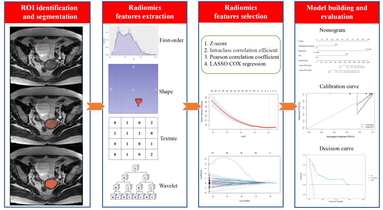
During January 2014 and August 2022, 165 patients with pathologically diagnosed EC were retrospectively enrolled in two centers (104 in the training cohort and 61 in the validation cohort). The patients preoperatively underwent magnetic resonance imaging (MRI), including axial T2-weighted imaging (T2WI) with and without fat saturation (FS) and DWI with a b value of 800 or 1000 s/mm2 on a 3T system (MAGNETOM Trio a Tim System, Siemens Healthcare, Erlangen, Germany) for center A and a 1.5T system (Achieva, Philips Healthcare, Best, the Netherlands) for center B. After DWI examination, ADC maps were generated. The radiomics features were extracted from multiparametric MR imaging and then selected by a least absolute shrinkage and selection operator (LASSO) Cox regression to develop a radiomics signature for PFS prediction. A nomogram was established using a multivariable Cox regression by incorporating significant clinical-pathological predictors and radiomics signatures. The performance was evaluated by an analysis of the receiver operating characteristic, calibration, and decision curves. The workflow of the model construction is shown in Figure 1.Results
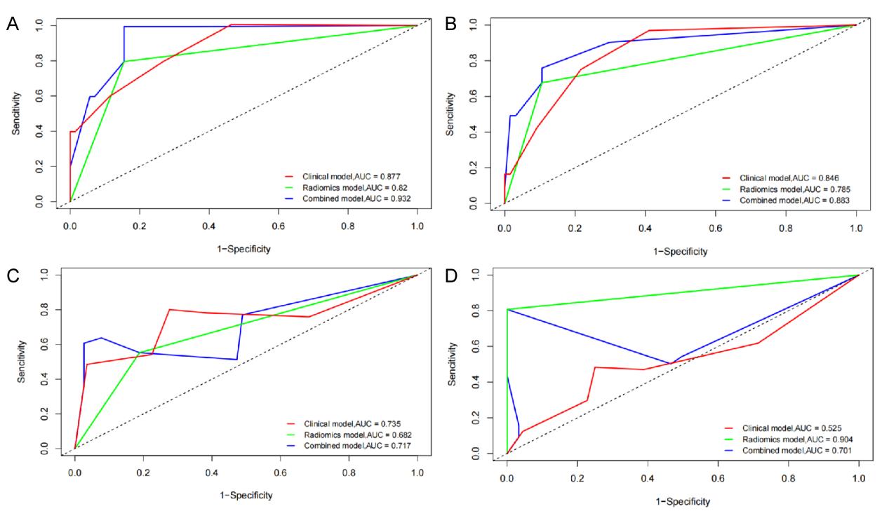
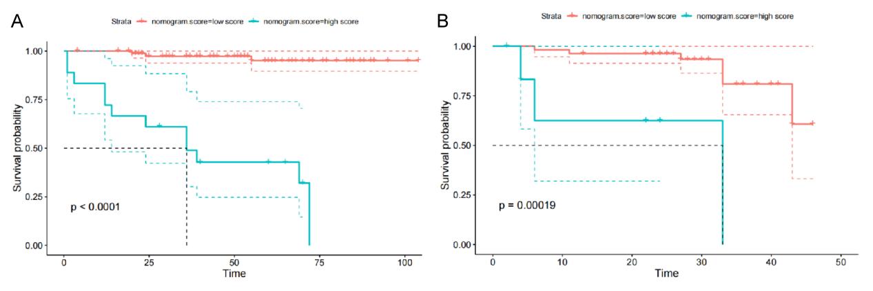
The radiomics signature was constructed using four significant features (Table 1). A multivariate Cox analysis revealed that parametrial invasion, the positivity rate of PR, and the radiomics score were independent risk variables associated with PFS, so they were incorporated into the construction of a nomogram (Table 2). The C-indexes of the nomogram were 0.877 (95% CI: 0.797–0.957) in the training cohort and 0.772 (95% CI: 0.641–0.903) in the validation cohort, which outperformed the clinical model and the radiomics signature (Table 3). The nomogram displayed good prediction performance in estimating PFS with AUC values of 0.932 and 0.883 at 1, and 3 years, respectively (Figure 2). A Kaplan–Meier survival analysis showed that the radiomics nomogram–defined high-risk groups of patients with EC had shorter PFS than the low-risk groups (Figure 3).Discussion
The present study primarily investigated the role of the radiomics nomogram in predicting the risk of progression and thus identified patients at high risk of early progression of EC. A radiomics nomogram, which was successfully developed and validated by integrating MRI radiomics features and clinicopathological characteristics, achieved good predictive value for predicting PFS. Furthermore, the radiomics nomogram–defined risk stratification was associated with PFS.Conclusion
The novel radiomics nomogram, which is based on combined preoperative MRI-based radiomics features and clinicopathological risk factors, may be considered a noninvasive prognostic index for predicting the individual PFS of patients with EC.Acknowledgements
We thank Xiangfeng Xu (Tianjin Central Hospital of Gynecology Obstetrics) for providing the data. We are also grateful to our team members for their help in data collection and thesis revision.References
[1] Crosbie EJ, Kitson SJ, McAlpine JN, Mukhopadhyay A, Powell ME, Singh N. Endometrial cancer. Lancet. 2022. 399(10333): 1412-1428.
[2] Fung-Kee-Fung M, Dodge J, Elit L, Lukka H, Chambers A, Oliver T. Follow-up after primary therapy for endometrial cancer: a systematic review. Gynecol Oncol. 2006. 101(3): 520-9.
[3] Sheikh MA, Althouse AD, Freese KE, et al. USA endometrial cancer projections to 2030: should we be concerned. Future Oncol. 2014. 10(16): 2561-8.
[4] Legge F, Restaino S, Leone L, et al. Clinical outcome of recurrent endometrial cancer: analysis of post-relapse survival by pattern of recurrence and secondary treatment. Int J Gynecol Cancer. 2020. 30(2): 193-200.
[5] Wang C, Tran DA, Fu MZ, Chen W, Fu SW, Li X. Estrogen Receptor, Progesterone Receptor, and HER2 Receptor Markers in Endometrial Cancer. J Cancer. 2020. 11(7): 1693-1701.
[6] Zhang S, Song M, Zhao Y, et al. Radiomics nomogram for preoperative prediction of progression-free survival using diffusion-weighted imaging in patients with muscle-invasive bladder cancer. Eur J Radiol. 2020. 131: 109219.
[7] Zhai Y, Bai J, Xue Y, et al. Development and validation of a preoperative MRI-based radiomics nomogram to predict progression-free survival in patients with clival chordomas. Front Oncol. 2022. 12: 996262.
[8] Liu D, Yang L, Du D, et al. Multi-Parameter MR Radiomics Based Model to Predict 5-Year Progression-Free Survival in Endometrial Cancer. Front Oncol. 2022. 12: 813069.
[9] Xiang F, Liang X, Yang L, Liu X, Yan S. Contrast-enhanced CT radiomics for prediction of recurrence-free survival in gallbladder carcinoma after surgical resection. Eur Radiol. 2022. 32(10): 7087-7097.
[10] Zhang W, Fang M, Dong D, et al. Development and validation of a CT-based radiomic nomogram for preoperative prediction of early recurrence in advanced gastric cancer. Radiother Oncol. 2020. 145: 13-20.
[11] Jacob H, Dybvik JA, Ytre-Hauge S, et al. An MRI-Based Radiomic Prognostic Index Predicts Poor Outcome and Specific Genetic Alterations in Endometrial Cancer. J Clin Med. 2021. 10(3).
[12] van der Putten L, Visser N, van de Vijver K, et al. Added Value of Estrogen Receptor, Progesterone Receptor, and L1 Cell Adhesion Molecule Expression to Histology-Based Endometrial Carcinoma Recurrence Prediction Models: An ENITEC Collaboration Study. Int J Gynecol Cancer. 2018. 28(3): 514-523.
Figures
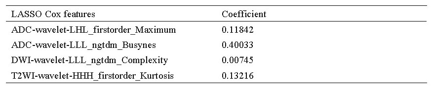
Table 1 LASSO Cox regression was used to finally filter the radiomics features used for calculating Rad-Scores
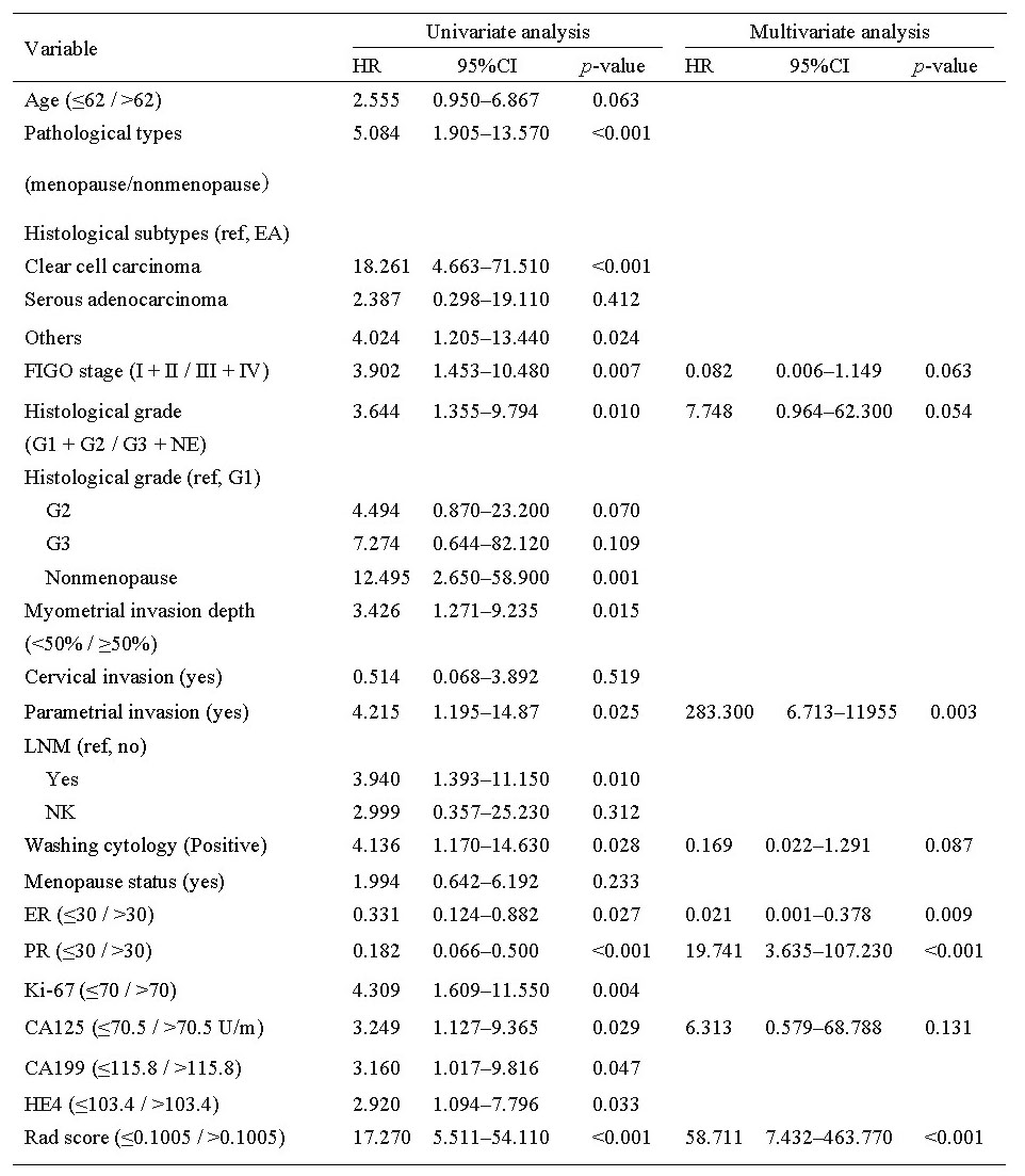
Table 2 Univariate and multivariate Cox analyses of risk factors for recurrence
HR, hazard ratio, CI, confidence interval.