3594
A new approach for automatic segmentation of prostate and its lesion regions on the magnetic resonance imaging1Baoji Central Hospital, Baoji, China, 2GE HealthCare MR Research, Beijing, China
Synopsis
Keywords: Diagnosis/Prediction, Machine Learning/Artificial Intelligence
Motivation: In recent years,many neural network models based on prostate lesion segmentation in magnetic resonance images have different stability and diagnostic efficiency.
Goal(s): we want to get an automatic segmentation model with high performance for the prostate and its lesion region.
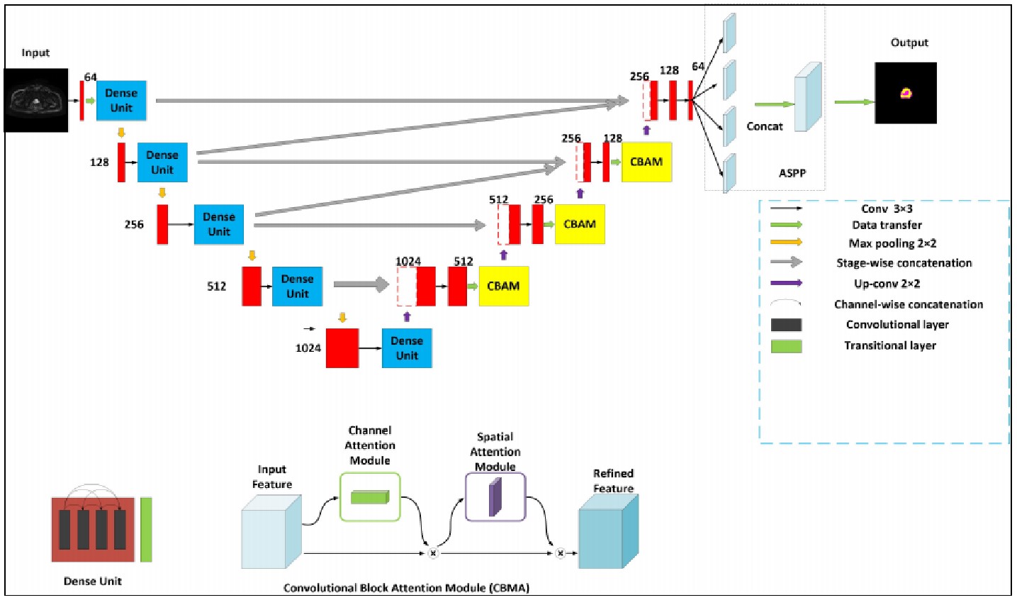
Approach: Our Network DCNN is inspired by the U-Net model with the encoding-decoding path as the backbone,importing dense block,attention mechanism techniques,and group norm-Atrous Spatial Pyramidal Pooling,these could be broadly used to improve the capability of CNN.
Results: Compared to the state-of-the-art models,FCN,U-Net,U-Net++,and ResUNet.The segmentation performance of DCNN for prostate lesions On the MR DWI image swas better than the other models.
Impact: The DCNN model with dense block, convolution block attention module, and group norm-Atrous Spatial Pyramid Pooling performed well in the segmentation of the prostate and its lesion regions. which supports its potential to assist prostate disease diagnosis in clinical medicine.
Introduction
The high-level spatial resolution and soft tissue conspicuity of MRI make it appropriate for prostate segmentation,staging and volume calculation of prostate cancer1.CNNs are obtaining a concern in the medical image field due to the state-of-the-art scores on plentiful image identification and segmentation tasks,U-Net is arguably an even hotter segmentation network.But the ambiguity of each tissue boundary inside the image makes it difficult to distinguish it from the heterogeneous tissue within the surrounding prostate, further resulting in under-segmentation or over-segmentation.Our Network DCNN is inspired by the U-Net model with the encoding-decoding path as the backbone, importing dense block, attention mechanism techniques,We want to develop an accurate and automatic segmentation model based on convolution neural network to segment the prostate and its lesion regions.Methods
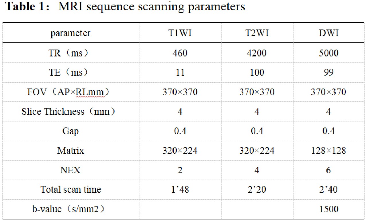
The study was approved by the Ethics Committee of Baoji Central Hospital and each subject signed informed consent.Of all 180 subjects, 122 healthy individuals and 58 patients with prostate cancer were included. For each subject, All patients were scanned on a 3.0T MR station (MR750w, GE Healthcare) scanner with a body phased array, which included T1WI, T2WI, DWI,All slices of the prostate were comprised in the DWIs,For the lesion extent of large sections of prostate pathology, the lesion boundary was delineated on DWI images,A novel DCNN is proposed to automatically segment the prostate and its lesion regions. This model is inspired by the U-Net model with the encoding-decoding path as the backbone, importing dense block, attention mechanism techniques, and group norm-Atrous Spatial Pyramidal Pooling. Data augmentation was used to avoid overfitting in training. In the experimental phase,the data set was randomly divided into a training (70%), testing set (30%). four fold cross-validation methods were used to obtain results for each metric. (Table 1,Figure 1)Results
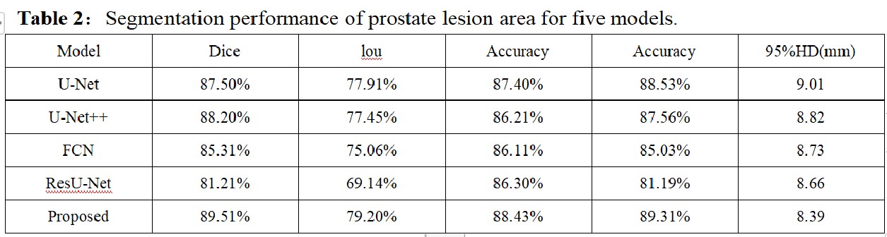
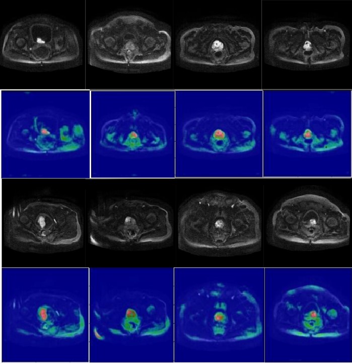
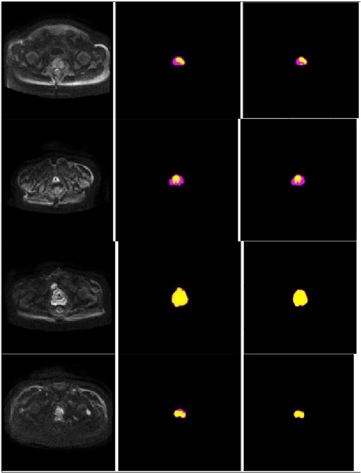
The proposed model achieved in terms of Iou, Dice score, accuracy, sensitivity, 95% Hausdorff Distance, 86.82%,93.90%, 94.11%, 93.8%,7.84 for the prostate, 79.2%, 89.51%, 88.43%,89.31%,8.39 for lesion region in segmentation. Compared to the state-of-the-art models, FCN, U-Net, U-Net++, and ResUNet, the segmentation model DCNN achieved more promising results. (Table 2,Figure 2,3)Discussion
In this study, we propose a novel DL-based architecture that utilizes the dense block and CBAM, as well as the GN-ASPP module, to fully leverage the complementary information encoded in different layers of the model.The segmentation output is obtained through an end-to-end approach.Finally,it was demonstrated that the proposed segmentation method outperformed the results of the state-of the-art methods for segmentation of the prostate and lesion region. Specifically, the proposed method exhibited excellent results,especially for the lesion region, which is of great significance for clinical diagnosis and treatment.Several studies demonstrates artifical intelligence is valid in urology works 2,3, especially using DCNN to segment the prostate or determine prostate cancer.Zhu et al.4 designed a DCNN model to segment the prostate zone and outer contour. The model was derived from a cascade of two models.One model was responsible for segmenting the prostate region and one for segmenting the prostate zone. However, an end-to-end model, like the one proposed in our study,is more efficient in reducing training time and facilitating clinical diagnosis. Duran et al.5 also developed a novel CNN model for PCa segmentation with an attention mechanism. This strategy is similar to our approach.our method has the same convergence effect as the classical U-Net because of the backbone of the model. Meantime, our model has also similar convergence rates and effects in both the prostate and lesion regions, which demonstrates that the model has generalization properties.Conclusion
The DCNN model with dense block, convolution block attention module, and group norm-Atrous Spatial Pyramid Pooling yielded excellent performance in accurate and automatic segmentation of the prostate and lesion regions, revealing that the novel deep convolutional neural network could be used in clinical disease treatment and diagnosis.Acknowledgements
No acknowledgement found.References
1.Zettinig O,Shah A,Hennersperger C,et al. Multimodal image-guided prostate fusion biopsy based on automatic deformable registration. Int J Comput Assisted Radiol Surg (2015) 10(12):1997–2007.
2.Checcucci E,Rosati S,De Cillis S,et al.Artificial intelligence for target prostate biopsy outcomes prediction the potential application of fuzzy logic. Prostate Cancer Prostatic Dis (2022) 25(2):359–62.
3.Wessels F, Kuntz S, Krieghoff-Henning E,et al.Artificial intelligence to predict oncological outcome directly from hematoxylin & eosin-stained slides in urology: a systematic review. Minerva Urol Nephrol(2022).
4.Zhu Y,Wei R,Gao G,et al.Fully automatic segmentation on prostate MR images based on cascaded fully convolution network.J Magn Reson Imaging(2019)49(4):1149–56.
5.Duran A,Dussert G,Rouvière O,et al.ProstAttention-net: A deep attention model for prostate cancer segmentation by aggressiveness in MRI scans.Med Image Anal(2022)77:102347.
Figures