3588
DCE-MRI radiomics models predict IDH mutation in adult diffuse gliomas1Department of Radiology, Department of Radiology, The Affiliated Drum Tower Hospital of Nanjing University Medical School, Nanjing, China, 2Department of Research and Development, Department of Research and Development, Shanghai United Imaging Intelligence Co., Shanghai, China, 3Department of Radiology, The Affiliated Drum Tower Hospital of Nanjing University Medical School, Nanjing University, Nanjing, China
Synopsis
Keywords: Tumors (Pre-Treatment), Tumor, DCE-MRI, IDH, Glioma, Machine learning
Motivation: IDH mutation status of glioma have important influence on its occurrence and prognosis.
Goal(s): To build radiomics models in DCE-MRI for predicting IDH mutation in adult diffuse gliomas.
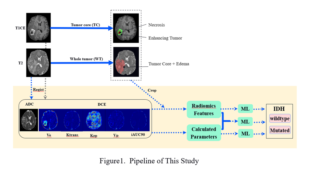
Approach: Several groups of features were extracted through multiparametric image: 1) Automatically calculated DCE-MRI metrics; 2) Structural MRI radiomics features, and 3) DCE-MRI radiomics features. Z-score normalization was used for feature normalization. Mann–Whitney U test was used for tumor selection. Stochastic gradient descent was used for machine learning classifier.
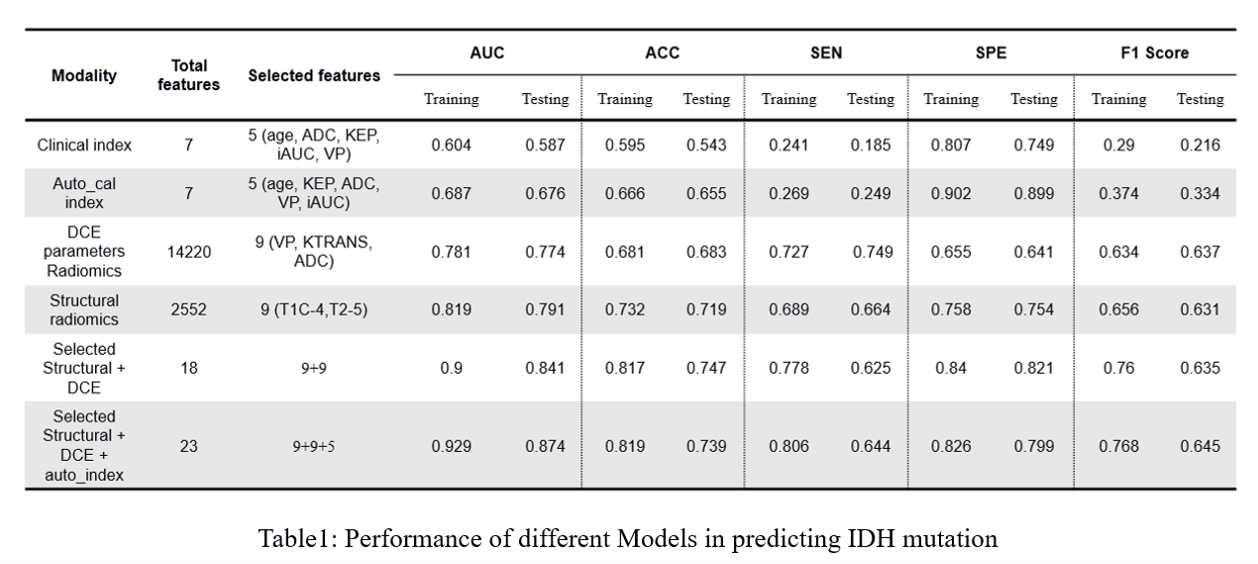
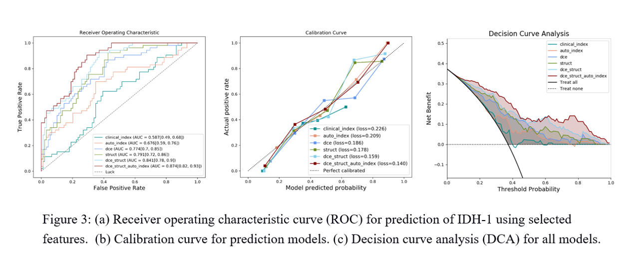
Results: We achived an AUC of 0.874 for model combing structural-MRI, DCE-MRI radiomics and automatically-calculated DCE-MRI metrics.
Impact: DCE-MRI radiomics models demonstrated great potential to predict IDH mutation status in Gliomas.
Background and Purpose
Gliomas are the most common type of primary intracranial tumors among the world1. In 2016, the 4th edition of World Health Organization Classification Criteria of Tumors of the Central Nervous System Tumor (WHO CNS4) first introduced iso-citrate dehydrogenase (IDH) mutation and 1p19q co-deletion status as diagnostic molecular markers for adult diffuse gliomas2. In the latest edition WHO CNS5, Adult diffuse gliomas consist of three subtypes: Glioblastoma, IDH wild type, Astrocytoma, IDH mutant and Oligodendroglioma, IDH mutant with 1p19q co-delated3. IDH mutation is important for patient management an prognoisis prediction.Dynamic contrast-enhanced MRI (DCE-MRI) can depict perfusion and microcirculation of tumors4. The aim of this research was to build radiomics models in DCE-MRI for predicting isocitrate dehydrogenase (IDH) mutation in adult diffuse gliomas.
Method
Patients diagnosed as adult diffuse glioma between September 2018 and September 2022 in our hospital were included. All patients underwent examination before surgery in a 3.0T MR scanner (uMR790, United Imaging Healthcare). IDH mutation status were determined by either immunohistochemistry or Sanger sequencing of tumor sample after surgery. DCE-MRI was performed after Gadodiamide injection (0.2mL/kg, 3.5mL/s).DCE-MRI metrics Ktrans, Kep, Ve, Vp, and iAUC were obtained through extended Tofts model. ADC map was obtained from DWI. Clinically obtained DCE-MRI and DWI metric values combined with Age and Sex were included as clinical indices.
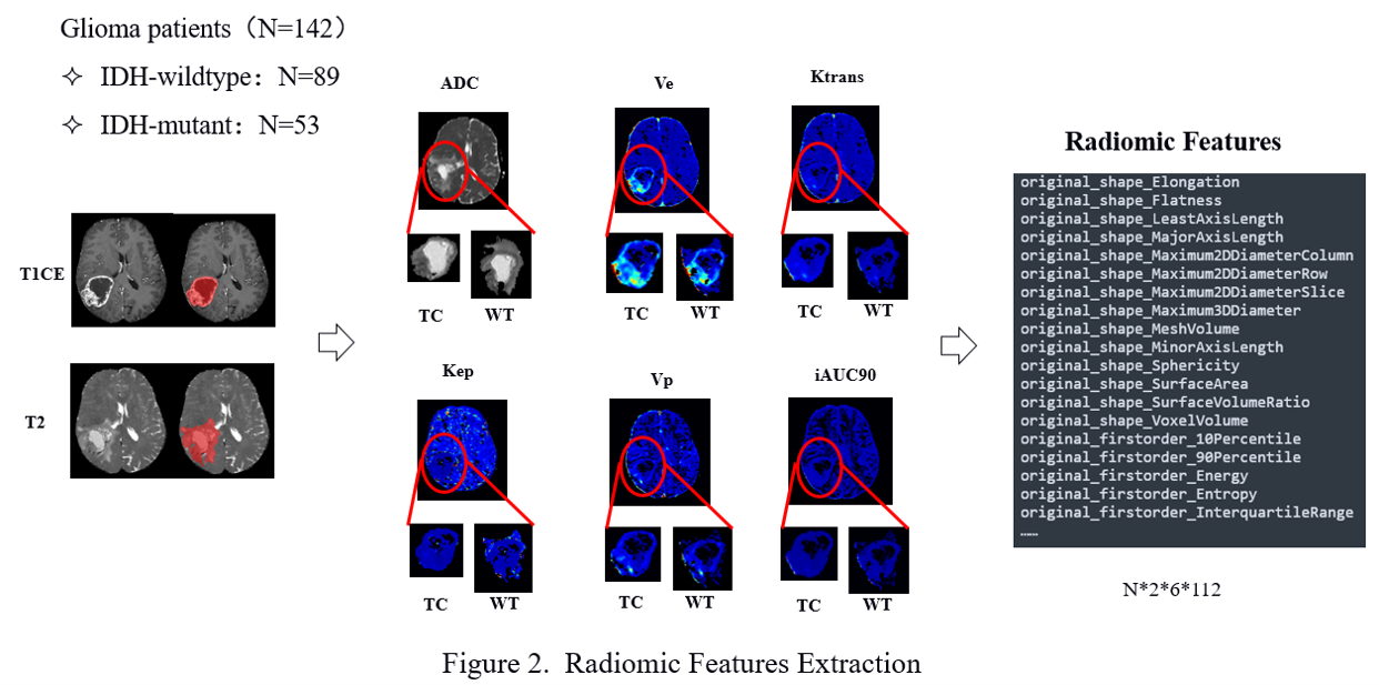
Region-of-interests (ROI) of Tumor Core (TC) and Whole Tumor (WT) were automatically delineated on T1CE and T2WI using a pretrained deep learning model. The ROI were registered to DCE-MRI and DWI metrics. Several groups of features were extracted through multiparametric image: 1) Automatically calculated DCE-MRI metrics; 2) Structural MRI radiomics features, and 3) DCE-MRI radiomics features. Z-score normalization was used for feature normalization. Mann–Whitney U test was used for tumor selection. Stochastic gradient descent was used for machine learning classifier. Performance of different models was quantified using area under receiver operating characteristic curve (AUC).
Results
A total of 142 patients were included, in which 89 patients are IDH-wildtype. Model-wise AUCs were 0.587 for clinical Index, 0.676 for automatically-calculated DCE metrics, 0.774 for structural-MRI radiomics, 0.791 for DCE-MRI radiomics, 0.841 for combined structural-MRI and DCE-MRI radiomics, and 0.874 for combined structural-MRI, DCE-MRI radiomics and automatically-calculated DCE-MRI metrics.Conclusion
DCE-MRI radiomics models demonstrated great potential to predict IDH mutation status in Gliomas.Acknowledgements
No acknowledgement found.References
1. SHOBEIRI P, SEYEDMIRZAEI H, KALANTARI A, et al. The Epidemiology of Brain and Spinal Cord Tumors [J]. Adv Exp Med Biol, 2023, 1394: 19-39.
2. LOUIS D N, PERRY A, REIFENBERGER G, et al. The 2016 World Health Organization Classification of Tumors of the Central Nervous System: a summary [J]. Acta Neuropathologica, 2016, 131(6): 803-20.
3. LOUIS D N, PERRY A, WESSELING P, et al. The 2021 WHO Classification of Tumors of the Central Nervous System: a summary [J]. Neuro Oncol, 2021, 23(8): 1231-51.
4. Sohn B, Park K, Ahn SS, Park YW, et al. Dynamic contrast-enhanced MRI radiomics model predicts epidermal growth factor receptor amplification in glioblastoma, IDH-wildtype. J Neurooncol. 2023 Sep;164(2):341-351.
Figures