3581
Triexponential multi-b-value diffusion-weighted imaging metrics may detect WHO grade and key molecular markers in glioma patients1Department of Radiology, The Affiliated Drum Tower Hospital of Nanjing University Medical School, Nanjing, China, 2Central Research Institute, United Imaging Healthcare Group Co., Ltd, Shanghai, China, Shanghai, China
Synopsis
Keywords: Tumors (Pre-Treatment), Brain, Ki67, IDH, Glioma
Motivation: WHO grade, Ki67 and IDH mutation are important for management and prognosis evaluation in glioma patients.
Goal(s): To investigate the value of Tri-exponential model (TEM) in preidcting WHO grade, Ki67 and IDH mutation of gliomas
Approach: 12 b-value DWI were obtained from glioma patients, TEM, SEM and IVIM model were analyzed for each patient. Univariate and Multivariate Logistic Regression were utilized to construct prediction model.
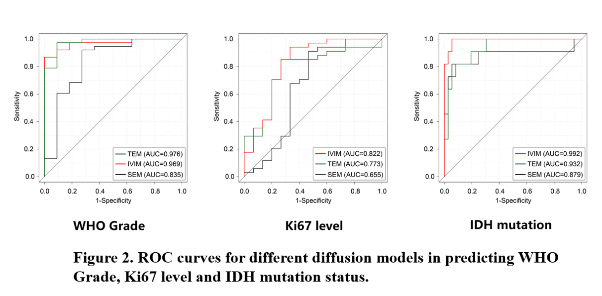
Results: TEM model achieved highest AUC on WHO Grade prediction task, while IVIM performed better on Ki67 and IDH mutation prediction task.
Impact: This study illustrated the potential of applying TEM model on predicting WHO grade Ki67 and IDH mutation in adult diffuse gliomas.
Intraduction
Glioma is the most common malignant brain tumor and can be classified into low-grade gliomas and high-grade gliomas. In the latest 2021 edition of WHO central nervous system tumor classification. Isocitrate dehydrogenase (IDH) was introduced as the key molecular marker to make an integrated genetic diagnosis[1].Diffusion weighted imaging (DWI) detects the motion of water molecules within tumor tissues and the tumor microenvironment. Apparent diffusion coefficient (ADC) value derived from a mono-exponential model is used clinically to quantify the water diffusion. In general, diffusion-weighted signal is assumed to follow an ideal exponential decay, and the resulting ADC should be independent of the chosen b-value. However, this is often inconsistent with what we actually observe in human tissue, which is often attributed to the non-Gaussian nature of diffusion. Dozens of diffusion models (e.g., diffusion kurtosis, fractional order calculus, continuous time random walk) have been created to address this problem [2-4].
Recently, a novel triexponential model (TEM) was established. The purpose of this study was to explore the diagnostic performance of TEM metrics in evaluating WHO grades and IDH mutation in glioma patients. We also compared the performance of TEM with Intro-Voxel Incoherent Movement (IVIM) and Stretched Exponential Model (SEM) in different prediction task.
Materials and Methods
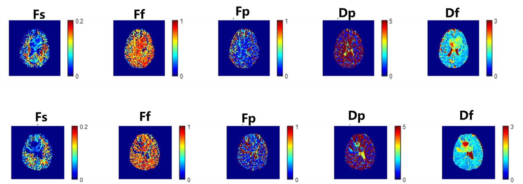
The prospective study was approved by our Institute Review Board. Patients diagnosed as adult diffuse glioma between September 2022 and September 2023 were included. WHO Grade of Tumor was determined by post-surgical pathological analysis according to 2021 5th edition of Central Nervous System Tumor Classification. Ki67 level was determined by immunochemical staining. IDH mutation were determined by Sanger sequencing of tumor sample after surgery.All patients underwent examination before surgery in a 3.0T MR scanner (uMR790, United Imaging Healthcare, Shanghai, China). Routine MRI sequences included: 3D T1-weighted imaging (T1WI), 3D T2-weighted imaging (T2WI), 3D T2 fluid-attenuated inversion-recovery (T2-FLAIR). Multi-b-value DWI was performed using a single-shot spin-echo echo planar imaging sequence (SS SE-EPI) with 12 b-values (0, 20, 50, 100, 200, 400, 1000, 1500, 2000, 2500, and 3000 s/mm2) in three orthogonal directions. All diffusion data were processed by using an in-house program implemented with MATLAB R2021b software. The intermediate and perfusion-related diffusion coefficients (Dint and Dp) and fractions of strictly limited, intermediate, and perfusion-related diffusion (Fvery-slow, Fint, and Fp) were obtained from triexponential multi-b-value DWI model:
$$S(b)= S_0*[F_s+F_f exp(-bD_f )+F_p exp(-bD_p ) ]$$,
$$F_s+F_f+F_p=1$$
where S0 and S(b) are signals at b-value = 0 s/mm2 and other b-values, respectively.
Region-of-interests of tumor and normal white matter (nWM) were delineated manually. Parameter values were normalized by nWM. Univariate and Multivariate Logistic Regression were used to select metrics and construct prediction model. For Univariate Logistic Regression, parameters with P<0.2 were selected and fused in Multivariate Logistic regression. For Multivariate Logistic regression, parameters with P<0.05 were considered as statistically significant. Performance of differential diagnosis was quantified using area under ROC curve (AUC).
Results
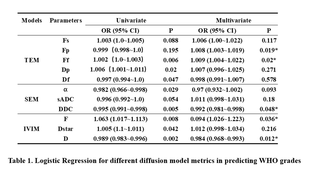
Forty-nine patients were included (mean age 60.67±13.75, 13 females) in this study, with 38 high-grade gliomas and 11 low-grade gliomas, 34 high Ki67 level and 15 low Ki67 level, 36 IDH-wildtype and 11 IDH mutant. Metrics of IVIM, SEM and TEM were obtained for each patient.For WHO Grades task, model-wise AUCs were 0.976 (TEM), 0.969(IVIM), 0.835 (SEM) respectively. Fp of TEM, Ff of TEM, DDC of SEM, F of IVIM and D of IVIM were independent predictive parameters in multivariate logistic regression.
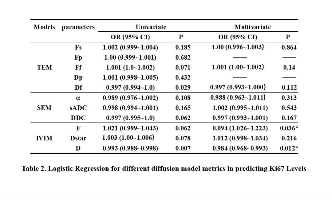
For Ki67 level task, model-wise AUCs were 0.822 (IVIM), 0.773(TEM), 0.655 (SEM) respectively. F of IVIM and D of IVIM were independent predictive parameters in multivariate logistic regression.
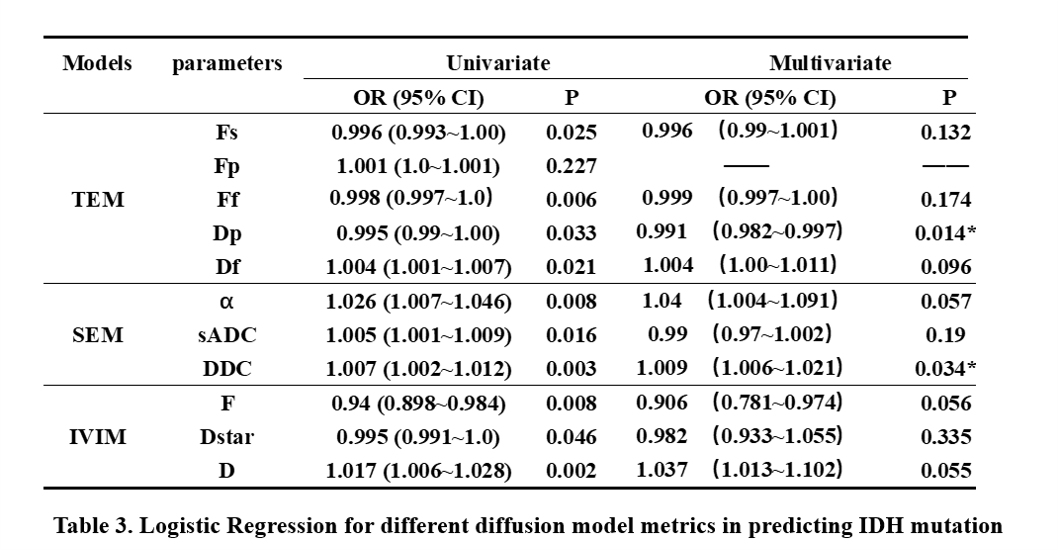
For IDH mutation task, model-wise AUCs were 0.992 (IVIM), 0.923(TEM), 0.879 (SEM) respectively. Dp of TEM and DDC of SEM were independent predictive parameters in multivariate logistic regression.
Discussion and conclusion
TEM model achieved highest AUC on WHO Grade prediction task, while IVIM performed better on Ki67 and IDH mutation prediction task.This study illustrated the potential of applying TEM model on predicting WHO grade Ki67 and IDH mutation in adult diffuse gliomas. Our following plan is a prospective study with large cohort to explore the diagnosis effect of TEM in predicting other key molecular markers in gliomas.
Acknowledgements
No acknowledgement found.References
1. SHOBEIRI P, SEYEDMIRZAEI H, KALANTARI A, et al. The Epidemiology of Brain and Spinal Cord Tumors [J]. Adv Exp Med Biol, 2023, 1394: 19-39.
2. Jensen JH, Helpern JA, Ramani A, Lu H, Kaczynski K. (2005) Diffusional kurtosis imaging: the quantification of non-Gaussian water diffusion by means of magnetic resonance imaging. Magn Reson Med, 53:1432–40.
3. Novikov DS, Fieremans E, Jensen JH, Helpern JA. (2011) Random walks with barriers. Nat Phys, 7(6):508–14
4. Zhou XJ, Gao Q, Abdullah O, Magin RL. (2010) Studies of anomalous diffusion in the human brain using fractional order calculus. Magn Reson Med 63:562–9.
Figures