3580
Multipool CEST MRI demonstrates good performance in predicting ATRX mutation status in IDH1-mutant lower-grade gliomas1Tongji Hospital, Tongji Medical College, Huazhong University of Science and Technology, Wuhan, China, 2Department of Clinical, Philips Healthcare, Wuhan, China
Synopsis
Keywords: Tumors (Pre-Treatment), Tumor, Brain, Glioma, CEST&MT
Motivation: ATRX gene mutation is correlated with better prognosis in IDH1-mutant lower-grade gliomas (LrGGs).
Goal(s): We aimed to investigate the predictability of ATRX mutation status using multipool CEST MRI.
Approach: The CEST Z-spectrum was fitted with 5 Lorentzian functions. We compared the differences in 5 metrics between ATRX mutant and wild-type gliomas. And ROC analyses were performed to evaluate predictive performances of metrics.
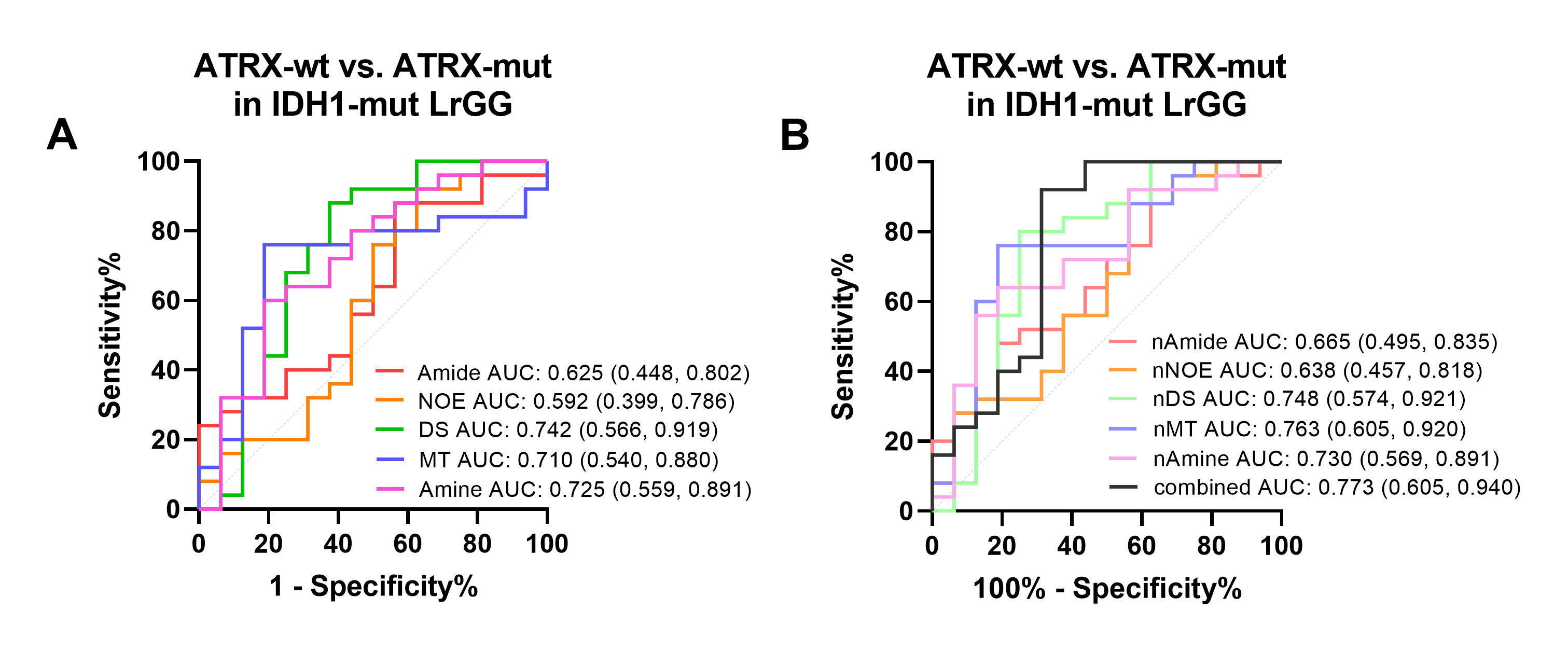
Results: ATRX mutant IDH1-mutant LrGGs showed significantly decreased direct saturated water (DS), semi-solid magnetization transfer (MT) signals and increased amine signal compared to ATRX wild-type group. The combination of metrics yielded the highest AUC of 0.773.
Impact: Multipool CEST MRI demonstrated good ability to distinguish ATRX mutant gliomas from wild-type gliomas, it may be as a useful imaging biomarker for precise prediction of ATRX mutation status and facilitate the diagnosis and prognosis of glioma patients.
Introduction
Gliomas are the most common primary malignant brain tumors1. The alpha thalassemia/mental retardation X-linked syndrome (ATRX) is a potent biomarker of gliomas, which is essential for maintenance of telomeres and chromatin modulation. Previous studies have demonstrated that ATRX mutation (ATRX protein expression loss) was correlated with better prognosis in IDH1-mutant lower-grade gliomas (LrGGs)2. Thus, precise diagnosis of ATRX status is of great importance for management of patients. Although histopathology analysis based on biopsy or surgery are still the golden standard, these methods are invasive and may confused by intra-tumoral heterogeneity. Chemical exchange saturation transfer (CEST) MRI is a novel technique. Furthermore, multipool Lorentzian fitting method was proposed to separately quantify each effect or signal of the full CEST Z-spectrum, may provide more mentalism information than conventional APTw-MRI3. Thus, we aimed to comprehensively evaluate the ability of multipool CEST MRI in predicting ATRX mutation status.Methods
Forty-one IDH1-mutant LrGG patients with confirmed ATRX mutation status were analyzed. All images were acquired on a 3.0T MRI scanner (Discovery MR750, GE Healthcare, Milwaukee, WI, USA) with a 32-channel head coil. After normalization to unsaturated image, B0 field correction was performed. And then, the pixel-wise B0 corrected Z-spectrum was fitted as a sum of five Lorentzian functions corresponding to amide pool, nuclear Overhauser enhancement (NOE) pool, semi-solid magnetization transfer (MT) pool, direct saturated water (DS) pool and amine pool. Peak areas were analyzed in this study instead of amplitudes4. Regions of interest (ROI) were defined as the contrast-enhanced regions in T1CE or the hyperintensity regions on T2FLAIR. The ROI of contralateral normal-appearing white matter (CNAWM) was also analyzed for normalization. Differences between continuous variables were evaluated using student-t or Mann-Whitney U test according to they were normal distribution or not. The receiver operating characteristic (ROC) analysis was performed to evaluate the diagnostic performances of each metric and their combinations. The area under the curve (AUC), sensitivity, specificity and cut-off value were also calculated.Results
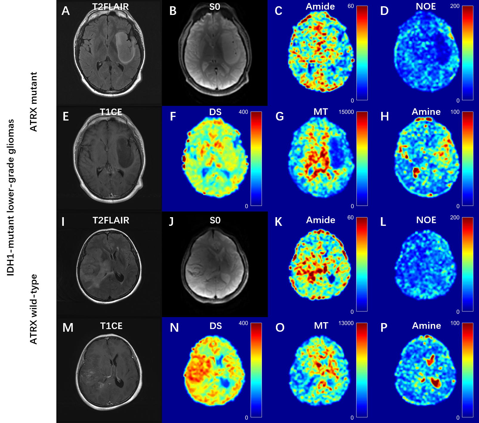
DS, MT and amine signals showed significant differences between ATRX wild-type and ATRX mutant patients in IDH1 mutant LrGGs. DS and MT were decreased, while amine effect was increased in ATRX mutant gliomas compared with those in ATRX wild-type gliomas (P = 0.015, 0.024 and 0.016, respectively). Besides, the normalized DS, MT and amine also showed significant differences (all P-values < 0.05). No significant differences were observed in amide and NOE and their normalized metrics. For metrics with significant differences, they had moderate diagnostic efficacies, showing AUCs from 0.710 to 0.760. Moreover, the combined models demonstrated the best performance, achieving an AUC of 0.773.Discussion
In our study, significant higher DS signal and lower amine signal were observed in ATRX wild-type IDH-mutant gliomas. The increased DS signal may be due to abnormal neovascularization and edema. ATRX wild-type IDH-mutant gliomas show higher extent of edema5. And perfusion MRI demonstrated that maintained ATRX expression was correlated with higher CBF value6,7, reflecting the neovascularization. These alterations may lead to an increased DS effect. Besides, Su et al8 found that 1p/19q codeletion gliomas had significantly lower CEST signal at 2ppm compared to 1p/19q non-codeletion gliomas. Considering that ATRX expression is mostly mutually exclusive with the 1p/19q codeletion, our findings are consistent with previous results. Furthermore, difference in MT signal between maintained ATRX and loss of expression was also detected. MT signal originates from semi-solid macromolecules, which can reflect the impairment of white matter9. However, further exploration is still needed to determine the pathophysiological mechanisms. In the ROC analysis, DS, amine and MT signal showed moderate performance in predicting ATRX mutation status, and their combinations reached the highest AUC, which indicating that multiple contrasts provided more comprehensive metabolic information.Conclusion
The multipool Lorentzian fitting method based on full Z-spectrum provided abundant contrasts, and demonstrated great potential for predicting of ATRX mutation status. Thus, the multipool CEST MRI may be used as a tool for more comprehensive glioma characterization.Acknowledgements
This study has received funding from the National Natural Science Foundation of China (Grant No. U22A20354).References
1. Louis DN, Perry A, Wesseling P, et al. The 2021 WHO Classification of Tumors of the Central Nervous System: a summary. Neuro Oncol 2021;23:1231-1251.
2. Haase S, Garcia-Fabiani MB, Carney S, et al. Mutant ATRX: uncovering a new therapeutic target for glioma. Expert Opin Ther Targets 2018;22:599-613.
3. Zhang XY, Wang F, Li H, et al. Accuracy in the quantification of chemical exchange saturation transfer (CEST) and relayed nuclear Overhauser enhancement (rNOE) saturation transfer effects. NMR Biomed 2017;30.
4. Cai K, Tain RW, Zhou XJ, et al. Creatine CEST MRI for Differentiating Gliomas with Different Degrees of Aggressiveness. Mol Imaging Biol 2017;19:225-232.
5. Yang X, Hu C, Xing Z, et al. Prediction of Ki-67 labeling index, ATRX mutation, and MGMT promoter methylation status in IDH-mutant astrocytoma by morphological MRI, SWI, DWI, and DSC-PWI. Eur Radiol 2023;33:7003-7014.
6. Lu D, Li Y, Lu H, Pillai JJ. Histogram-based analysis of cerebral blood flow using arterial spin labeling MRI in de novo brain gliomas: relationship to histopathologic grade and molecular markers. Neuroradiology 2021;63:751-760.
7. Brendle C, Hempel JM, Schittenhelm J, et al. Glioma Grading and Determination of IDH Mutation Status and ATRX loss by DCE and ASL Perfusion. Clin Neuroradiol 2018;28:421-428.
8. Su C, Xu S, Lin D, et al. Multi-parametric Z-spectral MRI may have a good performance for glioma stratification in clinical patients. Eur Radiol 2022;32:101-111.
9. Chan RW, Chen H, Myrehaug S, et al. Quantitative CEST and MT at 1.5T for monitoring treatment response in glioblastoma: early and late tumor progression during chemoradiation. J Neurooncol 2021;151:267-278.
Figures
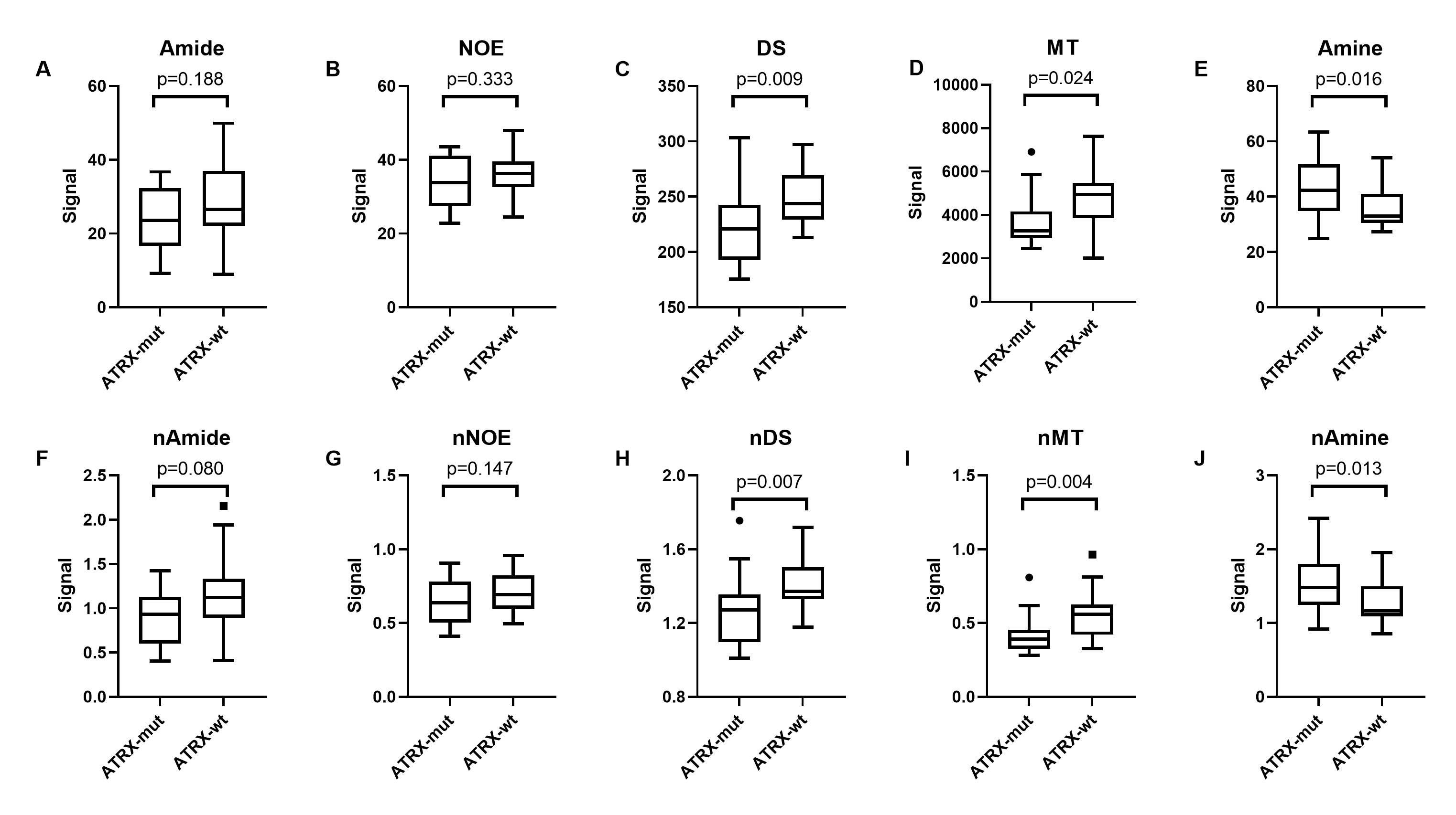
Box and whisker plots show differences in five Lorentzian fitted metrics and corresponding normalized metrics between ATRX mutation status of IDH1-mutant LrGGs.