3579
Fractality and Lacunarity of Tumor subcomponents is a Measure of Overall Survival: A novel approach to decipher Tumor Geometry and Survival1Biological Sciences, Indian Institute of Science Education and Research, Berhampur, Berhampur, India
Synopsis
Keywords: Tumors (Pre-Treatment), Multimodal, Glioma, Survival, presurgical
Motivation: Gliomas of similar histologic grade show a lot of difference in the growth and development. And the survival of patients with similar histologic grades also vary.
Goal(s): The shape variations of gliomas impact survival or not.
Approach: We calculated the fractal dimension and lacunarity of the subcomponents of gliomas and analyzed them along with survival data to obtain differences in overall survival.
Results: Variations in fractal Dimension and Lacunarity also present variations in overall survival. Subjects with higher enhancing fractal dimension had shortened survival and it was opposite for nonenhancing fractal dimension and enhancing lacunarity. Survival did not depend on edema subcomponent.
Impact: The study's results could revolutionize glioma patient care. Clinicians may integrate fractal dimension and lacunarity as prognostic markers for tailored treatment decisions. Scientists may explore their use in combination with genetic factors for accurate survival predictions and improving patient outcomes.
INTRODUCTION
In 2016, the World Health Organization (WHO) updated the classification of gliomas, introducing a molecular basis for categorization, distinguishing between IDH mutant and wild-type variants. Regardless of the histological grade, gliomas with mutations in isocitrate dehydrogenase (IDH) tend to have a favorable prognosis and increased survival compared to IDH wild-type ones. Furthermore, certain glioblastomas (GBMs) exhibit epigenetic methylation of the O6-methylguanine-DNA methyltransferase (MGMT) enzyme, while others have non-methylated MGMT. Gliomas with MGMT methylation generally have more prolonged overall survival and respond better to alkylating chemotherapy. Gliomas, even of the same histological type, display variations in their structure and shape. The fractal dimension measures the complexity of shapes, while lacunarity assesses the arrangement of substructures within the tumor mass. By analyzing these geometric parameters, we can analyze the survival differences in patients with different grades of gliomas and improve clinical management.METHODS
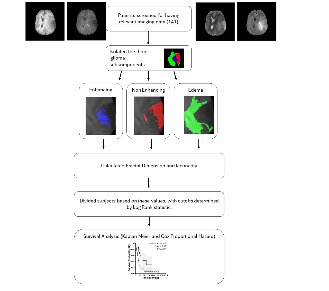
This study focused on individuals diagnosed with low-grade glioma or glioblastoma and used data from The Cancer Imaging Archive (TCIA), specifically the TCGA-LGG and TCGA-GBM repositories. The dataset included preoperative MRI scans with T1-weighted, T2-weighted, T2w-fluid attenuated inversion recovery (FLAIR), and contrast-enhanced T1-weighted (T1c) sequences. It also provided tumor segmentation masks and information about age at diagnosis, gender, and IDH mutation status. Tumor segmentation labels were generated following the GLISTRboost protocol and underwent manual corrections and expert neuroradiologist approval. Fractal dimension (FD) and lacunarity were computed using custom Python pipelines. FD was calculated using the 2D box-counting method applied to the binarized tumor mask. The estimated values were averaged for different tumor subcomponents (enhancing, non-enhancing, and edema). Lacunarity for each subcomponent was determined using the gliding box algorithm and the binned probability distribution method. It involved analyzing the intensity distribution of pixel values within each box. The mean lacunarity for each subcomponent was computed. Statistical analyses were conducted using Python and R, with a significance threshold of p<0.05, corrected for multiple comparisons. Maximally selected rank statistics determined optimal cutoff values for FD and lacunarity. Kaplan-Meier survival analysis and the log-rank test assessed overall survival based on these cutoff values. Univariate and multivariate Cox Proportional Hazard models explored the prognostic significance of FD and lacunarity for each tumor subcomponent.RESULTS
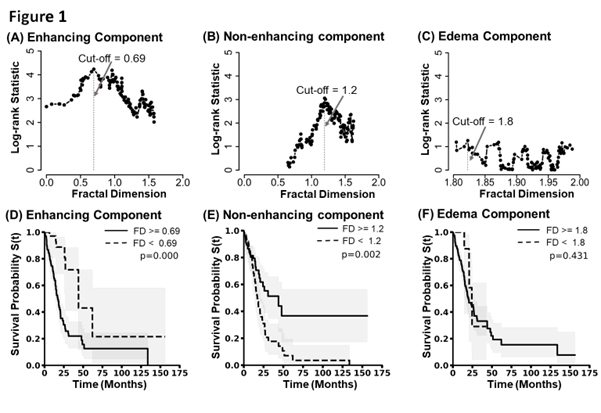
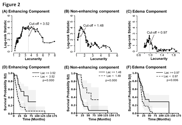
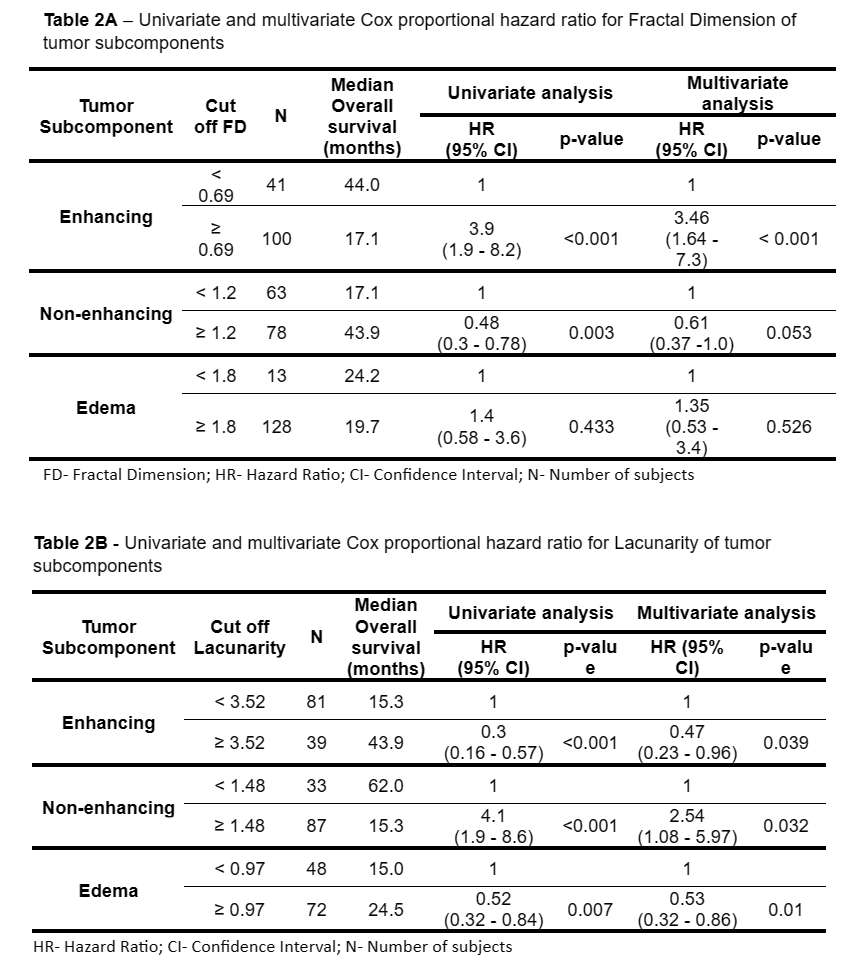
Cutoff values were employed to explore the relationship between fractality, lacunarity, and overall survival in distinct tumor subcomponents. In the enhancing subcomponent, a fractal dimension exceeding 0.69 was significantly associated with shortened overall survival (OS = 17.1 months, p < 0.0001), yielding a hazard ratio of 3.9 (95% CI, 1.9 - 8.2). Conversely, the non-enhancing subcomponent exhibited reduced survival with a fractal dimension below 1.2 (OS = 17.1 months, p = 0.002), resulting in a 52% reduction in the risk of death (HR = 0.48, 95% CI, 0.30 - 0.78). Notably, the edema subcomponent did not correlate significantly with overall survival (p = 0.43). Similarly, specific lacunarity cutoff values were established for each subcomponent. Within the enhancing subcomponent, lacunarity values falling below 3.52 were associated with shorter survival (OS = 15.3 months, p < 0.001) and a higher hazard ratio (HR = 4.1, 95% CI, 1.9 - 8.6). Likewise, the non-enhancing subcomponent showed a similar trend, with lacunarity levels above the threshold of 1.48 correlating with reduced survival (OS = 15.3 months, p < 0.001) and a higher hazard ratio. Lower lacunarity below the 0.97 threshold was associated with shorter survival (p = 0.006) and a decreased hazard ratio (HR = 0.52, 95% CI, 0.32 - 0.84) for the edema subcomponent. These findings underscore the prognostic significance of fractality and lacunarity in distinct tumor subcomponents.DISCUSSION
This pioneering study investigates glioma's three subcomponents (enhancing, non-enhancing, and edema) using fractal dimension and lacunarity calculations to correlate with patient survival. Higher fractal dimension indicates shorter survival in enhancing regions, reflecting tumor aggressiveness, while lower values are linked to reduced survival in non-enhancing areas. Lacunarity exhibits an opposing trend, with lower values in enhancing regions and higher values in non-enhancing areas associated with extended survival. These findings suggest the potential of fractal dimension and lacunarity as practical prognostic markers for gliomas.CONCLUSION
This study found a relationship between morphological variations within segmented tumor areas and patients' survival results. The fractal dimension and lacunarity, which serve as a measure of form heterogeneity, have revealed clear threshold values that can clearly distinguish the overall survival between patients. These findings highlight the potential of these morphological parameters to play an essential role in prognostic assessments and guide clinical decisions in cancer management.Acknowledgements
I acknowledge TCIA for the imaging and genomic data of Gliomas, and I also believe DST - SERB and ICMR India for the generous funding.References
1. Louis DN, Perry A, Reifenberger G, von Deimling A, Figarella-Branger D, Cavenee WK, Ohgaki H, Wiestler OD, Kleihues P, Ellison DW. The 2016 World Health Organization Classification of Tumors of the Central Nervous System: a summary. Acta Neuropathol. 2016 Jun;131(6):803-20. doi: 10.1007/s00401-016-1545-1. Epub 2016 May 9. PMID: 27157931.
2. Beiko J, Suki D, Hess KR et al. IDH1 mutant malignant astrocytomas are more amenable to surgical resection and have a survival benefit associated with maximal surgical resection.
3. Shyamala, K., H. C. Girish, and Sanjay Murgod. "Risk of tumor cell seeding through biopsy and aspiration cytology." Journal of International Society of Preventive & Community Dentistry 4.1 (2014): 5.
4. Clark K, Vendt B, Smith K, Freymann J, Kirby J, Koppel P, Moore S, Phillips S, Maffitt D, Pringle M, Tarbox L, Prior F. The Cancer Imaging Archive (TCIA): Maintaining and Operating a Public Information Repository, Journal of Digital Imaging, Volume 26, Number 6, December, 2013, pp 1045-1057. DOI: 10.1007/s10278-013-9622-7
5. Bakas S, Akbari H, Sotiras A, Bilello M, Rozycki M, Kirby J, Freymann J, Farahani K, Davatzikos C. (2017) Segmentation Labels and Radiomic Features for the Pre-operative Scans of the TCGA-LGG collection [Data Set]. The Cancer Imaging Archive. DOI: 10.7937/K9/TCIA.2017.GJQ7R0EF
6. Bakas S, Akbari H, Sotiras A, Bilello M, Rozycki M, Kirby J, Freymann J, Farahani K, Davatzikos C. (2017). Segmentation Labels for the Pre-operative Scans of the TCGA-GBM collection [Data set]. The Cancer Imaging Archive. DOI: 10.7937/K9/TCIA.2017.KLXWJJ1Q
7. Bakas S, Akbari H, Sotiras A, Bilello M, Rozycki M, Kirby J, Freymann J, Farahani K, Davatzikos C. (2017) Advancing The Cancer Genome Atlas glioma MRI collections with expert segmentation labels and radiomic features Nature Scientific Data, 4:170117 DOI: 10.1038/sdata.2017.117
8. Kroell, N., (2021). imea: A Python package for extracting 2D and 3D shape measurements from images. Journal of Open Source Software, 6(60), 3091, https://doi.org/10.21105/joss.03091
9. Plotnick, Roy E., Robert H. Gardner, and Robert V. O'Neill. "Lacunarity indices as measures of landscape texture." Landscape ecology 8 (1993): 201-211.
Figures