3574
Trade-off between Readout Duration and Concomitant Field Effect in 3D MRF at 0.55T1Ming Hsieh Department of Electrical and Computer Engineering, University of Southern California, Los Angeles, CA, United States, 2Afred E. Mann Department of Biomedical Engineering, University of Southern California, Los Angeles, CA, United States
Synopsis
Keywords: MR Fingerprinting, MR Fingerprinting
Motivation: The use of long readouts in 3D MRF improves SNR efficiency but also suffers from stronger concomitant field effects.
Goal(s): To evaluate the trade-off between readout duration and concomitant field effects in 0.55T 3D MRF and evaluate mitigation using the MaxGIRF framework.
Approach: We performed 0.55T 3D MRF scans of 14 readout durations on the NIST/ISMRM phantom and compared reconstructions with and without concomitant field effects compensation. We analyzed T1 array's standard deviations as a function of readout duration.
Results: With correction, MRF T1 standard deviation reduced to stability due to improved SNR efficiency of long readouts, however, broke due to uncorrectable blurring.
Impact: Practical trade-off between a readout duration and concomitant field effects are demonstrated. 0.55T 3D MRF benefits from a longer readout duration (6.1-14.7ms) provided a concomitant field effect correction.
Introduction
FISP-MRF1,2 has been extensively studied at 1.5T and 3T and has been recently demonstrated with good repeatability at 0.55T3,4 and even lower field strengths5. Improved field homogeneity enables SNR efficient longer readouts, which partially compensates for the reduced SNR due to equilibrium polarization (proportional to B0). When doing so, the concomitant field effect which is stronger at low field strengths and when using stronger gradients, requires mitigation. In this study, we evaluated 3D axial Stack-of-Spiral (SoS) FISP-MRF with 14 readout durations and 2 reconstruction approaches at 0.55T. We demonstrate an important performance trade-off by analyzing MRF T1 standard deviation as a function of readout duration without and with static off-resonance and concomitant field effect correction6.Methods
Experiments were performed using a whole body 0.55T system (prototype MAGNETOM Aera,Siemens Healthineers, Erlangen, Germany) equipped with high-performance shielded gradients (45mT amplitude, 200T/m/s slew rate). All data were reconstructed using the subspace and low-rank modeling7 with and without MaxGIRF6 encoding for simultaneous static off-resonance and concomitant field effect correction. For each result, MRF T1 standard deviation from vial #5 which has the most biologically relevant T1 values (~500ms) was compared and plotted as a function of readout duration to reflect SNR efficiency changes.Fourteen 3D (300mmx300mmx240mm FOV, 1.2mmx1.2mmx5mm resolution) axial SoS FISP-MRF sequences with different readout durations (2.9ms to 22.6ms with ~1ms increment) were implemented each with a constant TR and TE (1.4ms) in open-source PulSeq8. These sequences follow the same 3D acquisition strategy described in Campbell-Washburn et al3, with 48 encoding partitions. Gradient duty cycle was maximized in an effort to maximize SNR efficiency. We defined a continuous FA schedule for the smallest TR value and resampled it for others TR values. As a result, all sequences have a different TR and number of time frames but similar total scan time (7min 30±1sec). A separate static off-resonance map was acquired for correction.
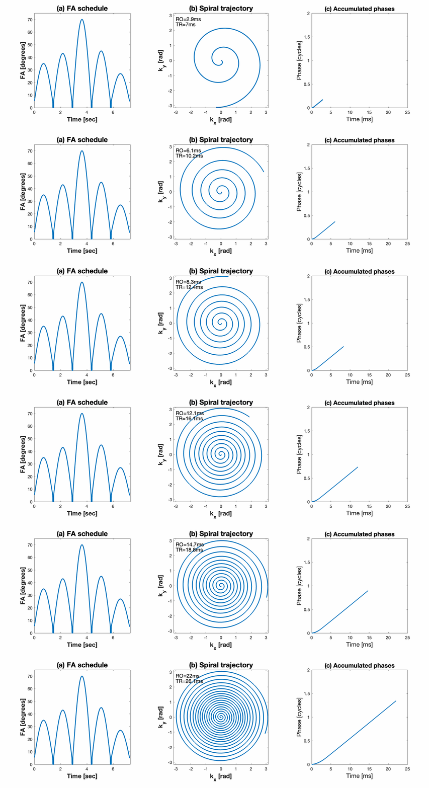
In axial MRI, readout gradients cause concomitant field inducing extra phase which is quadratic to slice offset, and the phase accrual causes spatial blurring in spiral imaging9. To demonstrate this effect, the ISMRM/NIST system phantom10 was rotated around the right-left axis such that its T1 array plate was perpendicualr to the main field direction (in the axial plane). Figure 1 demonstrates the continuous flip angle schedule, spiral readout trajectories and corresponding accumulated phase caused by concomitant field at Δz=±55mm. This slice offset matches the location of the T1 array.
Results
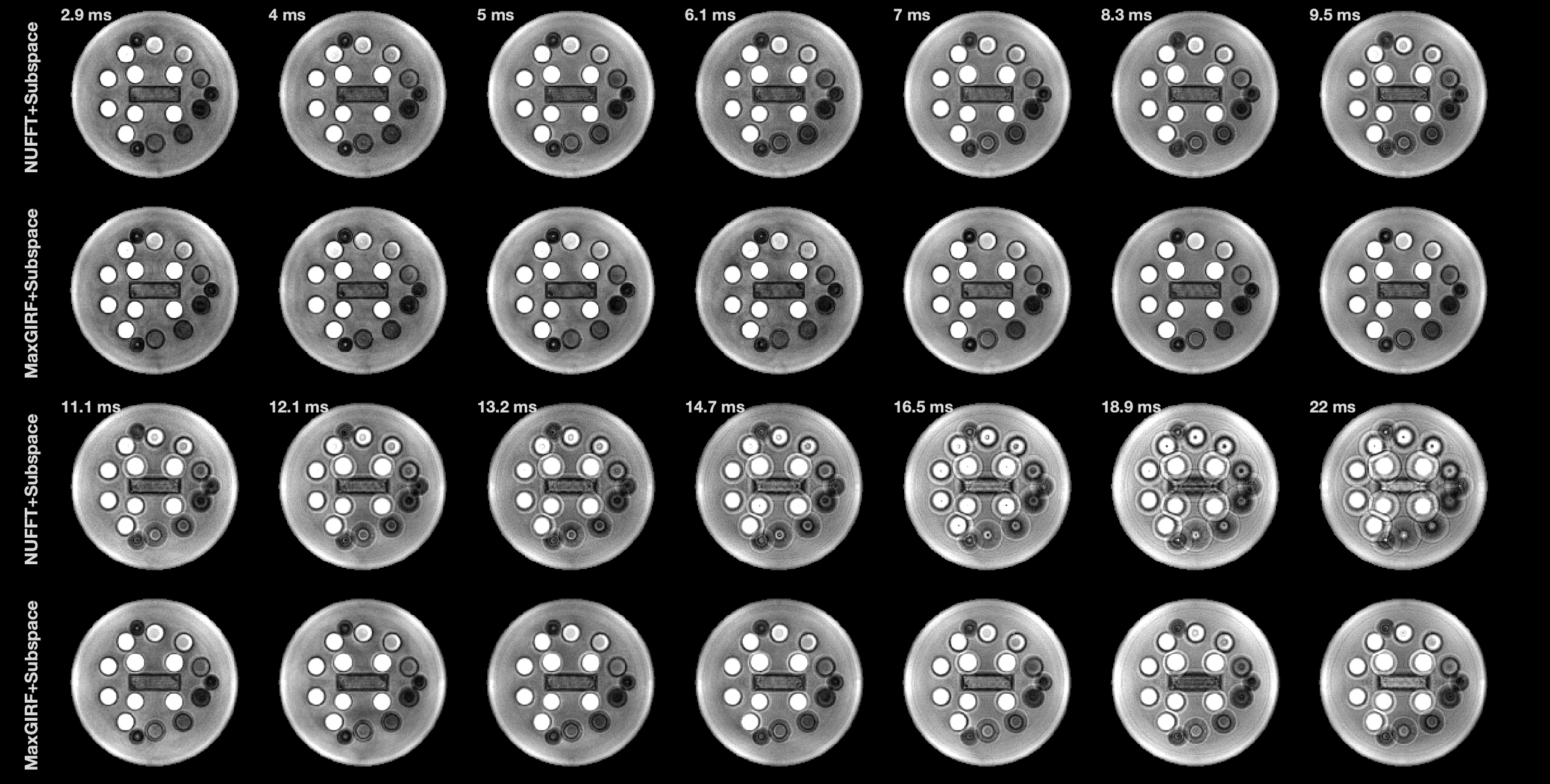
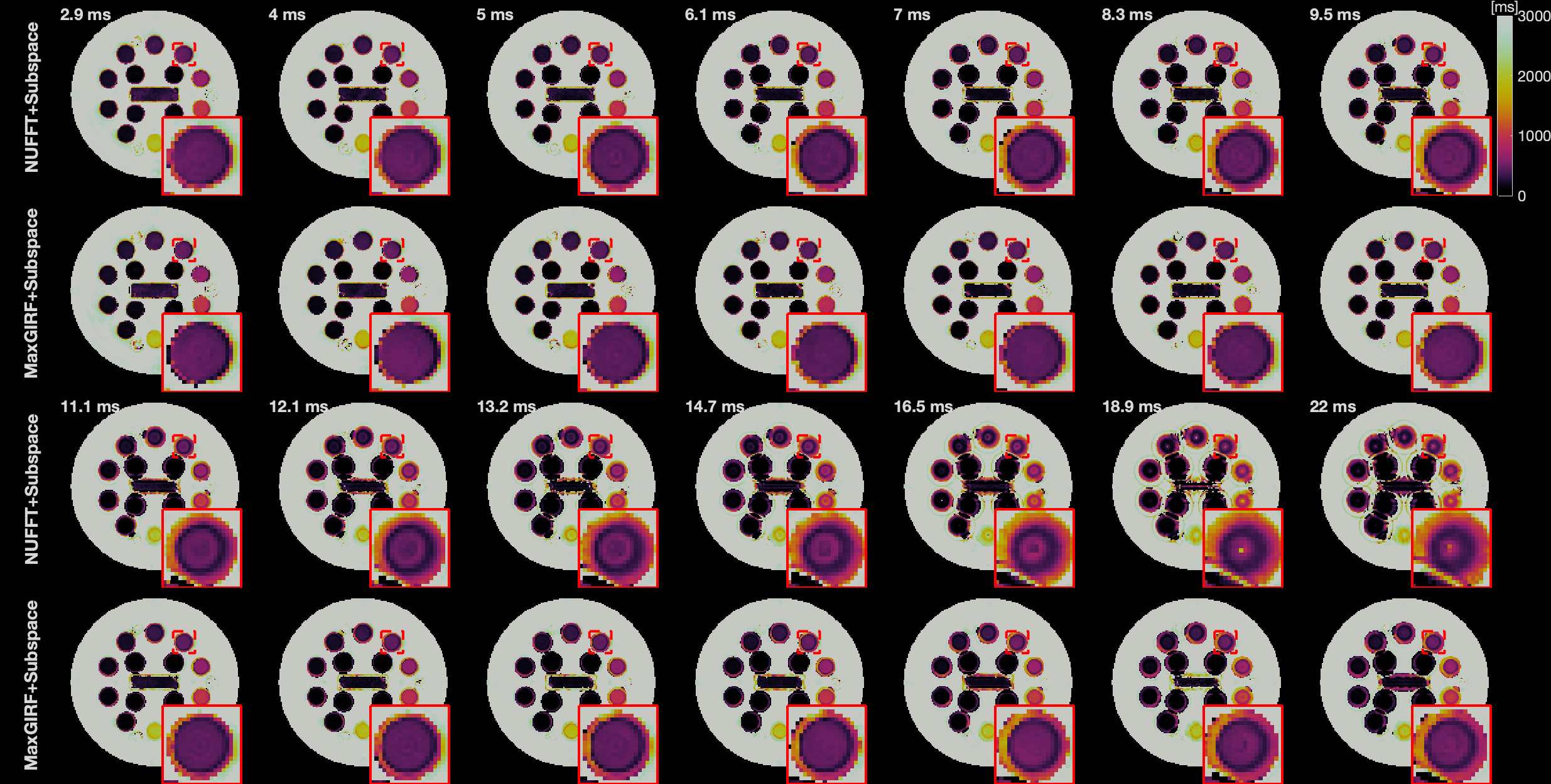
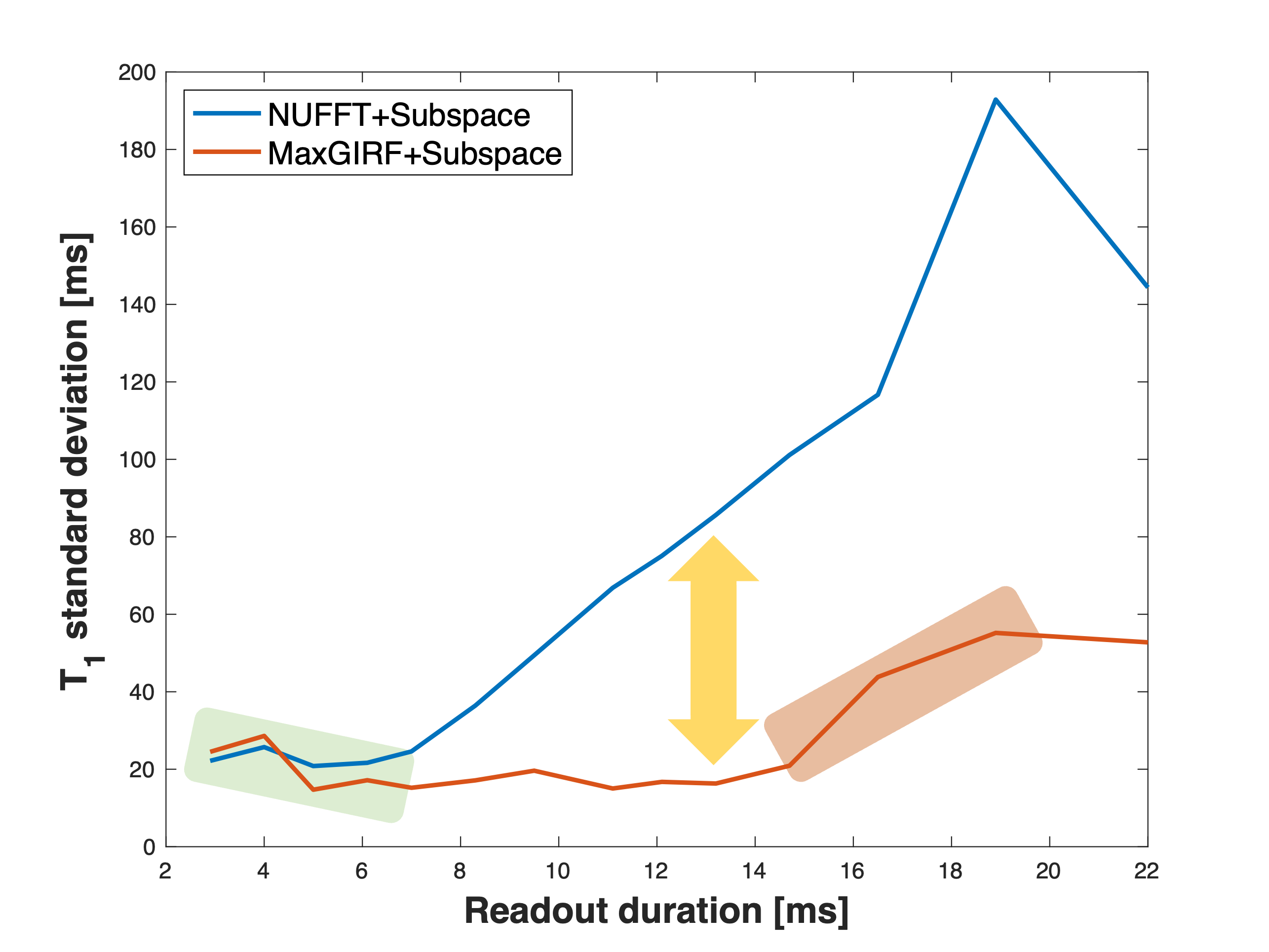
Figure 2 shows 14 reconstructed MRF images around 0.7sec in the flip angle schedule. Subspace reconstruction without concomitant field correction is shown in the 1st and 3rd rows, and with MaxGIRF correction is shown in the 2nd and 4th rows. Spatial blurring is stronger for longer readouts, and the blurring is largely mitigated by MaxGIRF correction. However, there is residual blurring when readout duration is >14.7ms. shows corresponding T1 maps that reflect the same spatial blurring.Figure 4 shows MRF T1 standard deviation from vial #5 as a function of readout duration. Results with and without concomitant field effect correction are in red and blue, respectively. T1 precision for the proposed reconstruction decreases up to 6.1ms, which we attribute to improved readout duty-cycle, and increases after 14.7ms, which we attribute to uncorrectable blurring due to off-resonance and/or concomitant field effect.
Discussion
We evaluated 0.55T 3D FISP MRF using 14 different readout durations using the ISMRM/NIST phantom. We found that spatial blurring became stronger as longer readout, and that this blurring impacts precision of MRF T1 maps. Blurring caused by static off-resonance and concomitant field effect can be largely mitigated using the MaxGIRF encoding, however, uncorrectable residuals exist for TR≥14.7ms.We indirectly evaluated SNR efficiency by analyzing MRF T1 standard deviation of the ISMRM/NIST phantom. Readout duty cycle increases with longer readout duration, resulting in improved SNR efficiency and decreased T1 standard deviation. With concomitant field effect correction, MRF T1 standard deviation was stable between 6.1ms and 14.7ms readout duration, indicating potential operational region for 0.55T 3D MRF.
There are limitations in this work. Only phantom experiments were performed due to long experiment time. The ISMRM/NIST phantom does not have realistic T1/T2 ratio and thus results might be different in in-vivo subjects, e.g., the readout threshold may arrive earlier due to shorter in-vivo T2 values.
Conclusion
This study shows that 0.55T 3D MRF produces T1 estimation with improved precision when using longer readout duration but is limited by uncorrectable spatial blurring. Thus, a potential operational readout region exists.Acknowledgements
We acknowledge grant support from the National Science Foundation (Award #1828736), and research support from Siemens Healthineers.References
1. Jiang Y, Ma D, Seiberlich N, Gulani V, Griswold MA. MR fingerprinting using fast imaging with steady state precession (FISP) with spiral readout. Magn Reson Med. 2015;74(6):1621-1631. doi:10.1002/mrm.255592.
2. Ma D, Jiang Y, Chen Y, et al. Fast 3D Magnetic Resonance Fingerprinting for a Whole-Brain Coverage. Magn Reson Med. 2018;79:2190-2197. doi:10.1002/mrm.268863.
3. Campbell-Washburn AE, Jiang Y, Körzdörfer G, Nittka M, Griswold MA. Feasibility of MR fingerprinting using a high-performance 0.55 T MRI system. Magn Reson Imaging. Published online June 8, 2021. doi:10.1016/j.mri.2021.06.0024.
4. Zhu Z, Lee NG, Tian Y, et al. Evaluation of MR Fingerprinting at 0.55T. In: ISMRM 31st Scientific Session. ; 2022:3492. Accessed November 6, 2022. https://cds.ismrm.org/protected/22MPresentations/abstracts/3492.html5.
5. Sarracanie M. Fast Quantitative Low-Field Magnetic Resonance Imaging With OPTIMUM-Optimized Magnetic Resonance Fingerprinting Using a Stationary Steady-State Cartesian Approach and Accelerated Acquisition Schedules. Invest Radiol. 2022;57(4):263-271. doi:10.1097/RLI.00000000000008366.
6. Lee NG, Ramasawmy R, Lim Y, Campbell-Washburn AE, Nayak KS. MaxGIRF: Image reconstruction incorporating concomitant field and gradient impulse response function effects. Magn Reson Med. 2022;88(2):691-710. doi:10.1002/MRM.292327.
7. Zhao B, Setsompop K, Adalsteinsson E, et al. Improved magnetic resonance fingerprinting reconstruction with low-rank and subspace modeling. Magn Reson Med. 2018;79(2):933-942. doi:10.1002/mrm.267018.
8. Layton KJ, Kroboth S, Jia F, et al. Pulseq: A rapid and hardware-independent pulse sequence prototyping framework. Magn Reson Med. 2017;77(4):1544-1552. doi:10.1002/mrm.262359.
9. King KF, Ganin A, Zhou J, Bernstein MA. Concomitant Gradient Field Effects in Spiral Scans. Magn Reson Med. 1999;41:103-112. doi:10.1002/(SICI)1522-2594(199901)41:110.
10. Russek SE, Boss M, Jackson EF, et al. Characterization of NIST/ISMRM MRI System Phantom. In: ISMRM 20th Scientific Session. ; 2012:2456. https://archive.ismrm.org/2012/2456.html
Figures