3566
Factorized Spatiotemporal Convolutions for Simultaneous Multislice Magnetic Resonance Fingerprinting1Radiation Oncology, Cleveland Clinic, Cleveland, OH, United States, 2Computer Science, University of North Carolina at Chapel Hill, Chapel Hill, NC, United States, 3Radiology, University of North Carolina at Chapel Hill, Chapel Hill, NC, United States, 4Radiology, Case Western Reserve University, Cleveland, OH, United States, 5Radiology, University Hospitals Cleveland Medical Center, Cleveland, OH, United States
Synopsis
Keywords: MR Fingerprinting, Quantitative Imaging
Motivation: Quantitative MR imaging with volumetric coverage is relatively slow, which hinders its clinical evaluation.
Goal(s): To leverage simultaneous multislice MR Fingerprinting (SMSMRF) and deep learning to achieve high multi-band factors for rapid quantitative imaging.
Approach: We introduced a Spatio-Temporal UNet (STUN) method to exploit both spatial and temporal correlations of signals in SMSMRF to largely accelerate data acquisition.
Results: High multi-band factors (3 and 4) with an additional 4x acceleration along the temporal dimension were achieved, providing rapid data sampling of 1.5~2 sec per slice for quantitative brain imaging.
Impact: The developed SMSMRF method holds great potential to accelerate quantitative imaging for challenging subjects, f.g. pediatric patients.
Introduction
MR Fingerprinting (MRF) is a quantitative imaging technique that enables rapid and simultaneous quantification of multiple tissue properties (1). Recent efforts have focused on expanding the spatial coverage of the technique, including simultaneous multislice (SMS) acquisitions (2-4). Due to the extremely high acceleration factor applied for in-plane encoding, dramatic aliasing artifacts exist which lead to significant challenges for unaliasing along the slice acquisition direction with SMS-MRF. Deep learning methods have been developed to replace the conventional template matching approach for improved tissue characterization and acquisition speed (5-7). Nevertheless, the majority of these methods focus solely on either the spatial or temporal aspects of the signal evolution, limiting its capability to extract information from the intricate MRF signals. In this study, we propose a Spatio-Temporal UNet (STUN) which exploits both spatial and temporal correlations of the signal to improve the performance of SMS-MRF.Methods
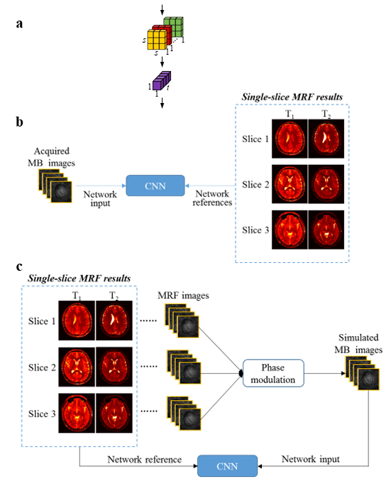
All MRI measurements were performed on a Siemens 3T scanner. SMS-MRF acquisitions with multi-band (MB) factors 3 and 4 were acquired from five normal subjects (10~20 scans per subject). Single-slice MRF acquisitions were also acquired from the corresponding slice locations to establish high-quality, reference T1 and T2 maps for training the network (described below). For both single-slice and SMS-MRF acquisitions, the acquisition time for each scan was 23 sec, encompassing a total of 2,304 MRF time frames. Multiband RF pulses generated from SINC waveforms with phase modulation were used to excite multiple slices simultaneously. The slice thickness was 5mm and the distance between adjacent slices was 10 mm. Each MRF time frame was highly undersampled in-plane by acquiring one spiral arm (R=48). Other imaging parameters included FOV, 30×30 cm; matrix, 256×256; flip angle, 5º∼12º; TR, 7 msec.A spatially-constrained quantification (SCQ) network was previously developed to accelerate data acquisition along the temporal dimension for single-slice 2D MRF (6). In this network, the MRF time frames are treated as channels in the neural network. As a result, the method is insensitive to the temporal ordering of the time frames. In order to exploit the signal temporal correlations, the temporal dimension of the input was treated as a new dimension in STUN instead of channels to allow for convolving jointly over time and space. This was performed using factorized spatio-temporal convolution with two separate and successive operations: a 2D spatial convolution and a 1D temporal convolution (Fig 1a). Factorized convolution is tailored to accommodate the distinctive nature of the MRF signal with different correlation patterns in the spatial and temporal dimensions.
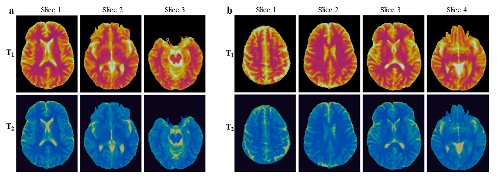
The STUN was first trained using the acquired SMS-MRF images (4x acceleration with only 576 time points) as the network input and high-quality T1&T2 maps from single-slice MRF (all 2304 time points) acquired at the corresponding slices as the network reference (Fig 1b). With the concern of potential head motions between the separated SMS-MRF and single-slice MRF acquisitions, the STUN network was also trained using SMS-MRF data generated through simulations based on single-slice MRF acquisitions (Fig 1c). Specifically, the same phase modulation used to generate the multiband RF was applied on the reconstructed single-slice MRF data to simulate an SMS-MRF dataset. The performance was compared to the standard template matching method and the SCQ network. Prospective acquisition of SMS-MRF data with 576 time points was also performed on another volunteer to evaluate the method. Acquisitions with a MB factor 3 and 4 were acquired in 6 sec, corresponding to 2 sec/slice and 1.5 sec/slice, respectively.
Results
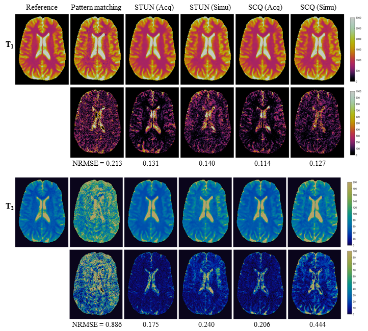
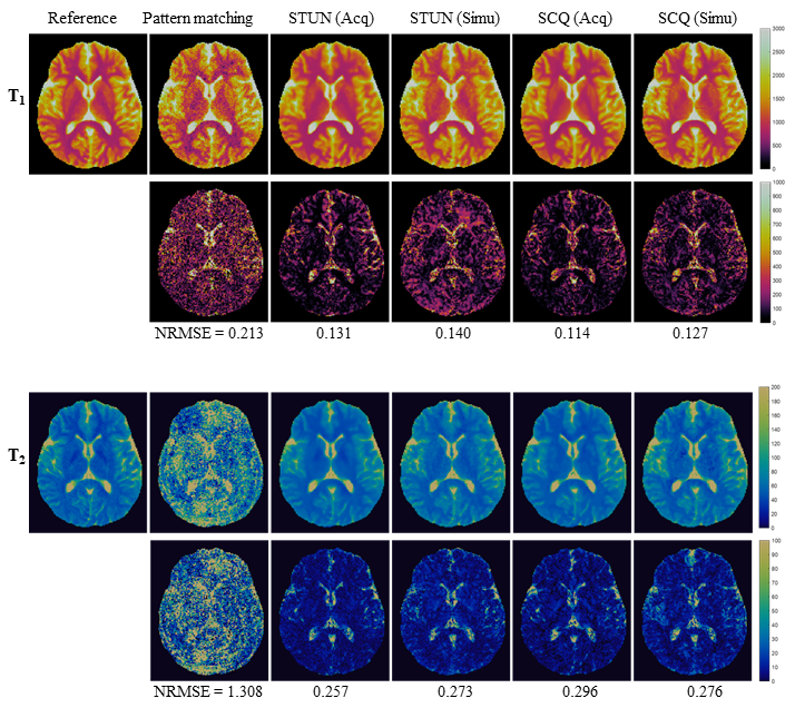
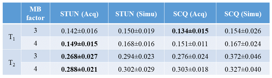
Figures 2&3 show representative T1 and T2 maps obtained with two different MB factors. The summary of cross-validation for all 5 subjects is presented in Table 1. Compared with the results from template matching, deep learning methods significantly reduced the amount of aliasing artifacts. The proposed STUN network also outperformed the SCQ network for most of the measurements, indicating more information is extracted with the proposed method. Both STUN and SCQ networks show better performance when trained using the acquired SMS-MRF data as compared to the simulated training data. This is likely due to imperfect SMS-MRF simulation, such as not accounting for the cross-talking effects between simultaneously excited slices. The prospectively acquired data shows similar map quality as compared to the results from retrospectively undersampled data for both MB factors (Fig 5).Discussion and Conclusion
In this study, a spatio-temporal U-Net was developed for efficient and accurate T1 and T2 quantifications for SMS-MRF. The same MRF acquisition patterns were applied to all excited slices in the experiment. Future work will focus on varying acquisition parameters across slices to achieve better image quality and MB factors (4).Acknowledgements
Siemens Healthineers and NIH grants 1R01 CA266702, and 1R01 CA282516.References
1. Ma D, et al. Nature, 2013; 187–192.
2. Hamilton, JI, et al. NMR Biomed, 2019; e4041.
3. Ye H, et al. MRM, 2016; 2078-2085.
4. Jiang Y, et al. MRM, 2017; 1870-1876.
5. Cohen O, et al. MRM, 2018; 885-894.
6. Fang Z, et al. TMI, 2019; 2364-2374.
7. Fang Z, et al. MRM, 2020; 579-591.
Figures