3561
Partial Diffusion-weighted MR Fingerprinting for the simultaneous quantification of relaxation time and diffusion coefficient1Department of Diagnostic Radiology, The University of Hong Kong, Hong Kong, China
Synopsis
Keywords: MR Fingerprinting, Image Reconstruction
Motivation: Subspace reconstruction has the potential to generate high-resolution images from highly undersampled data at each time point.
Goal(s): To develop an MRF-based approach to simultaneously measure relaxation time and diffusion coefficient.
Approach: This sequence was composed of two parts: conventional MRF and DW-SSFP. The central k-space was fully sampled, and the peripheral k-space was under sampled with 24 interleaves, using a dual density spiral trajectory. The subspace reconstruction was applied using the temporal basis obtained from central k-space data.
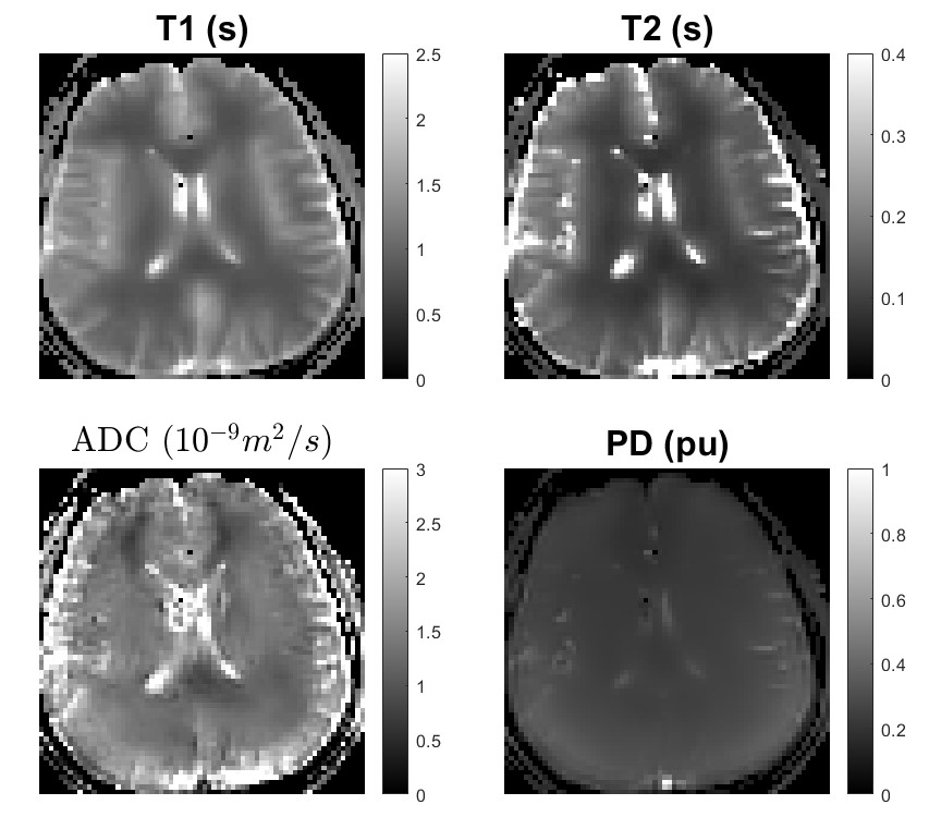
Results: T1, T2, PD, and ADC can be accurately quantified for both phantom and in-vivo scans.
Impact: This work indicate that full-resolution DWI can be reconstructed at each time point for a multi-shot multi-dynamic sequence using the subspace reconstruction. Our proposed sequence can be used to estimate relaxation time, proton density and diffusion coefficient simultaneously.
Introduction
MR fingerprinting (MRF) is a technique used to measure multiple quantitative MRI parameters at the same time, by varying scanning parameters in each time point. Previous studies integrated diffusion measurement into the MRF framework, demonstrated on phantom or using diffusion preparation for in vivo measurements1,2. In comparison, estimation of the apparent diffusion coefficient (ADC) was challenging for the diffusion-weighted SSFP (DW-SSFP) sequence, since the relaxation quantification was also required3. To address this, we propose a partial DW-SSFP sequence to simultaneously quantify relaxation time, proton density (PD), and ADC. To avoid the problem of shot-to-shot phase variations caused by motion during diffusion encoding, subspace reconstruction was applied by estimating the temporal basis from fully sampled central k-space data4,5.Methods
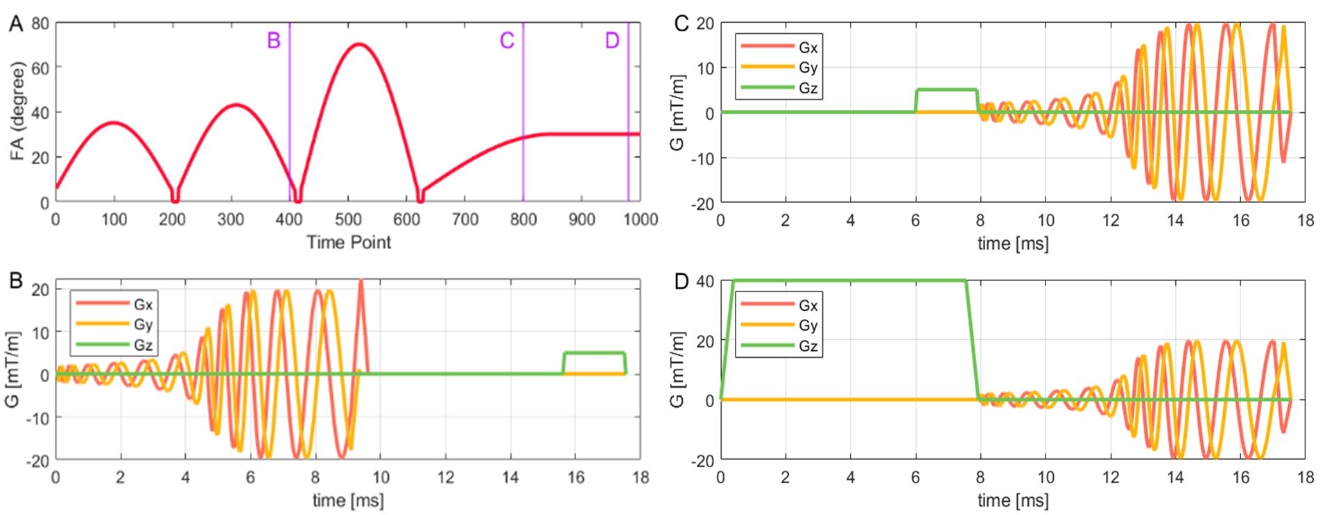
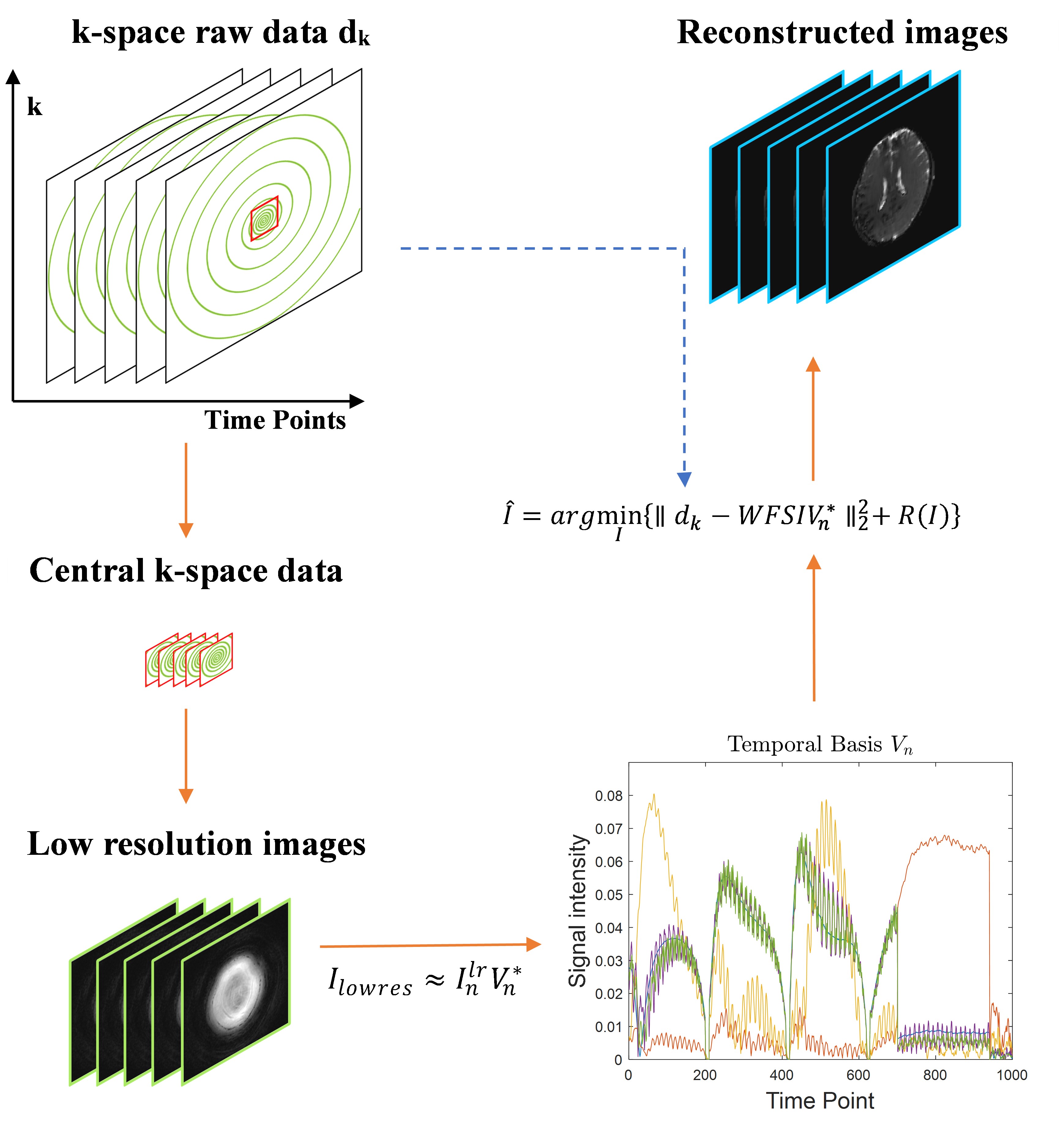
All experiments were performed on a 3T MRI (GE Signa Premier) scanner with a 48-channel head coil. As shown in Figure 1, the sequence was composed of two parts: conventional MRF for the first 800 time points, and DW-SSFP for the last 200 time points. Diffusion gradient pulse (with gradient strength of 40 mT/m and duration of 5 ms) was used for the phantom scan, while diffusion gradient pulse with strength of 5 mT/m and duration of 1.9 ms for b0, and gradient strength of 40 mT/m and duration of 7.5 ms for b1 were used for the in-vivo scans. The central k-space with a matrix size of 12 × 12 was fully sampled, and the peripheral k-space was under sampled with 24 spiral interleaves, i.e., 24 times acceleration, using a dual density spiral (DDS) trajectory. Variable flip angles were applied in the conventional MRF acquisitions, while a constant flip angle of 37 degree was applied for DW-SSFP acquisitions. Other scan parameters were: TE of 2.3 and 10.0 ms for the FISP and DW-SSFP partitions, TI=18 ms, slice thickness=5 mm, FOV=30 cm × 30 cm. The TR was 16 ms, and matrix size was 256×256 for phantom experiment. For in-vivo experiment, TR was 21 ms and matrix size was 128×128. To simulate the MRF dictionary, the extended phase graph algorithm was used. The algorithm selected the following ranges of parameters: 0.3 < T1 < 3.0 s, 0.03 < T2 < 1.8 s, and 0.2 < ADC < 3.0 × 10⁻⁹ m2/s. All quantitative values were estimated using conventional dictionary matching. This method was evaluated on a NIST/QIBA diffusion phantom and a healthy volunteer. The reference values of T1, T2, PD, and ADC for the 13 tubes in the phantom were measured by a conventional MRF and EPI-based DWI scan, respectively. Figure 2 shows the subspace reconstruction process. First, low-resolution images were reconstructed from fully sampled central k-space data. The temporal basis was then estimated using SVD decomposition for the first 800 and last 200 time points separately from the low-resolution images. Then, using the temporal basis for the two parts of this sequence separately, a full-resolution image was reconstructed at each time point.Results
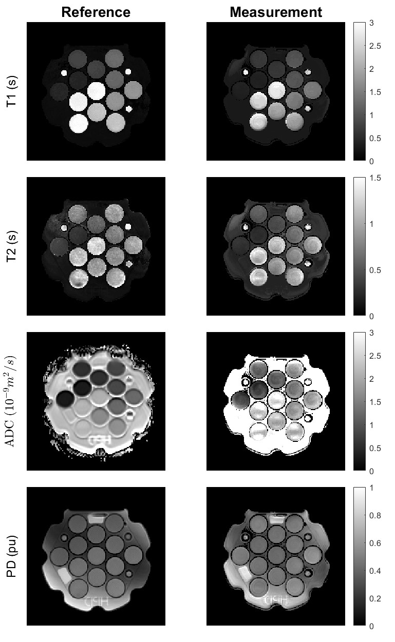
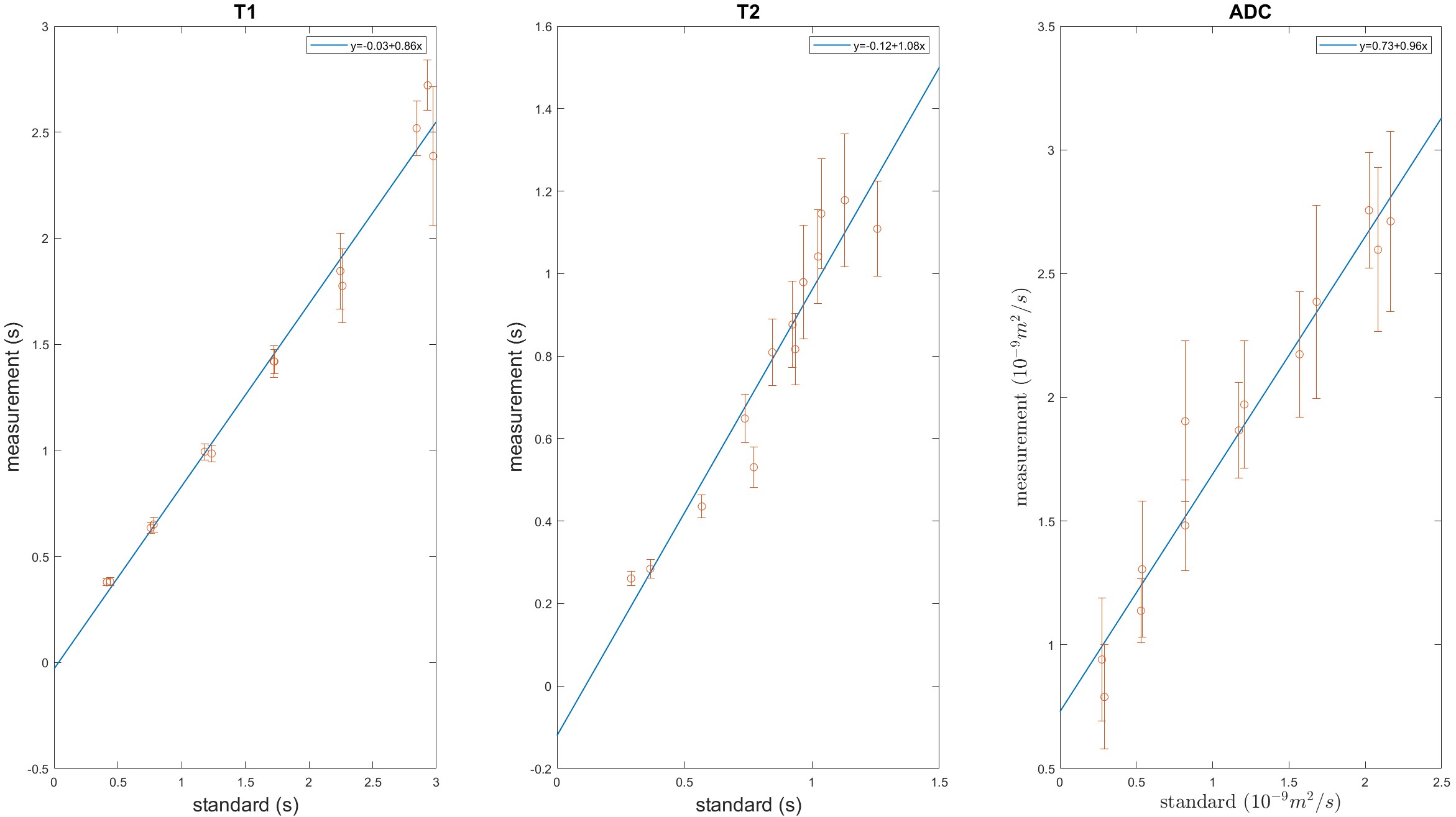
Figure 3 displays the reference and measured quantitative maps in the phantom scan. While Figure 4 compares the measured parameters with the reference values in each tube of the phantom. Additionally, Figure 5 exhibits the measured quantitative maps for the in-vivo scan. The results demonstrate that T1, T2, PD, and ADC can be accurately quantified using this sequence and the subspace reconstruction for both phantom and in-vivo scans. However, T2 and ADC measurements were overestimated compared to the reference values due to their joint contribution to the signal attenuation during the DW-SSFP acquisition.Discussion
We discovered that our sequence design can be used to estimate relaxation time, proton density and diffusion coefficient simultaneously. By using subspace reconstruction based on the temporal basis estimated from the central k-space data, we can generate high-resolution images and avoid the impact of shot-by-shot phase variations.Conclusion
The quantitative maps obtained from the phantom and in-vivo scans demonstrate the potential applications of this diffusion weighted MRF design.Acknowledgements
Not applicableReferences
1. Afzali M, Mueller L, Sakaie K, et al. MR Fingerprinting with b-Tensor Encoding for Simultaneous Quantification of Relaxation and Diffusion in a Single Scan. Magn Reson Med 2022;88(5):2043-2057.
2. Qiu Z, Hu S, Zhao W, et al. Simultaneous Quantification of Relaxation and Diffusion using MR Fingerprinting with Self-Calibrated Subspace Reconstruction. Proceedings of the 2023 ISMRM & ISMRT Annual Meeting & Exhibition. Toronto, Canada; 2023.
3. McNab JA, Miller KL. Steady-state diffusion-weighted imaging: theory, acquisition and analysis. NMR Biomed 2010;23(7):781-793.
4. McGivney DF, Pierre E, Ma D, et al. SVD compression for magnetic resonance fingerprinting in the time domain. IEEE Trans Med Imaging 2014;33(12):2311-2322.
5. Lu H, Ye H, Zhao B. Improved Low-Rank and Subspace Reconstruction for Magnetic Resonance Fingerprinting with Self-Navigating Acquisitions. Proceedings of the 2023 ISMRM & ISMRT Annual Meeting & Exhibition. Toronto, Canada; 2023.
Figures