3560
Unsupervised individual-specific in vivo parcellation of the human cerebral cortex using magnetic resonance fingerprinting1Australian Institute for Bioengineering and Nanotechnology, The University of Queensland, Brisbane, Australia, 2ARC Training Centre for Innovation in Biomedical Imaging Technology, The University of Queensland, Brisbane, Australia
Synopsis
Keywords: MR Fingerprinting, Tissue Characterization
Motivation: In vivo MR image resolution limitations hinder precise characterization of individual gray matter microstructure. Addressing this unmet need can particularly enhance neurosurgery precision and assist neurological disorder monitoring.
Goal(s): We aimed to investigate the feasibility of developing an unsupervised method for individual-specific voxel-wise cortical mapping by extending the MR fingerprinting (MRF) residual analysis framework.
Approach: We employed k-shape clustering to leverage the wealth of cortical area-specific microstructural information in voxel-wise MRF residual timeseries for training an unsupervised model.
Results: High sensitivity (75.7%) and specificity (88.3%) of our method demonstrated the feasibility of unsupervised in vivo microstructural cortical mapping using on MRF residual signals.
Impact: Our unsupervised MR fingerprinting residual-based method offers a transformative approach to individual-specific human cortical mapping, potentially enhancing neurosurgical precision and neurological disorder diagnosis. This lays the foundation for exploring novel questions in microstructural neuroimaging and cerebral cortex developments in individuals.
Introduction
Accurate microstructural characterization of the human cerebral cortex is essential for understanding its structure-function relationships,1 supporting precise localization of abnormal tissue in neurosurgical applications,2 detecting age-related microstructural changes,3 and monitoring the progression of neurological disorders.4 MRI has facilitated observer-independent individual-specific cortical parcellation. However, the limitations in achievable in vivo MRI resolution prevents direct mapping of individual microscopic tissue properties.5 Previously, using MR fingerprinting (MRF)6 as an efficient multi-parametric tissue property mapping method, we introduced a novel method for cortical gray matter characterisation through MRF residual signals.7 These signals in conventional MRF are discarded, and mostly the MR relaxometric tissue properties (i.e., T1, T2) corresponding with the best matching dictionary signal are retained. We established that MRF residual signals are influenced by microstructural differences between cortical regions,7-9 which are not fully explained by MR relaxometry parameters. This led to the development of an automated machine learning classification model for MRF-based in vivo voxel-wise microstructural cortical parcellation,10 wherein voxels within distinct cortical regions were annotated using a probabilistic histological atlas of the cortex.11 We aim to extend this framework by creating an automated atlas-free method for in vivo individual-specific cortical parcellation via MRF residual signal analysis.Methods
We acquired 1000 frames of 3D EPI-MRF scans from six healthy participants aged 31±4 years using a Siemens 7T MRI scanner, as detailed previously.10 After MRF dictionary matching, voxel-wise MRF residuals were calculated as previously described.12 We used the maximum probability maps of cortical regions obtained from the Juelich brain atlas,11 to identify the primary somatosensory and primary motor cortical regions.Building on MRF residual signals from microarchitectonically distinct cortical regions having unique shape characteristics,10 we employ the k-shape clustering method13 to develop an unsupervised cortical parcellation, considering the rich spatio-temporal information captured in the MRF residual timeseries. The k-shape clustering method is an extension of the k-means algorithm tailored for high-dimensional timeseries data, aiming to group timeseries into distinct clusters based on their temporal pattern dissimilarities. Unlike the traditional k-means algorithm, k-shape employs normalized cross-correlation as the distance metric, considering both amplitude differences and temporal alignments for an iterative optimisation of the alignment between each timeseries and a cluster centroid. We trained a k-shape model (iterations=100 and inertia_variation_threshold=1e-15) using a combined dataset of voxel-wise timeseries from all cortical regions-of-interest (ROI).
We evaluated our model by measuring sensitivity (true positive rate) and specificity (true negative rate) of the cluster predictions for the cortical ROI voxels. Additionally, an iterative cluster evaluation analysis was performed to determine the optimal number of clusters (indicated by the highest Silhouette score, representing the highest cluster separability) found by the k-shape algorithm in the input dataset.
Results and Discussion
Figure 1 depicts the qualitative evaluation of the predictive performance of the k-shape clustering model in parcellating the three cortical ROI from one example participant. The average actual MRF residual signals closely aligned with the cluster centroid predicted by our k-shape model for all cortical regions.Across all participants, our k-shape clustering model achieved an average sensitivity of 75.7% and specificity of 88.3% for parcellation of three cortical ROI. Table 1 demonstrates the quantitative evaluation of the model performance in parcellating each cortical ROI across all subjects. Figure 1 and Table 1 suggest that the k-shape model is less effective in accurate parcellation of the primary motor region BA4a, when performance is compared to that achieved for the BA2 and BA6 regions. This might be explained by the relatively high microstructural similarity between BA4a and BA6 and the fact that a lower number of samples were available for BA4a in the training set, because of the smaller physical size of the BA4a region compared to the other two regions. Future work may further improve the model effectiveness by generating a balanced training dataset in view of cortical region size.
The cluster evaluation analysis shown in Figure 2 confirmed that the optimal number of clusters with the highest cluster separability was achieved by the k-shape algorithm when it was trained to group the input cortical MRF residual signals into three clusters. This agrees with the actual number of clusters (i.e., cortical regions) presented to the algorithm for training.
Figure 3 depicts the spatial distribution of vertex-wise k-shape clustering predictions for timeseries from an example participant. Our findings support previously reported observations presented in our MRF residual-based cortical dissociation studies.7,10
Conclusion
Employing the sensitivity of MRF residual signals to microstructural variations in the human gray matter, we demonstrated the feasibility of developing an unsupervised clustering model for accurate atlas-free microstructural parcellation of the cerebral cortex in individuals in vivo.Acknowledgements
This research was conducted at the Australian Research Council Training Centre for Innovation in Biomedical Imaging Technology (IC170100035) and funded by the Australian Government. The authors also acknowledge the facilities and scientific and technical assistance of the National Imaging Facility, a National Collaborative Research Infrastructure Strategy (NCRIS) capacity, at the Centre for Advanced Imaging, The University of Queensland.References
- Wahl, M.; Li, Y. O.; Ng, J.; LaHue, S. C.; Cooper, S. R.; Sherr, E. H.; Mukherjee, P., Microstructural correlations of white matter tracts in the human brain. Neuroimage 2010, 51 (2), 531-541.
- Awad, I. A.; Rosenfeld, J.; Ahl, J.; Hahn, J. F.; Luders, H., Intractable epilepsy and structural lesions of the brain: mapping, resection strategies, and seizure outcome. Epilepsia 1991, 32 (2), 179-86.
- Gong, N.-J.; Wong, C.-S.; Chan, C.-C.; Leung, L.-M.; Chu, Y.-C., Aging in deep gray matter and white matter revealed by diffusional kurtosis imaging. Neurobiology of aging 2014, 35 (10), 2203-2216.
- Rose, S. E.; Janke Phd, A. L.; Chalk, J. B., Gray and white matter changes in Alzheimer's disease: a diffusion tensor imaging study. Journal of Magnetic Resonance Imaging: An Official Journal of the International Society for Magnetic Resonance in Medicine 2008, 27 (1), 20-26.
- Cercignani, M.; Bouyagoub, S., Brain microstructure by multi-modal MRI: Is the whole greater than the sum of its parts? Neuroimage 2018, 182, 117-127.
- Ma, D.; Gulani, V.; Seiberlich, N.; Liu, K.; Sunshine, J. L.; Duerk, J. L.; Griswold, M. A., Magnetic resonance fingerprinting. Nature 2013, 495 (7440), 187-192.
- Moinian, S.; Vegh, V.; O'Brien, K.; Reutens, D., Magnetic resonance fingerprinting residual signals can disassociate human grey matter regions. Brain Struct Funct 2022, 227 (1), 313-329.
- Bagheri, S. M.; Vegh, V.; Reutens, D. C. In Magnetic Resonance Fingerprinting (MRF) Can Reveal Microstructural Variations in the Brain Gray Matter, Proceedings of the International Symposium in Magnetic Resonance in Medicine (ISMRM), Paris, France, 2018; pp 17-21.
- Vegh, V.; Moinian, S.; Yang, Q.; Reutens, D. C., Fractional order magnetic resonance fingerprinting in the human cerebral cortex. Mathematics 2021, 9 (13), 1549.
- Moinian, S.; Vegh, V.; Reutens, D., Towards automated in vivo parcellation of the human cerebral cortex using supervised classification of magnetic resonance fingerprinting residuals. Cerebral Cortex 2023, 33 (5), 1550-1565.
- Amunts, K.; Mohlberg, H.; Bludau, S.; Zilles, K., Julich-Brain: A 3D probabilistic atlas of the human brain's cytoarchitecture. Science 2020, 369 (6506), 988-992.
- Moinian, S.; Vegh, V.; O'Brien, K.; Reutens, D., Magnetic resonance fingerprinting residual signals can disassociate human grey matter regions. Brain Structure & Function 2021.
- Paparrizos, J.; Gravano, L. In k-shape: Efficient and accurate clustering of time series, Proceedings of the 2015 ACM SIGMOD international conference on management of data, 2015; pp 1855-1870.
Figures
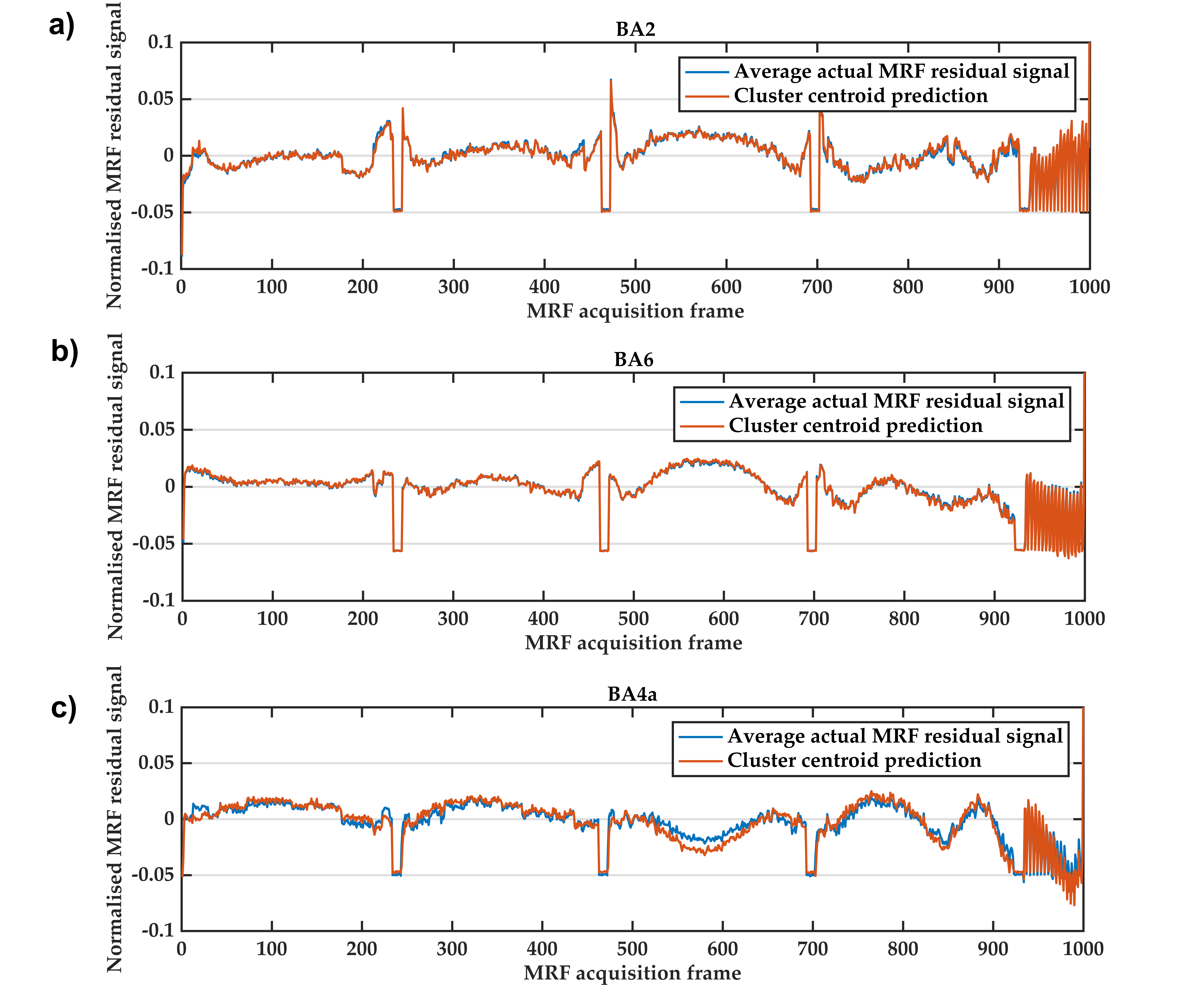
Figure 1 Qualitative evaluation of our k-shape clustering model predictions for a) primary somatosensory region BA2, b) premotor cortical region BA6 and c) primary motor cortical BA4a in an example participant. The blue line represents the average actual MRF residual signal of all voxels from each region of interest extracted from the histological atlas of the human cerebral cortex. The red line depicts the cluster centroid for each region of interest predicted by our k-shape clustering model.

Table 1 Quantitative evaluation of the predictive performance of our k-shape clustering model in parcellating three cortical regions: BA2, BA6 and BA4a.
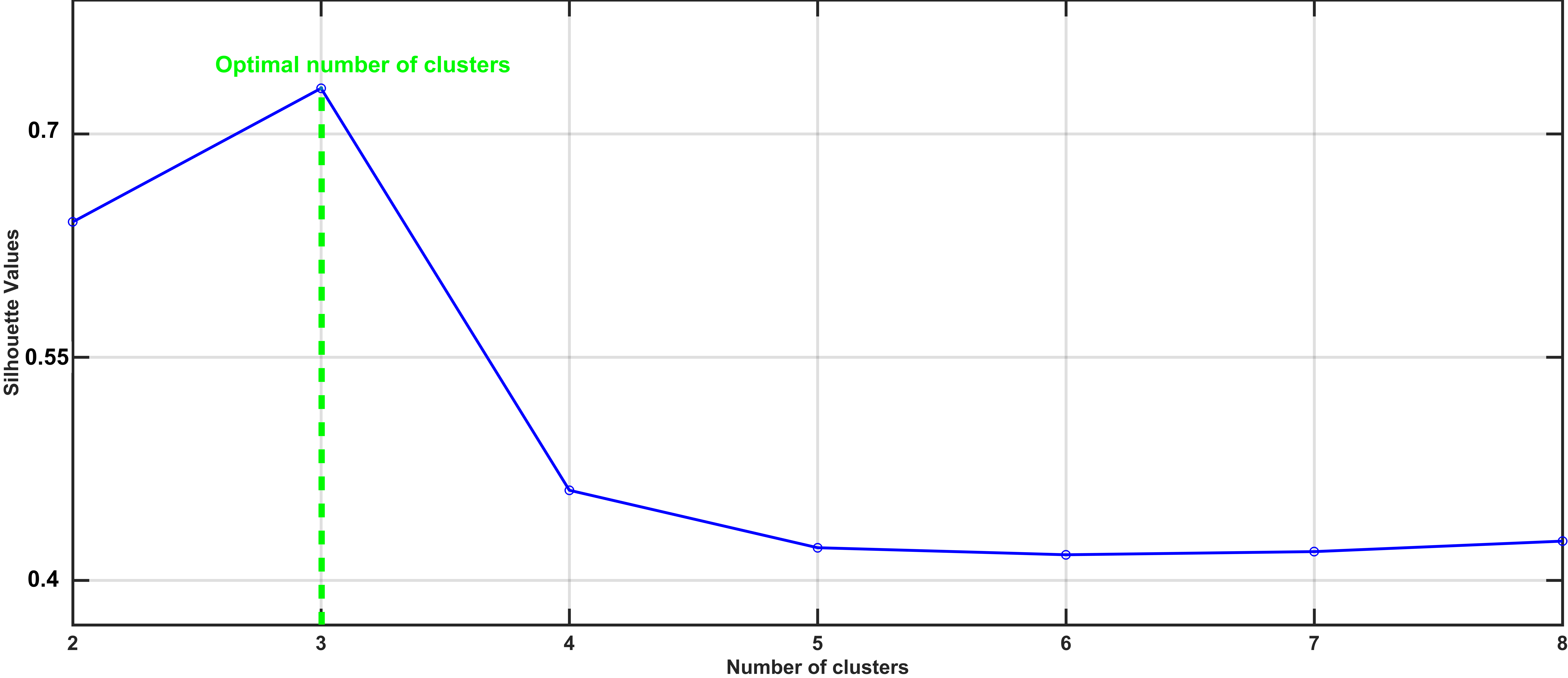
Figure 2 Cluster evaluation to assess cluster separability as measured by the Silhouette scores for different number of clusters provided to the k-shape clustering as a priori information. The number of clusters associated with the highest Silhouette score (indicated by dashed green line) defines the optimal number of clusters with the highest cluster separability found by the k-shape clustering model.
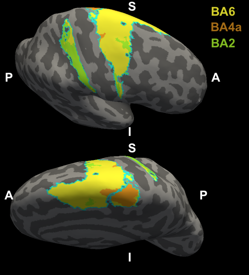
Figure 3 Unsupervised cortical parcellation results for premotor area BA6 (yellow), primary motor area BA4a (orange) and primary somatosensory area BA2 (green) an example participant, using the k-shape clustering model trained on voxel-wise MRF residual signals. The cyan solid lines around each area show the true class borders obtained from a histological atlas of the human cerebral cortex. The predicted area labels are overlaid on an inflated model of the right hemisphere cortical surface of the participant, generated by FreeSurfer.