3556
Motion-robust Three-dimensional Magnetic Resonance Fingerprinting (MR-3DMRF) for Quantitative Abdominal Imaging1Department of Health Technology and Informatics, The Hong Kong Polytechnic University, Hong Kong, China, 2Department of Diagnostic Radiology, The University of Hong Kong, Hong Kong, China
Synopsis
Keywords: MR Fingerprinting, MR Fingerprinting, Motion Correction
Motivation: Current abdominal magnetic resonance fingerprinting (MRF) techniques require breath-hold acquisition to avoid motion artifacts, which is difficult for patients with breathing difficulties.
Goal(s): This study aims to develop a motion-robust three-dimensional MRF (MR-3DMRF) technique for abdominal patients and evaluate the repeatability of tissue property quantification.
Approach: MR-3DMRF incorporates a non-smooth motion modeling method and a dynamic weighting strategy to allow free-breathing MRF acquisition and motion-resolved reconstruction.
Results: Full-width half-maximum of organ boundaries in MR-3DMRF-derived tissue maps is 3.1mm±1.8mm, significantly lower than motion-blurred tissue maps (10.1mm±4.3mm). The linearity of test-retest MRF measurement for T1, T2, and PD was 0.982, 0.894, 0.925, respectively.
Impact: This study, for the first time, investigates the feasibility of free-breathing MRF acquisition and motion-resolved MRF reconstruction on abdominal patients. The MR-3DMRF-derived tissue map are highly repeatable and accurate, potentially contribute to abdominal disease diagnosis and treatment assessment.
Introduction
Magnetic resonance fingerprinting (MRF) has demonstrated promising diagnostic performance in abdominal diseases1-3 with high tissue quantification repeatability3 and reproducibility4. However, all current clinical MRF studies require breath-hold acquisition to avoid the motion artifacts, which is difficult for patients with breathing difficulties. Recently, several motion- resolved MRF techniques have been developed. These methods retrospectively sorted MRF signals into different motion states based on respiratory signals and reconstruct tissue maps for each state separately5,6. Besides, motion modeling has been used to eliminate the motion artifacts by deforming the MRF signals acquired from different respiratory motion states into one target motion state7,8. Despite promising results, all current motion-resolved techniques are only validated on health volunteers, lacking clinical validation on abdominal patients. In this prospective study, we developed a novel motion-robust three-dimensional magnetic resonance fingerprinting (MR-3DMRF) technique for improved T1, T2, and PD property mapping in human abdominal imaging.Methods
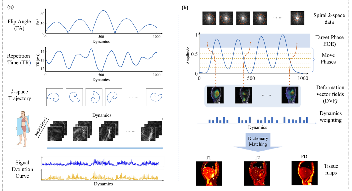
MRF Acquisition:MR-3DMRF was validated in thirteen liver cancer patients and two health volunteers. The MRF experiments were performed on a 3.0T-MRI scanner (SIGNA Premier, GE Healthcare) with 50-channel body coils. Participants were scanned using a fast acquisition with steady-state precession (FISP) sequence customized for MRF (MRF-FISP)9. A schematic illustration of the MRF-FISP sequence and the acquisition scheme is shown in Figure 1(a). To evaluate repeatability of tissue property quantification with varying respiratory condition, ten consecutive MRF scans were performed on two health volunteers and one patient. Besides, scan-rescan experiment was performed on 12 patients.
MR-3DMRF image reconstruction:
MR-3DMRF incorporates multiple motion-robust features that allow free-breathing MRF acquisition, including a non-smooth motion modeling method and a dynamic weighting strategy. As shown in Figure 1(b), the k-space data was sorted to six motion states, including one target state and five moving states. The anatomical position of each motion phase was determined by dictionary-based low-rank projection10. The deformation vector fields (DVFs) between target phase and move phases were calculated by a state-of-the-art non-rigid registration algorithm. This algorithm adopts isotropic TV-regularization for the non-smooth sliding motion between the liver and its surroundings11. The dynamics-weighting strategy divided dynamics into four groups: target dynamics, dictionary-sensitive dynamics, organ-sensitive dynamics, and non-sensitive dynamics based on Minkowski distance (MD) and Derivatives of magnetization (DOM)12, with the weighting of 1, 0.95, 0.9, and 0.85, respectively, for each group.
MR-3DMRF Evaluation:
The variation of tissue property quantitation was assessed by same-day test-retest repeatability and 10-repetition repeatability using coefficient of variation (CV). The performance of motion correction was assessed by the sharpness of the organ boundaries and was quantified by the full-width half maximum (FWHM) of the lung-liver boundary13.
Results
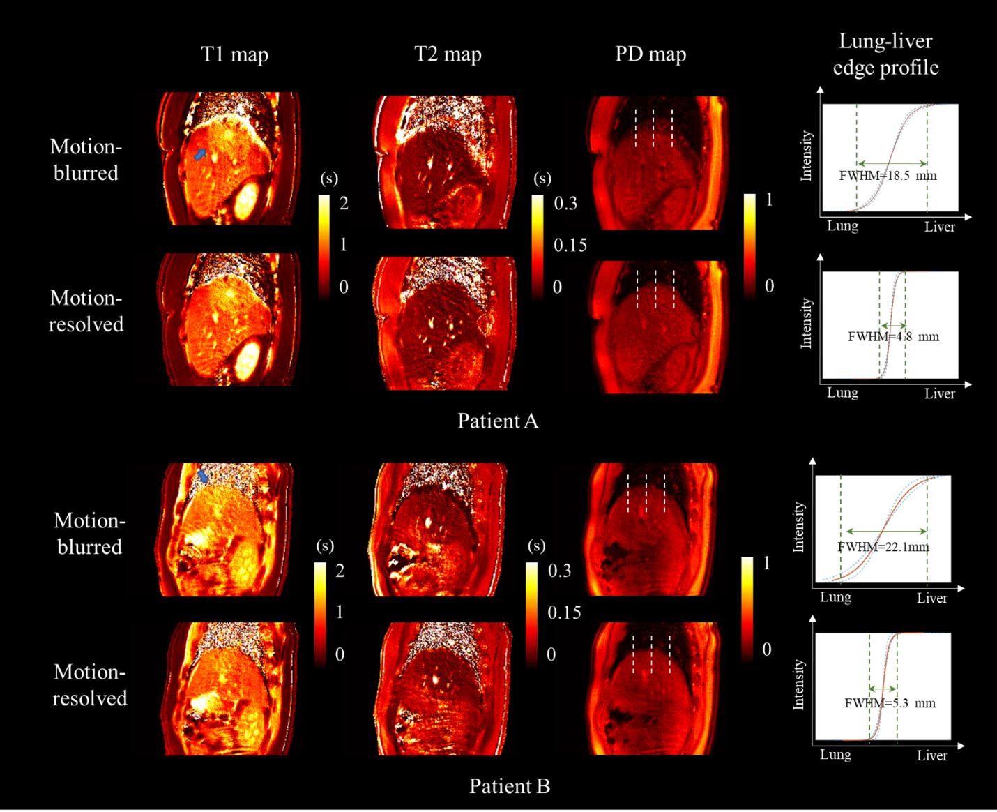
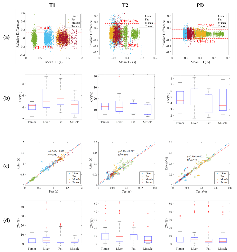
The T1, T2, and PD maps of two representative patients are shown in Figure 2. As shown, significant motion artifacts observed in the motion-blurred tissue maps are eliminated in the motion-resolved tissue maps reconstructed from MR-3DMRF. On average, the Mean ± SD FWHM was 3.1mm ± 1.8mm in the motion-resolved tissue map, significantly lower than motion-blurred tissue map (10.1mm ± 4.3mm, p-value=6.3e-7). Figure 3 presents the 10-repetition repeatability results (Figure 3(a) and Figure 3(b)) and test-retest repeatability results (Figure 3(c) and Figure 3(d)). The Bland-Altman plots in Figure 3(a) presents the tissue property measurement agreement among 10 repeated MRF scans. T2 has the highest CV values (12.0% ±2.2%). In contrast, the mean CV value in T1 and PD are within 4.0% to 5.5% and 3.6% to 5.1%, respectively, for different tissues. The test-retest results demonstrate good repeatability, achieving coefficients of determination (R^2) of 0.982, 0.894, and 0.925, respectively, for T1, T2, and PD. The T1 has the lowest CV values (4.7%±1.3%), whereas the mean CV results of T2 and PD range from 6.9% to 11.8% and 7.1% to 9.8%, respectively.Discussion and Conclusion
MR-3DMRF provided repeatable and accurate tissue property maps from the free-breathing MRF acquisition scheme. Respiratory motion-induced artifacts were eliminated from the free-breathing k-space data, resulting in clear organ position and shape. The FWHM of the liver-lung edge in the motion-resolved tissue maps was three times smaller than the motion-blurred tissue maps. The MR-3DMRF-derived tissue maps demonstrated promising image quality for abdominal disease diagnosis and treatment assessment, especially suitable for patients who could not perform breath-hold scans. Future studies should include histopathologic studies to investigate the correlation between tissue values and disease biomarkers.Acknowledgements
This research was partly supported by General Research Funds (GRF 15102219, GRF 15104822, GRF15104323) and National Natural Science Foundation of China (NSFC) Young Scientist Fund (8220294).References
- Chen, Y., et al., MR Fingerprinting for Rapid Quantitative Abdominal Imaging. Radiology, 2016. 279(1): p. 278-286. 2.
- Jaubert, O., et al., Multi-parametric liver tissue characterization using MR fingerprinting: Simultaneous T1, T2, T2*, and fat fraction mapping. Magnetic Resonance in Medicine, 2020. 84(5): p. 2625-2635.
- Fujita, S., et al., MR Fingerprinting for Liver Tissue Characterization: A Histopathologic Correlation Study. Radiology, 2022. 0(0): p. 220736.
- MacAskill, C.J., et al., Rapid B1-Insensitive MR Fingerprinting for Quantitative Kidney Imaging. Radiology, 2021. 300(2): p. 380-387.
- Li, T., et al., Time-resolved magnetic resonance fingerprinting for radiotherapy motion management. Medical Physics, 2020. 47(12): p. 6286-6293.
- Huang, S.S., et al., Free-Breathing Abdominal Magnetic Resonance Fingerprinting Using a Pilot Tone Navigator. Journal of Magnetic Resonance Imaging, 2021. 54(4): p. 1138-1151.
- Cao, P., et al., Motion-resolved and free-breathing liver MRF. Magn Reson Imaging, 2022. 91: p. 69-80.
- Cruz, G., et al., Generalized low-rank nonrigid motion-corrected reconstruction for MR fingerprinting. Magnetic Resonance in Medicine, 2021.
- Jiang, Y., et al., MR fingerprinting using fast imaging with steady state precession (FISP) with spiral readout. Magnetic Resonance in Medicine, 2015. 74(6): p. 1621-1631.
- McGivney, D.F., et al., SVD compression for magnetic resonance fingerprinting in the time domain. IEEE transactions on medical imaging, 2014. 33(12): p. 2311-2322.
- Vishnevskiy, V., et al., Isotropic Total Variation Regularization of Displacements in Parametric Image Registration. IEEE Transactions on Medical Imaging, 2017. 36(2): p. 385-395.
- Stolk, C.C. and A. Sbrizzi, Understanding the Combined Effect of k -Space Undersampling and Transient States Excitation in MR Fingerprinting Reconstructions. IEEE Trans Med Imaging, 2019. 38(10): p. 2445-2455.
- Xiao, H., et al., A dual-supervised deformation estimation model (DDEM) for constructing ultra-quality 4D-MRI based on a commercial low-quality 4D-MRI for liver cancer radiation therapy. Med Phys, 2022. 49(5): p. 3159-3170.
Figures