3542
Assessment of cardiac structural and functional changes in patients with Parkinson's disease by CMR-FT: a pilot study1West China Hospital, Sichuan University, Chengdu, China, 2Radiology, West China Hospital, Sichuan University, Chengdu, China
Synopsis
Keywords: Heart Failure, Parkinson's Disease, Cardiac magnetic resonance, feature tracking, left heart
Motivation: To evaluate the changes of cardiac structure and function in patients with Parkinson's disease (PD).
Goal(s): To evaluate the structural and functional changes of left heart in PD patients by cardiac magnetic resonance feature tracking (CMR-FT), and to explore the influencing factors of left heart structure and function.
Approach: Cardiac function and strain parameters of left heart were obtained, and the correlation between CMR parameters and motor dysfunction and autonomic dysfunction was evaluated.
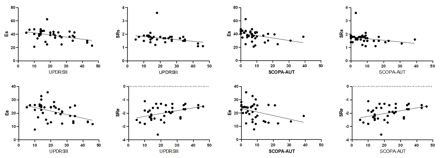
Results: Left atrium reserve and conduction function are related to the severity of motor dysfunction and autonomic dysfunction. CMR-FT can detect the early changes of cardiac function in PD patients.
Impact: The prevalence of heart failure has been twice among Parkinson's Disease patients compared with overall population. Cardiac magnetic resonance feature tracking technology can be used as an effective tool for early diagnosis of cardiac dysfunction in Parkinson's Disease patients.
Purpose:
Cardiac changes in PD patients include cardiac autonomic dysfunction, cardiomyopathy, coronary heart disease, arrhythmia or sudden cardiac death 1,2,3. Therefore, early diagnosis and intervention of cardiac abnormalities in PD patients is particularly important to reduce the incidence of adverse cardiovascular events. The aim of this study is to evaluate the changes of left ventricular structure and function in patients with Parkinson's disease (PD) using cardiac magnetic resonance feature tracking technology, to explore whether there is early subclinical cardiac dysfunction in PD patients, and to correlate the cardiac changes in PD patients with the severity of the disease.Materials and methods:
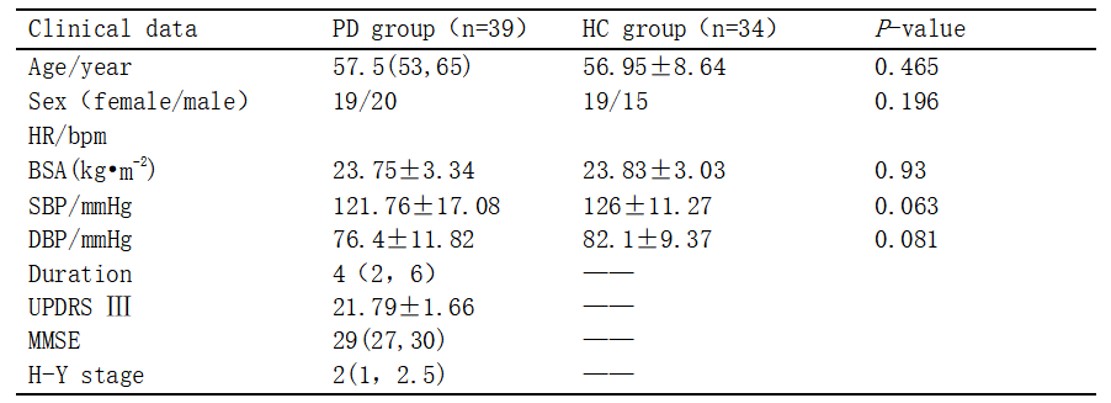
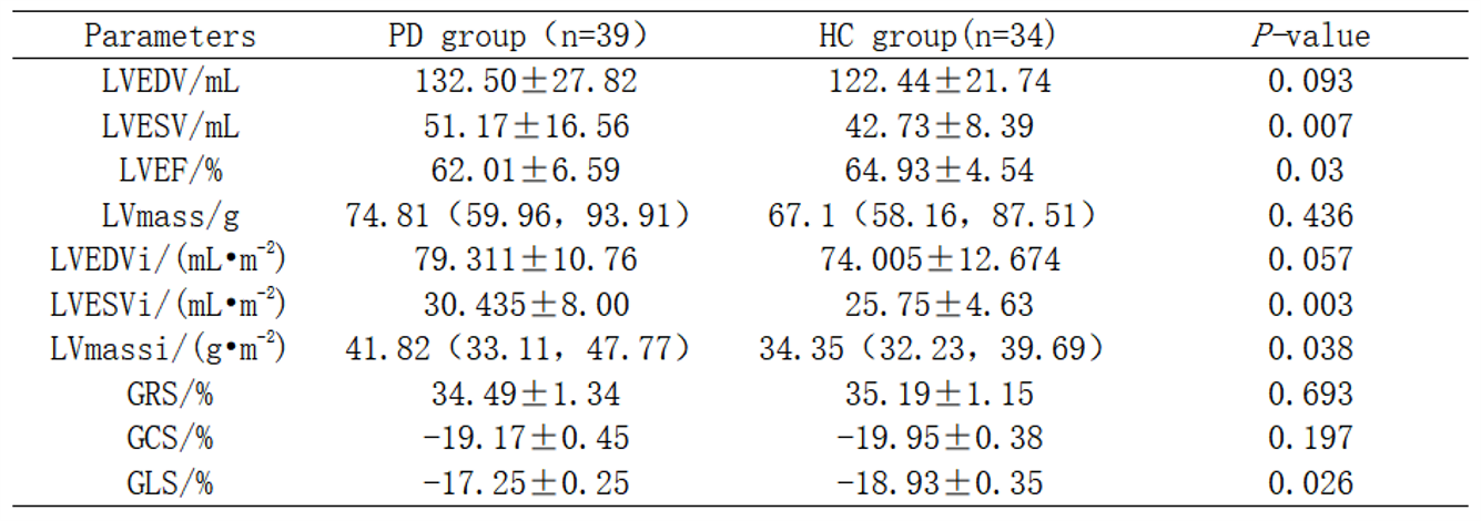
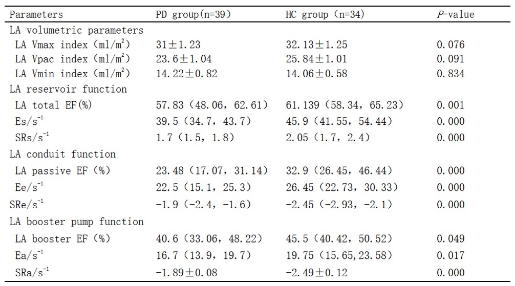
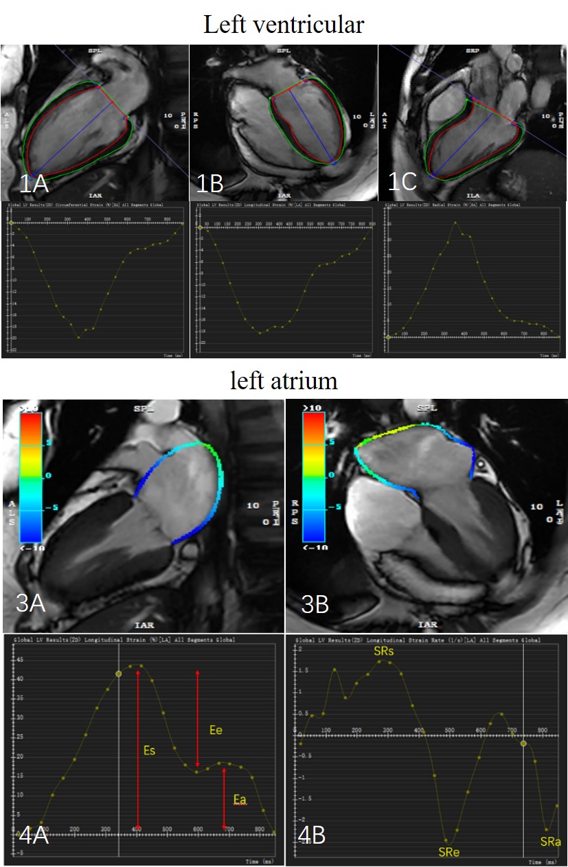
Thirty-nine cases (mean age: 57 years) of PD patients (PD group) and 34 cases (mean age: 56 years) of healthy controls (control group), all of whom had completed routine cardiac magnetic resonance examinations (uMR 790, United Imaging Healthcare, Shanghai, China), were included in this study. LV function and myocardial strain were analyzed using the specialized post-processing software CVI42 (version5.13.5, Circle Cardiovascular Imaging, Canada). Left ventricular (LV) function parameters included: left ventricular end-diastolic volume index (LVEDVi), left ventricular end-systolic volume index (LVESVi), left ventricular ejection fraction (LVEF), and left ventricular myocardial mass index (LVMi); and myocardial strain parameters included: overall radial strain (GRS), overall circumferential strain (GCS), and overall longitudinal strain (GLS) of the LV. Left atrial (LA) functional parameters included: LA reserve function (total ejection fraction [EF total], total strain [Es], peak positive strain rate [SRs]), conduit function (passive ejection fraction [EF passive], passive strain [εe], early peak negative strain rate [SRe]), and pump function (active ejection fraction [EF booster], active strain [Ea], late peak negative strain rate [SRa]). Comparisons between the two groups were performed using the independent samples t test or Mann-Whitney U test. Pearson or Spearman correlation coefficient models were used to analyze the correlation of cardiac function and strain parameters with motor dysfunction (UPDRS III) and autonomic dysfunction (SCOPA-AUT), and to explore the factors affecting cardiac function and strain parameters.Results:
Analysis of left ventricular function and myocardial strain showed that LVEF and GLS in the PD group were lower than those in the control group, and LVESV, LVESVi, and LVMi were higher than those in the control group, and the differences were statistically significant (all P < 0.05). Analysis of left atrial function and myocardial strain showed that the differences in left atrial volumes LA Vpaci, LA Vmini, and LA Vmaxi in the PD group were not statistically significant compared with those in the control group (P > 0.05). Left atrial reserve function (EF total, Es, SRs) and left atrial catheterization function (EF passive, Ee, SRe) in PD patients and pump function (EF booster, Ea, SRa) were significantly impaired compared with controls (all P < 0.05). Correlation analysis showed that left atrial reserve function Es and SRs were negatively correlated with UPDRS III (r= -0.409, -0.355, P< 0.05). Left atrial conduit function LAEF passive, Ee were negatively correlated with UPDRS III (r=-0.326, -0.445, P<0.05), SRs were positively correlated with UPDRS III (r=0.417, P<0.05). Left atrial reserve function Es and SRs were negatively correlated with SCOPA-AUT (r=-0.535, -0.319, both P<0.05). Left atrial conduit function Ee was negatively correlated with SCOPA-AUT (r= -0.316, P< 0.05), and SRe was positively correlated with SCOPA-AUT (r= 0.359, P< 0.05).Conclusion:
Strain and strain rate parameters obtained by CMR-FT allow early assessment of cardiac impairment in PD patients. Left ventricular systolic function and left atrial reserve, conduction, and pump function are impaired in PD patients; left atrial reserve function and conduction correlate with the severity of motor dysfunction and autonomic dysfunction.Acknowledgements
This study was supported by Key Research and Development Projects in Sichuan Province, China (grant Number: 2020YFS0123).References
1. Tysnes O B, Storstein A. Epidemiology of Parkinson's disease[J]. J Neural Transm (Vienna), 2017,124(8):901-905.
2. Tysnes O B, Storstein A. Epidemiology of Parkinson's disease[J]. J Neural Transm (Vienna), 2017,124(8):901-905.
3. Piqueras-Flores J, López-García A, Moreno-Reig Á, et al. Structural and functional alterations of the heart in Parkinson's disease[J]. Neurol Res, 2018,40(1):53-61.
Figures