3535
Accelerated ferumoxytol-enhanced free-running acquisitions for whole-heart angiography in congenital heart disease patients.1Department of Radiology, Lausanne University Hospital and University of Lausanne, Lausanne, Switzerland, 2Advanced Clinical Imaging Technology, Siemens Healthineers International AG, Lausanne, Switzerland, 3Center for Biomedical Imaging (CIBM), Lausanne, Switzerland, 4Division of Pediatric Cardiology, Woman-Mother-Child Department, Lausanne University Hospital and University of Lausanne, Lausanne, Switzerland, 5Service of Cardiology, Lausanne University Hospital and University of Lausanne, Lausanne, Switzerland, 6University Lyon, INSA-Lyon, University Claude Bernard Lyon 1, UJM-Saint Etienne, CNRS, Inserm, CREATIS, Villeurbanne, France, 7Department of Radiology, Louis Pradel Hospital, Bron, France
Synopsis
Keywords: Vascular, Cardiovascular, Rapid MRI
Motivation: Angiography using ferumoxytol-enhanced free-running MRI can be obtained within an acquisition window of 6 minutes, which could be further reduced for faster clinical workflows.
Goal(s): To determine the lower bounds of scan time for fast free-running whole-heart MRI using a data-driven reconstruction (SIMBA).
Approach: Fifteen datasets were retrospectively undersampled and image quality metrics were determined as a function of scan time reduction.
Results: A 3-minute acquisition provides comparable image quality to that of its original 6-minute counterpart, and when applying compressed sensing we can confidently further reduce the acquisition time of 3D MR angiography with high resolution to 2 minutes.
Impact: Free-running whole-heart MRI acquisitions can be greatly sped-up by exploiting ferumoxytol contrast enhancement, in conjunction with a data-driven reconstruction, which facilitates fast whole-heart angiography in congenital heart disease patients.
Background
One of the major challenges in CMR is to abbreviate scan times for shorter protocols compatible with clinical workflows1. In 3D MR angiography, several strategies have been proposed to shorten scan times by exploiting temporal redundancies as in XD-GRASP2, or spatial redundancies as in 3D-PROST3. However, these approaches use motion management methods, e.g. ECG-triggering or self-navigation, susceptible to heart rate variabilities or respiratory drifts. A similarity-driven multi-dimensional binning algorithm (SIMBA)4 was introduced to reconstruct free-running whole-heart MRI data without explicitly defining physiological phases, but by combining similar motion states occurring over a long enough 6-minute acquisition. Together with ferumoxytol enhancement, SIMBA allows for a comprehensive angiographic evaluation. The goal of this work was to take advantage of the high blood-pool signal from ferumoxytol and explore its potential for scan time reduction of free-running MRI using SIMBA, and its compressed sensing extension XD-MC-SIMBA5, while preserving image quality.Methods
Fifteen congenital heart disease patients were scanned after injection of ferumoxytol on a 1.5T clinical MRI scanner (MAGNETOM Sola, Siemens Healthineers AG, Erlangen, Germany) using a free-running GRE research imaging sequence6 with a 3D radial phyllotaxis trajectory7. Each dataset was retrospectively undersampled, starting from 6 minutes (full acquisition $$$I_{6min}$$$) and taking each time 1 minute less of data until $$$I_{1min}$$$ . For $$$I_{1min}...I_{6min}$$$, SIMBA was applied as in Heerfordt et al.4, and reconstruction was performed with both a 3D gridded reconstruction (SIMBA) and a CS motion-resolved reconstruction along the SIMBA cluster dimension (XD-MC-SIMBA)5. The SIMBA clustering and data selection were categorized by using ECG and self-gating as references. Moreover, we computed the percentage of overlap between the selected data for each undersampling level and that of $$$I_{6min}$$$. For both SIMBA and XD-MC-SIMBA, the blood to myocardium interface sharpness6 and the structural similarity index measure (SSIM)7 were computed between $$$I_{6min}$$$ and all the other images. Statistical significance was defined by two-sided paired sample t-tests with p<0.01 considered statistically significant, after Bonferroni correction for multiple comparisons.Results
Comparison of SIMBA clustering (Fig1) from $$$I_{6min}$$$ to $$$I_{1min}$$$ shows how the clustering remains consistent in the low-dimensional space and the location of the selected resting phase is stable across undersampling levels. On average, there is an overlap in data selection compared to $$$I_{6min}$$$ of 93$$$\pm$$$5% for $$$I_{5min}$$$, 91$$$\pm$$$3% for $$$I_{4min}$$$, 89$$$\pm$$$3% for $$$I_{3min}$$$, 87$$$\pm$$$2% for $$$I_{2min}$$$, and 85$$$\pm$$$2% for $$$I_{1min}$$$ (Fig2A). Nonetheless, there are 2 cases for which we observe differences in the reconstructed phases (e.g., swap between systolic and diastolic phase; arrow Fig3). Fig4 and Fig5 show example datasets reconstructed with SIMBA and XD-MC-SIMBA: we can observe how images $$$I_{6min}$$$ to $$$I_{1min}$$$ have very similar image quality, and how, using XD-MC-SIMBA, we compensate for the strong undersampling in $$$I_{2min}$$$ and $$$I_{1min}$$$. Comparison of the blood-myocardium sharpness shows only a statistically significant difference when comparing the SIMBA reconstructions for $$$I_{1min}$$$ and $$$I_{6min}$$$ (Fig2B). Additionally, we observe higher sharpness values for $$$I_{5min}$$$vs.$$$I_{6min}$$$. Compared to $$$I_{6min}$$$, the SSIM rapidly decreases from $$$I_{5min}$$$ to $$$I_{1min}$$$ but at a reduced rate for XD-MC-SIMBA. The mean of SSIM is significantly greater (p<0.01) than 0.95 for $$$I_{5min}$$$ to $$$I_{3min}$$$ for SIMBA, and for $$$I_{5min}$$$ to $$$I_{2min}$$$ for XD-MC-SIMBA (Fig2C).Discussion and conclusion
This study supports the hypothesis that SIMBA and XD-MC-SIMBA can be used to significantly abbreviate scan time of ferumoxytol-enhanced free-running acquisitions at no or minor expense in image quality. Analysis of data selection, blood-myocardium sharpness, and SSIM suggest that a 3-minute scan (1/2 of the original time) results in concordant image quality relative to the full 6-minute acquisition, when reconstructed with SIMBA. With XD-MC-SIMBA and according to the same metrics, we can further reduce scan time to 2 minutes (1/3 of the original time). The increased blood-myocardium sharpness for $$$I_{5min}$$$ suggests that less data may be more favorable for the current version of SIMBA, which should be further investigated. The SIMBA clustering may be unstable in a few instances, resulting in the choice of a different physiological phase for the different undersampling levels at no visible expense of image quality. In the future, we plan to add a blind consensus reading to assign diagnostic quality scores. Another limitation lies in the retrospective nature of the data selection, as the retrospectively undersampled data may not have the same trajectory uniformity as that of an acquisition with abbreviated scan time. We will address this by prospectively acquiring datasets at these newly defined reduced scan times, confirming the hypothesis that SIMBA and XD-MC-SIMBA can be used to accelerate 3D acquisitions of the heart with ferumoxytol while keeping a high conspicuity of relevant anatomical structures.Acknowledgements
No acknowledgement found.References
1. Munoz, C., Fotaki, A., Botnar, R.M. and Prieto, C. Latest Advances in Image Acceleration: All Dimensions are Fair Game. J Magn Reson Imaging. 57: 387-402 (2023).
2. Piccini D, Feng L, Bonanno G, et al. Four-dimensional respiratory motion-resolved whole heart coronary MR angiography. Magn Reason Med. 77:1473-1484 (2017).
3. Bustin A, Ginami G, Cruz G, et al. Five-minute whole-heart coronary MRA with sub-millimeter isotropic resolution, 100% respiratory scan efficiency, and 3D-PROST reconstruction. Magn Reson Med. 81:102-115 (2019).
4. Heerfordt, J. et al. Similarity-driven multi-dimensional binning algorithm (SIMBA) for free-running motion-suppressed whole-heart MRA. Magn. Reson. Med. 86, 213–229 (2021).
5. Romanin L, Milani B, Roy CW, Bustin A, Si-mohamed S, Prsa M, et al. Similarity-driven motion-resolved reconstruction for ferumoxytol-enhanced whole-heart MRI of congenital heart disease patients. In: Proc Intl Soc Mag Reson Med 31 (2023).
6. Roy CW, Di Sopra L, Whitehead KK, Piccini D, Yerly J, Heerfordt J, et al. Free-running cardiac and respiratory motion-resolved 5D whole-heart coronary cardiovascular magnetic resonance angiography in pediatric cardiac patients using ferumoxytol. J Cardiovasc Magn Reason. 24(1):1–12 (2022).
7. Piccini, D., Littmann, A., Nielles-Vallespin, S. & Zenge, M. O. Spiral phyllotaxis: The natural way to construct a 3D radial trajectory in MRI. Magn. Reson. Med. 66, 1049–1056 (2011).
8. Ahmad R, Ding Y, Simonetti OP. Edge Sharpness Assessment by Parametric Modeling: Application to Magnetic Resonance Imaging. Concepts Magn Reson Part A Bridg Educ Res [Internet]. 2015/09/28. 44(3):138–49 (2015).
9. Wang Z, Bovik AC, Sheikh HR, Simoncelli EP. Image quality assessment: from error visibility to structural similarity. IEEE Trans Image Process. 13(4):600–12 (2004).Figures
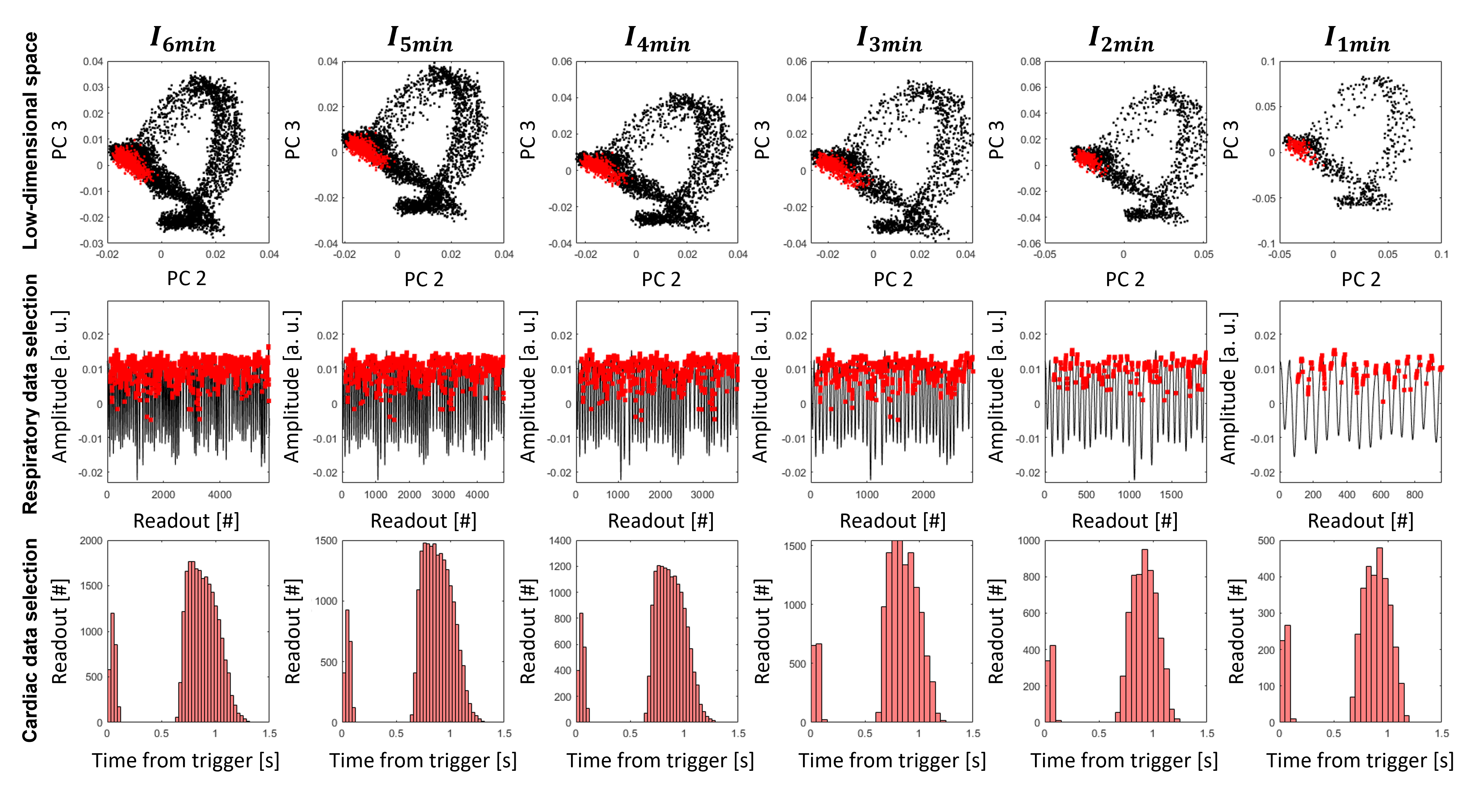
Fig1. Comparison of the SIMBA clustering and data selection (red) for different levels of undersampling. For the low-dimensional space, two of the first principal components computed in SIMBA (PC2 & PC3) are projected against each other to graphically display the distribution of the acquired data. The respiratory signal is obtained from self-gating. The cardiac data selection is obtained using the ECG as reference.
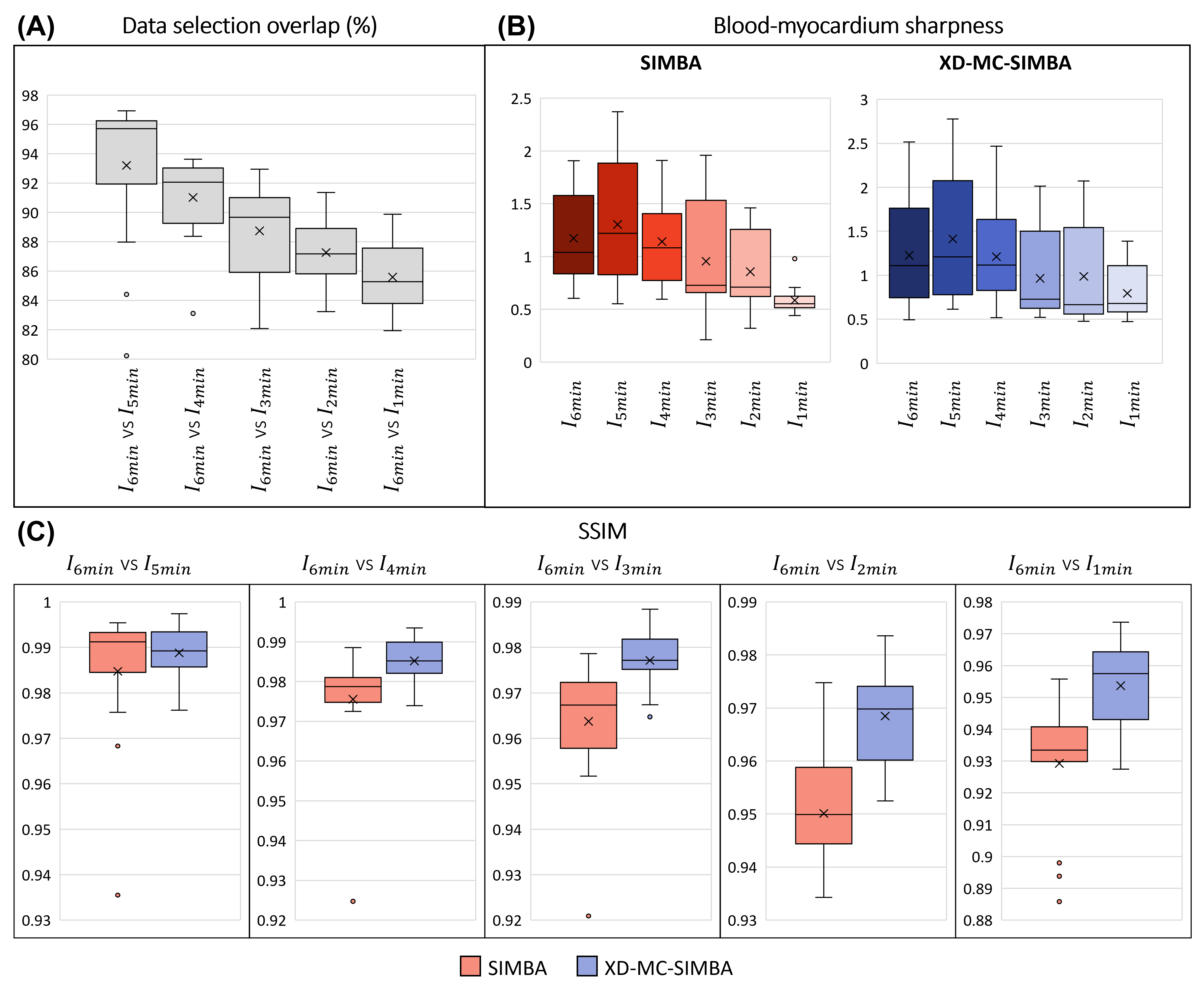
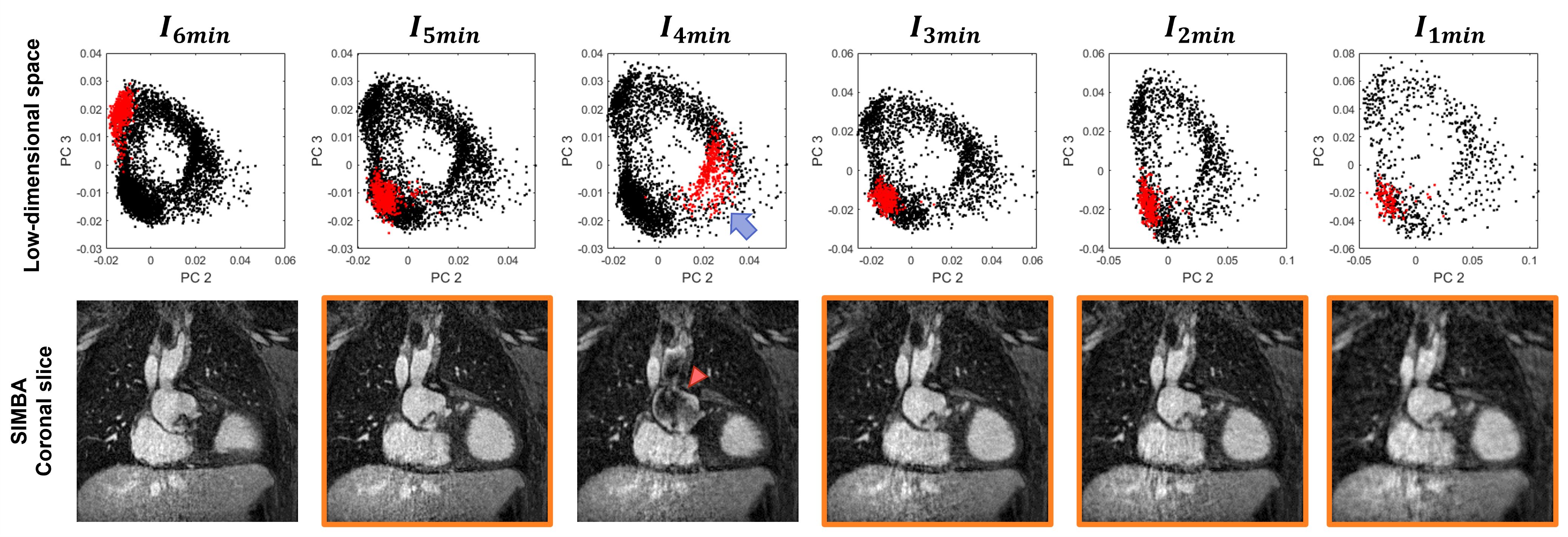
Fig3. Example of dataset for which the SIMBA clustering results in the selection of a different cardiac phase: $$$I_{6min}$$$ is in a mid-diastolic phase, $$$I_{5min}$$$ together with $$$I_{3min}$$$, $$$I_{2min}$$$ and $$$I_{1min}$$$ in an end-diastolic phase (highlighted in orange), while $$$I_{4min}$$$ (blue arrow) in a systolic phase (observable signal void artefact due to high blood flow velocity; red arrow).
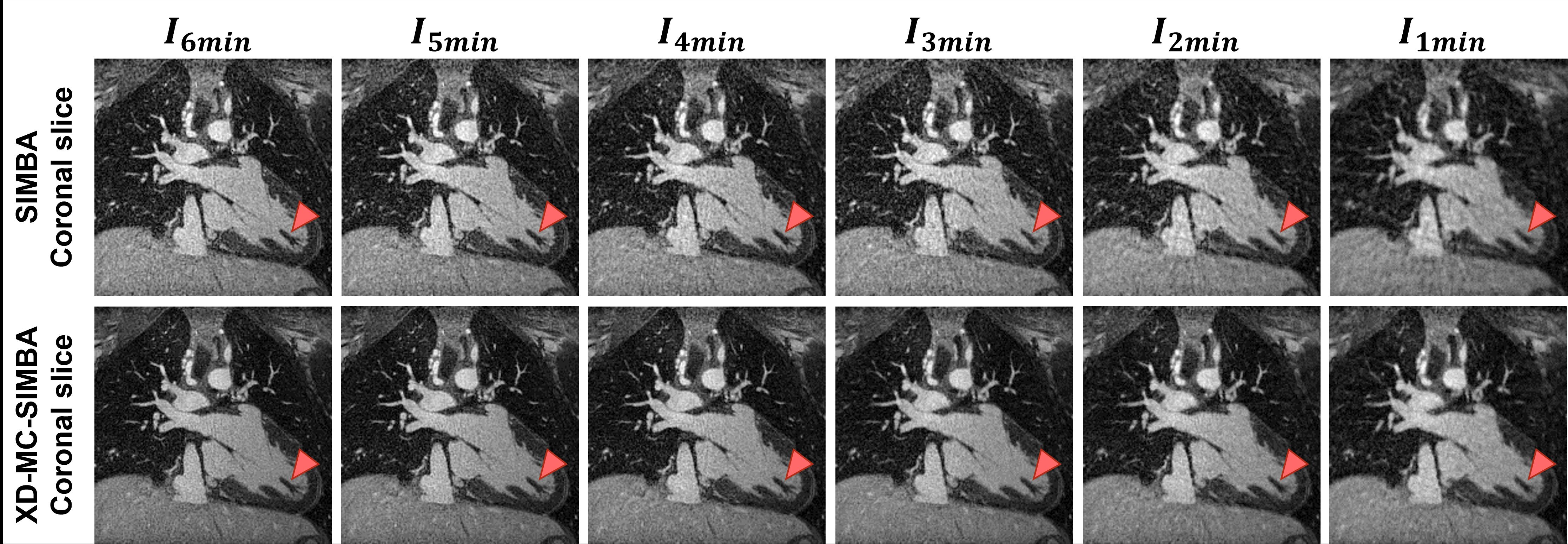
Fig4. Example of ferumoxytol-enhanced GRE acquisition of a CHD patient (male; 20 years old) after a Fontan procedure. Major anatomical features are visible in all images, even at very high undersampling (1min) rates, but with XD-MC-SIMBA these features are much sharper and more clearly delineated, compared to SIMBA (e.g. papillary muscles, red arrow).
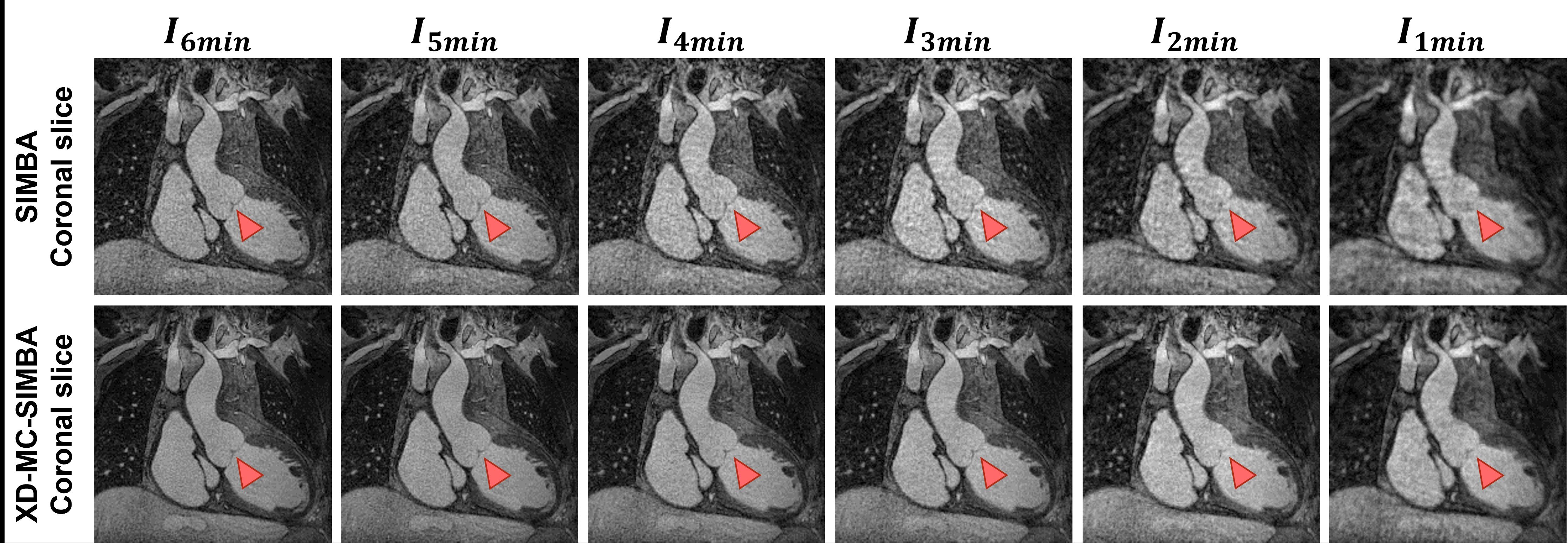
Fig5. Example of ferumoxytol-enhanced GRE acquisition of a CHD patient (male; 39 years old) after a Fontan procedure. As in Fig4, major anatomical features are visible in all images, but in this case high undersampling levels (2min and 1min) greatly benefit from using XD-MC-SIMBA (e.g. better visible valve leaflets, red arrows).