3532
Effects of RAFF2 periodicity on contrast between infarct and remote myocardium1A.I. Virtanen Institute, Kuopio, Finland, 2University of Minnesota, Minneapolis, MN, United States, 3University of Oulu, Oulu, Finland
Synopsis
Keywords: Myocardium, Preclinical, RAFF2, Rotating frame relaxations, myocardial infarct
Motivation: Current MRI techniques are insufficiently sensitive to determine specific physiological changes in the myocardium after infarction.
Goal(s): To optimize sensitivity of RAFF2 technique to specific tissue changes of the myocardium after myocardial infarct (MI) by modulating the durations of P-packets of RAFF2 pulses.
Approach: Mice hearts are imaged ex vivo 7 days after the factitious myocardial infarction by using different durations of P-packets of the RAFF2 along with other rotating frame relaxation techniques.
Results: Duration of the RAFF2 pulse have an impact on relaxation maps and contrast of MI area compared to the remote area. Increased rotating frame relaxation times were also detected.
Impact: This study presents novel application of RAFF2 technique for quantitative ex vivo assessment of mice myocardium after infarction. This method allows to study physiological changes in the myocardium with high sensitivity.
Introduction
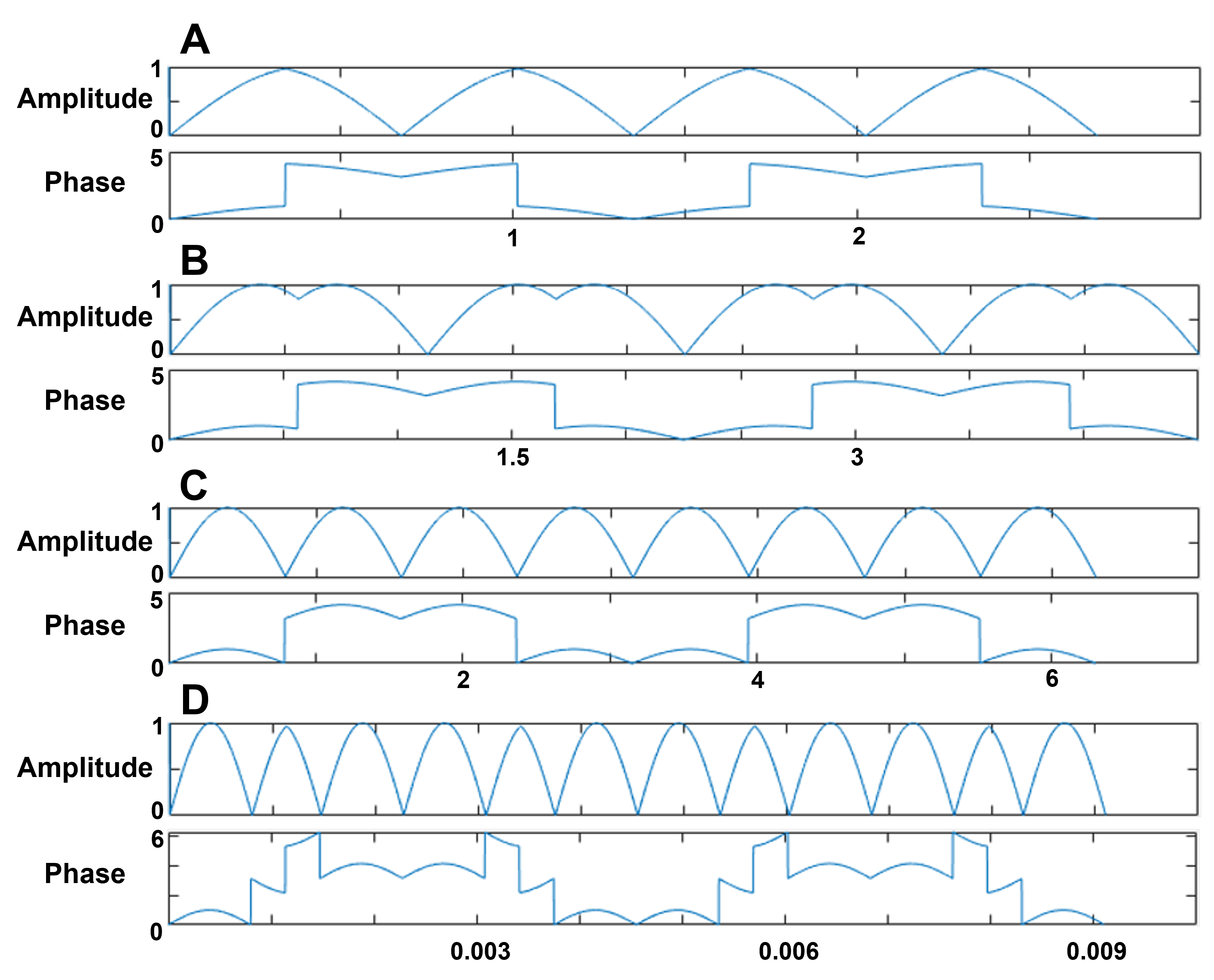
Cardiovascular diseases (CVD) are the leading cause of death in the Western countries [1]. One dangerous disease of the CVD is myocardial infarct (MI). MI is caused by blockage of coronary artery, which initiate an ischemic reaction in the myocardium leading eventually to fibrosis and scar formation [2]. Relaxation Along a Fictitious Field in the rotating frame of rank n (RAFFn), especially RAFF2, have potential to characterize fibrosis-related myocardial diseases [3]. The RAFFn with rank 2 can be tuned to specific tissue changes of the myocardium by changing the durations of P-packets in RAFF2 pulses. Periodicity of radiofrequency (RF)-irradiation can be modified by changing the duration of Sine/Cosine amplitude and frequency modulated RAFFn pulses used in the pulse train as presented in [4] and in Figure 1. A significant increase in exchange-induced relaxation rate constants had been shown at the side bands generated during the periodic irradiation with RAFFn [4]. In this work, we embarked on tuning the periodicity of irradiation with modifying the duration of P-packages in RAFF2 pulse to determine the alterations of the physiological conditions during MI.Purpose
To study the effect of periodicity of irradiation by changing the durations of P-packets of RAFF2 as defined by the stretching factor (TL). Additionally, the purpose was to compare relaxation times and contrast between infarct and remote areas 7 days after MI ex vivo.Materials and Methods
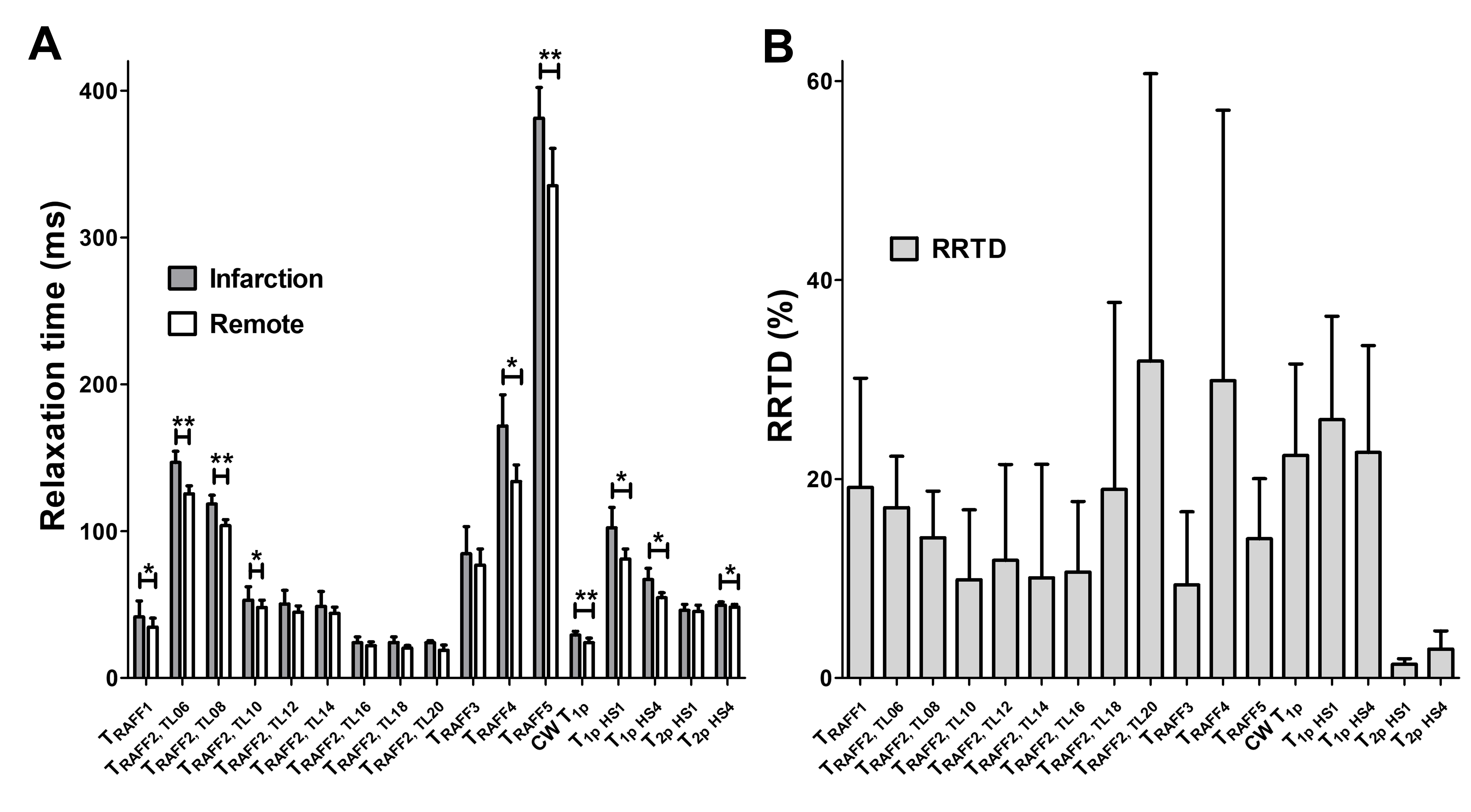
The hearts of C57BL (n = 5) mice were collected 7 days after occlusion of Left Anterior Descending (LAD) Coronary Artery, fixed 4% PFA in PBS, stored in 15% sucrose and imaged in Galden with 9.4 T. Relaxation studies were performed by measuring 4 images with different pulse train or spin lock durations. Stretching factor (TL) defines how long period of Sine/Cosine modulation is included in a P-package for ampitude and phase modulations (Figure 1) [4]. The original P-package in RAFF2 pulse is marked as TL1.0 (Figure 1). Due to variation in RAFF2 pulse duration, maximum number of pulses in pulse train varied from 4 to 32. RAFFn measurements were performed by detecting SI evolution to the steady state from +Z and -Z. 2-D FISP readout with 1 mm thickness was used for selection of the lower mid level of the heart. Relaxation time constant maps were calculated using monoexponential. MI and remote areas were assessed using regions of interests (ROIs) analysis. The ROIs were then used to average the data in relaxation time maps. The contrast between remote and infarcted area was measured as Relative Relaxation Time Difference (RRTD). The RRTD was calculated as (T(infarct) - T(remote)) / T(remote)*100%, where T refers for the relaxation time in specific area. The ROI analysis was performed using MATLAB R2019b (MathWorks Inc., Natick, CA, USA) and Aedes Software (http://aedes.uef.fi).Results
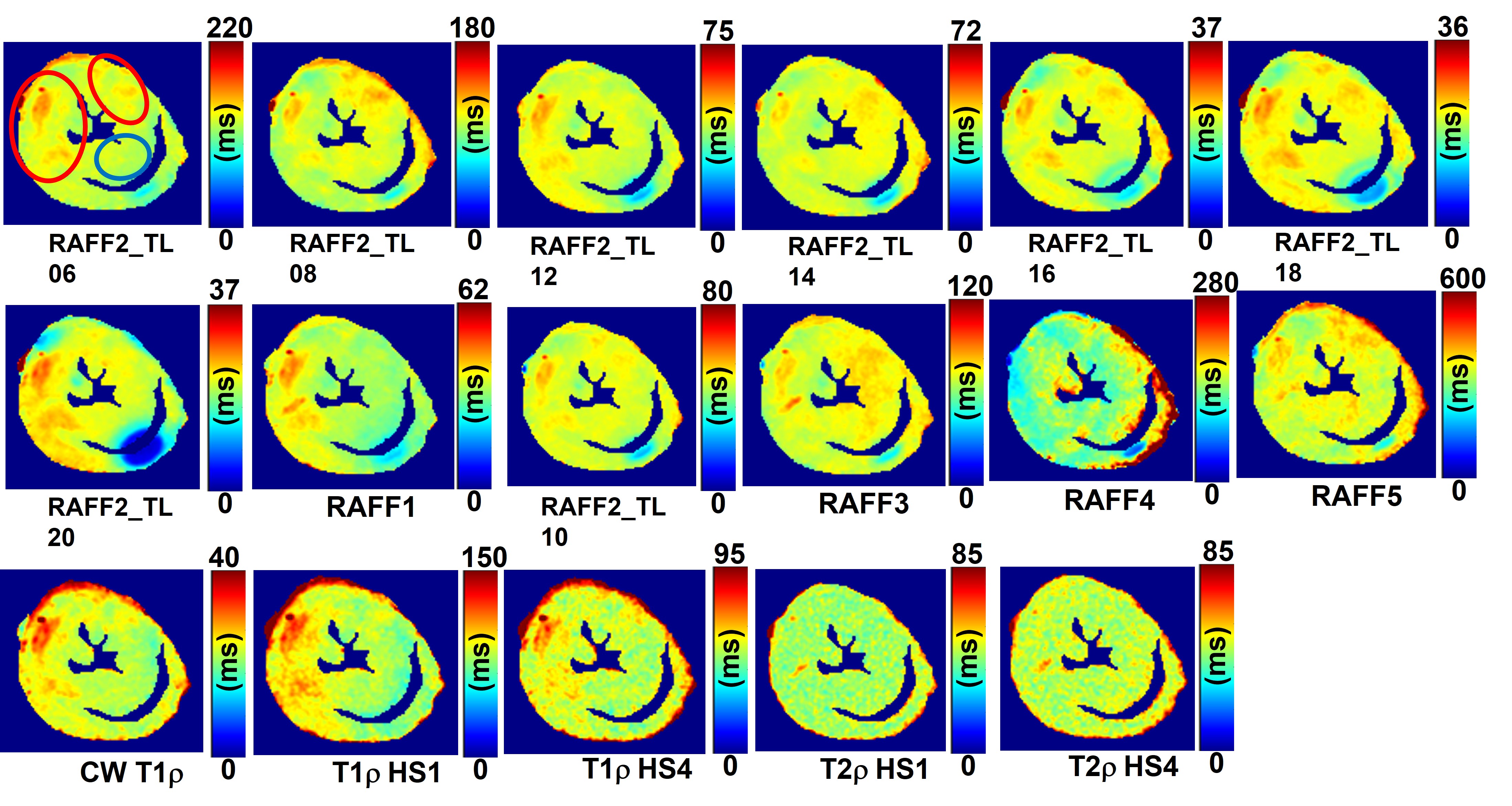
Significant differences between infarct and remote areas were detected with RAFF1, RAFF2 TL0.6, RAFF2 TL0.8, RAFF2 TL1.0, RAFF3, RAFF5, adiabatic T1rho, adiabatic T2rho, and continuous wave spin lock T1rho (Figure 2A). The contrast differences between MI and remote areas can also be seen in RRTD-values (Figure 2B). Out of RAFF2 versions, the highest RRTDs were found with TL1.8 and 2.0 (Figure 2B). Also, high RRTDs were detected with RAFF4, CW T1rho and adiabatic T1rho (Figure 2B). In all rotating relaxation time maps, higher relaxation times are associated with MI (indicated as a red oval) as compared to the rest of the myocardium (Figure 3).Discussion
Increase of stretching factor used in RAFF2 is affecting the relaxation times and the contrast between infarct and remote myocardium. The lowest RRTDs were found with TLs 1.0 - 1.6, while in TLs 1.8 and 2.0 maps contained B0 artifacts. The RAFF2 with TL 0.6 exhibited the highest relaxation time from other TLs and it had high RRTD value and did not have any artifacts. Further development of pulse design along with dedicated shimming solutions is required to reduce B0 and B1 artifacts, along with histological validations of infarcted tissue. The rotating relaxation time maps RAFF4, adiabatic and continuous wave spin lock T1rho showed also increased relaxation times at the same areas as detected with RAFF2 with different TLs. These rotating frame relaxation times agrees with relaxation times found in the literature [5].Conclusion
By altering stretching factor the sensitivity of RAFF2 in MI area can be improved.Acknowledgements
Authors thank for Svetlana Laidinen for doing infarct operations and taking the heart out of the mice. Authors also want to thank Kuopio biomedical imaging unit (BIU) for using their magnet. Authors thank GeneCellNano flagship, Finnish Cultural Foundation, Finnish Foundation for Cardiovascular Research, Valtion tutkimusrahoitus (VTR) also known as government funding for research, Mauri and Sirkka Wiljasalo Foundation, Paavo Nurmi Foundation, Emil Aaltonen Foundation, Matti and Vappu Maukonen Foundation, Maud Kuistila Memorial Foundation and NIH grant P41 EB027061 for supporting this research project.References
[1] Mozaffarian D, et al. Circulation. 2015; 131:e29-322., [2], Ertl G, Frantz S. Healing after myocardial infarction. Cardiovasc Res. 2005;66:22–32., [3] Ylä-Herttuala E et al. J Cardiovasc Magn Reson. 2018;30:34., [4] Liimatainen T et al. J Magn Reson. 2018;296:79-84. [5] Ylä-Herttuala E et al. under a review in J Cardiovasc Magn Reson.Figures