3528
Exploring the additive effects of hypertension on left cardiac structure and deformation in type 2 diabetes mellitus patients using CMR-FT1Department of Radiology, the Second Hospital of Dalian Medical University, Dalian, China, 2MR Research Collaboration, Siemens Healthcare, Beijing, China
Synopsis
Keywords: Myocardium, Cardiovascular, CMR-FT, Hypertension, T2DM
Motivation: Hypertension and type 2 diabetes mellitus (T2DM)-related cardiac damage have a partial common pathological mechanism[1].
Goal(s): To investigate the effects of hypertension on left cardiac structure and deformation in patients with T2DM using cardiac magnetic resonance feature tracking.
Approach: The balanced steady-state free precession cine sequence and commercial software, cvi42, were used for image acquisition and post-processing. Covariance and multivariate linear regression were used for statistical analysis.
Results: Hypertension has additive effects on left atrial and left ventricular geometry and strain in patients with diabetes.
Impact: Our findings regarding the additive effect of hypertension in patients with diabetes provides a basis for clinical management and treatment of these comorbidities.
Introduction
Hypertension (HT) and type 2 diabetes mellitus (T2DM) are well-established risk factors for cardiovascular disease morbidity and mortality [2]. Several studies have elaborated the independent effects of HT [3] or T2DM [4] separately on left atrial (LA) and left ventricular (LV) structure and function. However, few studies have analyzed the combined effects of HT and T2DM on the left cardiac system. Cardiovascular magnetic resonance feature tracking (CMR-FT), which measures subclinical myocardial function, is a post-processing technique based on balanced steady-state free precession (b-SSFP) sequences [3,5]. Compared with the left ventricular ejection fraction (LVEF), CMR-FT enables better detecting subtle changes in the early stages of cardiac functional impairment [3,5]. Thus, we conducted a CMR-FT study in individuals with normal LVEF to determine the additive effects of HT on LA and LV structure and strain in patients with T2DM.Methods
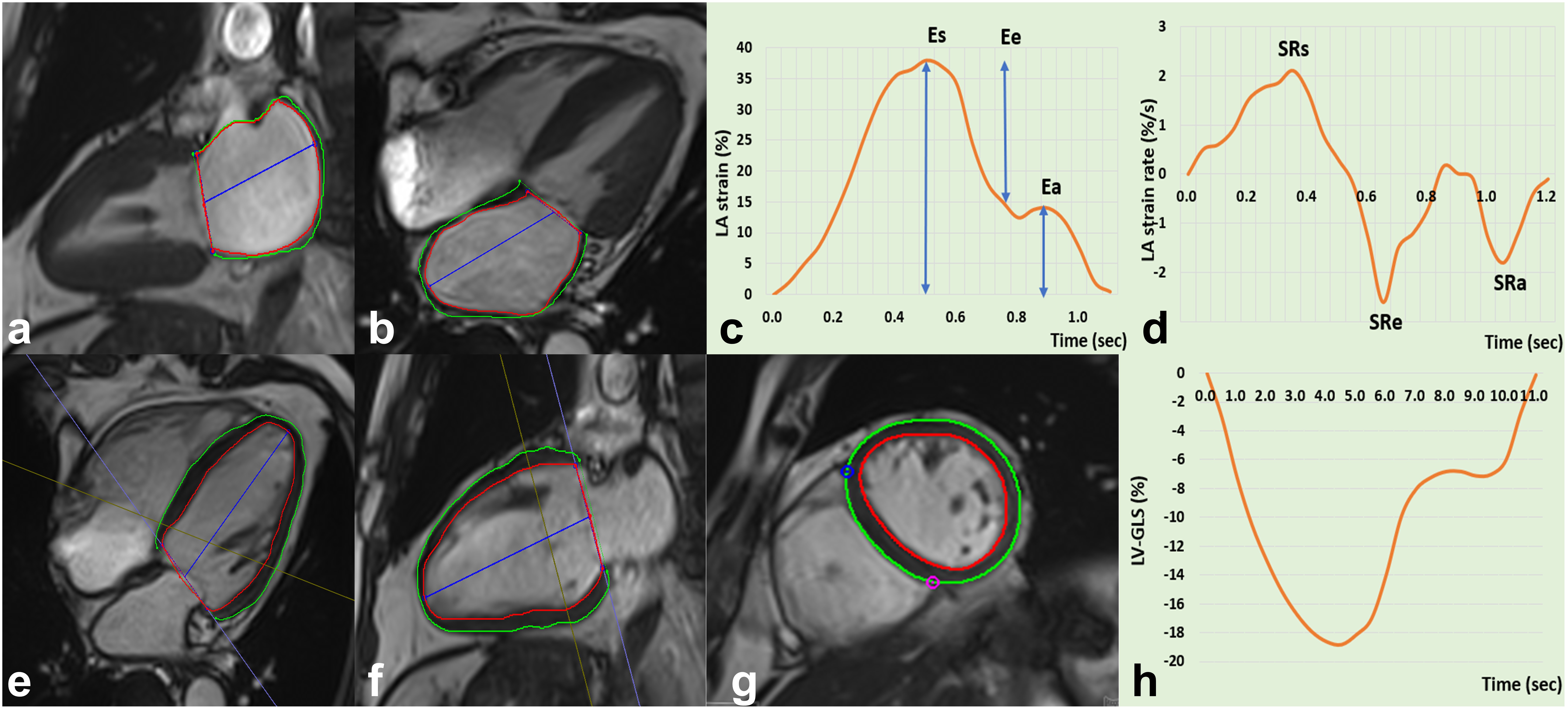
Ninety-four controls, 46 patients with T2DM (T2DM-only) and 83 patients with T2DM and HT (T2DM+HT) were included. The exclusion criteria were LVEF <50%, estimated glomerular filtration rate <60 mL/min/1.73 m2, and presence of organic heart disease. All individuals underwent cardiac magnetic resonance imaging on a MAGNETOM Skyra 3T MRI scanner (Siemens Healthineers AG, Erlangen, Germany). The b-SSFP cine sequence (TR: 39.2 ms; TE: 1.43 ms; pixel size: 1.6*1.6*6mm; flip angle: 39°; slice thickness: 8 mm; matrix size: 208×139; FOV: 234 mm×280 mm) was performed from the base to apex level on the short-axis view and long-axis view for continuous cine imaging. The commercial software, cvi42 (Circle Cardiovascular Imaging, Calgary, Canada), was used for post-processing (Figure 1). Covariance was used to assess the differences in LA and LV geometry and strain parameters between the three groups. Multivariate linear regression analysis was used to analyze the independent factors influencing myocardial strain, and p<0.05 was considered statistically significant.Results
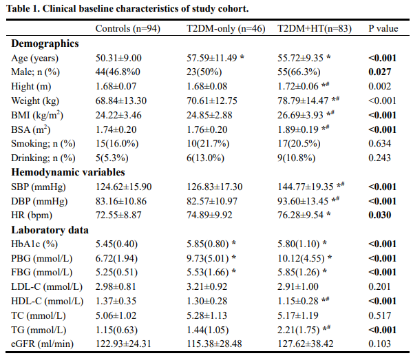
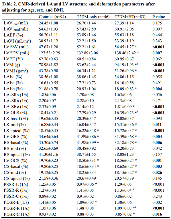
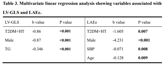
After adjusting for covariates (age, sex, and body mass index [BMI]; Table 1), LA conduit strain (LAEe, p=0.004) and LA early negative peak strain rate (LA-SRe, p<0.001) were significantly decreased in the T2DM+HT group but preserved in the T2DM-only group compared with those of the controls (Table 2). LA structural parameters did not significantly differ between the control, T2DM-only and T2DM+HT groups. For the LV myocardial strain, LV global longitudinal strain (LV-GLS, p<0.001) was reduced in the T2DM+HT group compared with those of the control and T2DM-only groups. LV global radial strain (p=0.004) and LV global circumferential strain (p=0.001) were decreased in both the T2DM-only and T2DM+HT groups. For segment strain, LV apical longitudinal strain (p<0.001), LV basal radial strain (p=0.006) and circumferential strain (p=0.003) were impaired in both the T2DM-only and T2DM+HT groups. LV mid-longitudinal (p=0.011) and circumferential (p=0.026) strain were decreased in the T2DM+HT group but not in the T2DM-only group. For LV structural parameters, end-diastolic LV volume (p=0.007) and end-systolic LV volume (p<0.001) were increased significantly compared with those of the controls. The radial LV peak diastolic strain rate (p<0.001) and circumferential LV peak diastolic strain rate (p=0.016) were also reduced in patients with T2DM+HT. Multivariate linear regression analysis (Table 3) showed that T2DM+HT (b=−0.86, p<0.001), the male sex (b=−0.87, p<0.001), and triglycerides (b=−0.35, p<0.001) were independent influencing factors for LV-GLS, and T2DM+HT (b=−1.61, p=0.007), the male sex (b=−4.23, p<0.001), age (b=−0.13, p =0.009), and systolic pressure (b=−0.07, p=0.008) were independent influencing factors for LAEe.Discussion and Conclusion
HT has additive effects on LA and LV structure and strain in patients with T2DM and can be detected at an early stage via CMR-FT. First, we found that HT further impairs LA myocardial strain in patients with T2DM, even if the LA structural parameters are within the normal range. Second, LV global and segment strain were impaired in patients with T2DM+HT, especially for longitudinal and circumferential strain. Finally, further multivariable linear regression analysis indicated that in patients with diabetes, HT was significantly associated with LV-GLS and LAEe.Acknowledgements
We thanks the Siemens Research Team, the scientific adviser, for sorting and guiding the abstract’s content. Their contributions helped improve the abstract.References
[1] Russo C, Jin Z, Homma S, et al. Effect of diabetes and hypertension on left ventricular diastolic function in a high-risk population without evidence of heart disease. Eur J Heart Fail 2010; 12:454-461.
[2] de Boer IH, Bangalore S, Benetos A, et al. Diabetes and Hypertension: A Position Statement by the American Diabetes Association. Diabetes Care 2017; 40:1273-1284.
[3] Li L, Chen X, Yin G, et al. Early detection of left atrial dysfunction assessed by CMR feature tracking in hypertensive patients. Eur Radiol 2020; 30:702-711.
[4] Liu JH, Chen Y, Yuen M, et al. Incremental prognostic value of global longitudinal strain in patients with type 2 diabetes mellitus. Cardiovasc Diabetol 2016; 15:22.
[5] Liu H, Wang J, Pan Y, et al. Early and Quantitative Assessment of Myocardial Deformation in Essential Hypertension Patients by Using Cardiovascular Magnetic Resonance Feature Tracking. Sci Rep 2020; 10:3582.
Figures