3525
Association between neutrophil/lymphocyte ratio and MR myocardial first pass perfusion in patient with coronary microvascular dysfunction1First Affiliated Hospital of Henan University of Traditional Chinese Medicine, Zhengzhou, China, 2Philips Healthcare, Beijing, China
Synopsis
Keywords: Inflammation, Infiltration, Inflammation
Motivation: The study on correlation between blood neutrophil-to-lymphocyte ratio (NLR) and cardiac magnetic resonance perfusion imaging (CMRI) quantitative indexes in patients with cardiac syndrome X (CSX) is few.
Goal(s): To investigate the relationship between the blood neutrophil/lymphocyte ratio (NLR) and the mean value and coefficient of variation of the magnetic resonance myocardial first pass perfusion parameters in patients with cardiac syndrome X.
Approach: A total of 53 patients with CSX and 43 patients with unstable angina pectoris (UAP) were evaluated using MR myocardial perfusion and blood routine examination.
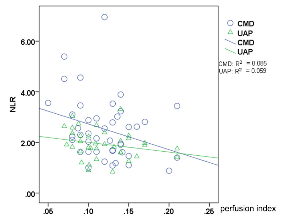
Results: A negative correlation was observed between the myocardial perfusion index in CSX and blood NLR.
Impact: It is provided further evidence for the potential utility of NLR as a diagnostic marker for CSX.
Aim
To investigate the relationship between the blood neutrophil/lymphocyte ratio (NLR) and the mean value and coefficient of variation of the magnetic resonance myocardial first pass perfusion parameters in patients with cardiac syndrome X (CSX).Materials and methods
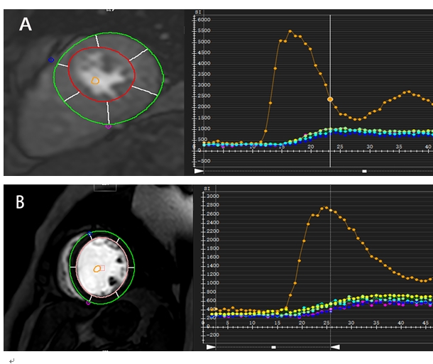
A total of 53 patients diagnosed with cardiac syndrome X (CSX) and 43 patients diagnosed with unstable angina pectoris (UAP) were evaluated using resting magnetic resonance myocardial perfusion examination and blood routine examination. The analysis involved the examination of myocardial perfusion parameters, including the perfusion index, slope/baseline signal intensity, peak time, 20%-80% peak time, and maximum signal intensity/baseline signal intensity. Additionally, the mean and coefficient of variation were calculated for each of the 16 segments of the left ventricle. Intergroup differences in myocardial perfusion parameters and blood neutrophil-to-lymphocyte ratio (NLR) were explored, and the correlations between these variables were analyzed.Results
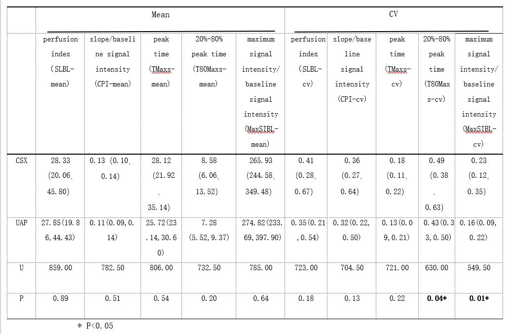
There were no significant differences in myocardial perfusion parameters and mean values among different segments in the CSX group compared to the UAP group. Nevertheless, the coefficient of variation for 20% to 80% peak time and the coefficient of variation for the maximum signal intensity/baseline signal intensity, consisting of 16 segments, were found to be significantly higher in the CSX group (P<0.05). Additionally, the levels of blood neutrophils (N) and NLR were also observed to be higher in CSX group (P<0.05). There was a negative correlation between the blood NLR and the average myocardial perfusion index in the CSX group, and no significant correlation was identified between blood NLR and myocardial perfusion parameters in UAP group.Conclusion
This study revealed that the heterogeneity of myocardial first-pass perfusion indices was greater in CSX patients compared to UAP patients. Additionally, a negative correlation was observed between the myocardial perfusion index in CSX patients and the blood NLR, providing further evidence for the potential utility of NLR as a diagnostic marker for CSX.Acknowledgements
No acknowledgement found.References
No reference found.Figures