3521
MRI and fluorescence imaging of perfluorohexane loaded nanoparticles targeting atherosclerosis and their effects on activated macrophages1Renmin Hospital of Wuhan University, Wuhan, China
Synopsis
Keywords: Atherosclerosis, Molecular Imaging
Motivation: Cardiovascular disease is the main cause and disease of morbidity and mortality in the world, and atherosclerosis causes more than 90% of cardiovascular diseases.
Goal(s): Early diagnosis and timely treatment of atherosclerosis to reduce the morbidity and mortality of residents.
Approach: In the development of atherosclerosis, reducing the content of macrophages which derived foam cells can reverse the formation of atherosclerotic plaque.
Results: The dextran sulfate in the nanoparticles can target SR-A of activated macrophages in atherosclerosis, and then the nanoparticles undergo phase transformation in macrophages to induce apoptosis.
Impact: The nanoparticles have multi-mode molecular imaging capabilities, and have excellent targeting performance. Under LIFU, it can undergo phase transformation and induce apoptosis of macrophages, providing a promising strategy for the diagnosis and treatment of atherosclerotic plaque in the future.
INTRODUCTION
Atherosclerosis is an important pathological basis for the occurrence and development of cardiovascular diseases.1 Vulnerable atherosclerotic plaques are prone to sudden rupture, leading to fatal events.2 The rupture of atherosclerosis plaque is closely related to the composition of plaque. Therefore, non-invasive detection of the composition of atherosclerosis plaque will have a very important clinical application value for the treatment and prognosis of atherosclerosis patients. In this study,we prepare a magnetic resonance imaging (MRI)/near-infrared fluorescence dual-mode molecular probe targeting scavenger receptor A with phase change material-perfluorohexane (PFH) and dextran sulfate (DS), and study its effect on activated macrophages, aiming at early intervention of vulnerable atherosclerotic plaques at the molecular level and evaluating the therapeutic effect.METHODS
Nanoparticles (PFH-Fe/DiR-DS) were prepared using an improved double emulsion method and electrostatic adsorption method.3 The average particle size, polydispersity indexes, and surface potential of the nanoparticles were measured using the Marvin particle size analyzer. The morphology and internal structural characteristics were observed using scanning electron microscopy and high-resolution transmission electron microscopy. Study the in vitro MRI and near-infrared fluorescence imaging of nanoparticles and their phase transition performance. Cultivate mouse macrophages RAW 264.7 in vitro, study the targeting effect of nanoparticles on activated macrophages, conduct cell viability experiments, and analyze the induction of macrophage apoptosis by nanoparticles phase transition using biological electron microscopy.RESULTS
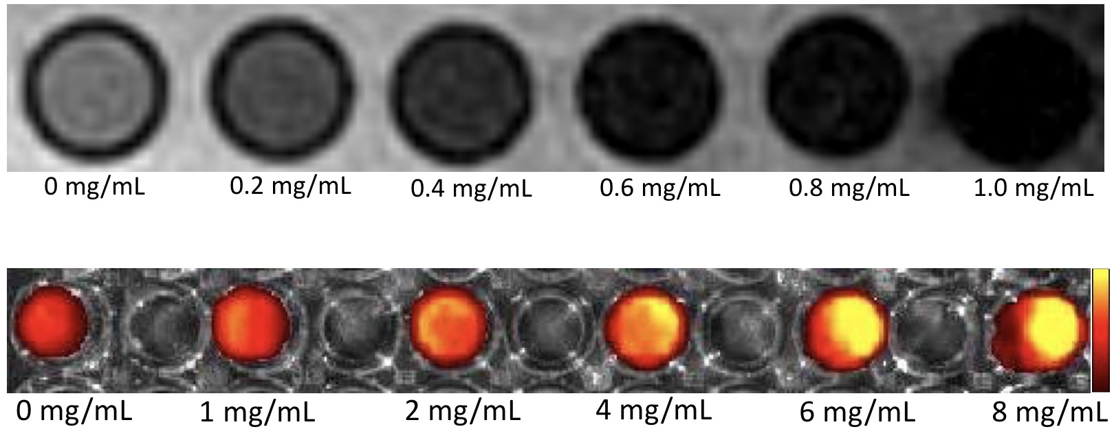
The final size of the prepared PFH-Fe/DiR-DS nanoparticles is (286.30 ± 16.82) nm, with a polydispersity indexes of (0.102 ± 0.057) and a surface potential of (-18.65 ± 1.09) mV. They have a shell core structure and a smooth three-dimensional spherical surface. In the element mapping images collected under high-resolution transmission electron microscopy, it can be seen that the fluorine element of PFH and the iron element of Fe3O4 are concentrated inside the nanoparticles, while the sulfur element of DS exhibits a clear circular structure distributed outside the nanoparticles. The PFH-Fe/DiR-DS nanoparticles can be used as ideal MRI contrast agents and have good near-infrared fluorescence imaging ability. Fluorescence signals are enhanced in a concentration dependent manner. Under the irradiation of low intensity focused ultrasound (LIFU), PFH-Fe/DiR-DS nanoparticles can undergo phase transition. Nanoparticles have good targeting properties towards activated macrophages and are time dependent. After being internalized by macrophages and irradiated by LIFU, they can induce macrophage apoptosis through sound induced phase transition effect.CONCLUSION
In this study, we successfully constructed multimodal multifunctional nanoparticles (PFH-Fe/DiR-DS) targeting atherosclerotic plaques, and induced macrophage apoptosis under LIFU irradiation. The application of magnetic resonance and near-infrared fluorescence molecular imaging technology to monitor and evaluate the status of plaque is expected to be able to carry out specific diagnosis and targeted treatment of atherosclerotic plaque. So as to effectively prevent cardiovascular and cerebrovascular events and reduce medical costs. Significantly improve the quality of life of patients and alleviate the contradiction between doctors and patients, which not only provides an effective means for non-invasive, early assessment, treatment and efficacy evaluation of vulnerable atherosclerosis plaque, but also has important significance for promoting social harmony and stability.Acknowledgements
No acknowledgement found.References
1. An J, Zhang YY, Zhou H, et al. Incidence of Atherosclerotic Cardiovascular Disease in Young Adults at Low Short-Term But High Long-Term Risk. J Am Coll Cardiol.2023;81(7):623-632.
2. Patterson MT, Williams JW. Metabolic regulation of macrophage proliferation and function in atherosclerosis. Curr Opin Lipidol.2021;32(5):293-300.
3. Hou J, Zhou J, Chang M, et al. LIFU-responsive nanomedicine enables acoustic droplet vaporization-induced apoptosis of macrophages for stabilizing vulnerable atherosclerotic plaques. Bioact Mater. 2022; 16:120-133.
Figures
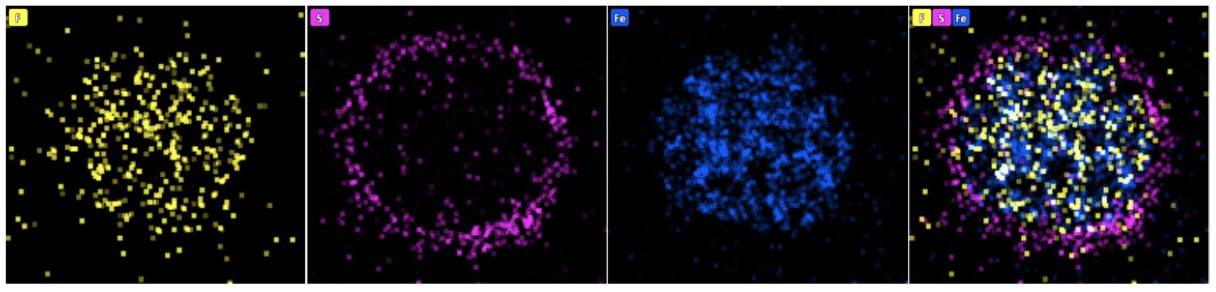
Figure 1. The corresponding elemental mapping F, S and Fe, as well as the merged HRTEM images from PFH-Fe/DiR-DS nanoparticles.