3516
Detecting Response of Metastatic Prostate Cancer to Chemotherapy in PDX Models using Hyperpolarized 13C MRI1Department of Radiology and Biomedical Imaging, University of California San Francisco, San Francisco, CA, United States, 2Department of Hematology/Oncology, University of California San Francisco, San Francisco, CA, United States, 3Department of Medicine, University of California San Francisco, San Francisco, CA, United States
Synopsis
Keywords: Prostate, Hyperpolarized MR (Non-Gas), Prostate cancer, Preclinical models
Motivation: Diagnosis and treatment assessment of aggressive small cell neuroendocrine prostate cancer (SCNC) is challenging due to its admixture presence in conjunction with adenocarcinoma phenotype and differential response to treatments subjective to the metastatic sites.
Goal(s): This study aims to assess the therapeutic efficacy of platinum-based chemotherapy in preclinical SCNC metastatic models using patient derived xenografts.
Approach: Hyperpolarized 13C MRI was used to measure the apparent rate of change in glycolysis (kPL) in PDX models of SCNC in metastatic sites.
Results: kPL values clearly demonstrated a decrease upon treatment concordant to the change in tumor volume in both the liver and bone metastatic models.
Impact: kPL, measured by hyperpolarized MRI, can be used to assess treatment efficacy yielding a non-invasive, potentially early biomarker readily translatable for use in patients with metastatic tumors for optimal therapeutic approaches.
Introduction
SCNC prostate cancer has risen as a consequence of second-line anti-androgen therapy1. This trans-differentiation commonly found in metastatic castration-resistant prostate cancer (mCRPC) is very aggressive and associated with poor prognosis2,3. SCNC tumors respond differently to therapy compared to the adenocarcinoma counterpart and has limited treatment options. Patients with metastases in the liver have a particularly poor prognosis relative to those with bone metastases alone. New clinical trial findings revealed that patients with mCRPC and liver metastases benefited from certain therapies such as androgen receptor signaling inhibitors and docetaxel, whereas those with bone metastasis did not4. These findings underscore the need to be able to assess the response to therapy in a timely manner in metastatic sites which are generally considered unevaluable by RECIST criteria. We demonstrate the use of hyperpolarized (HP) [1-13C]pyruvate MRI to assess response to chemotherapy in clinically pertinent models of SCNC using patient-derived xenograft (PDX) propagated in the bone and liver.Methods
LTL6105 PDX of the SCNC tumor was propagated under the renal capsule of male NSG mice. Tumors were resected and after digestion to single cells were injected into the tibia6 (500,000 cells/mice in 10uL) and liver7 (1M cells/mice in 20uL). All procedures were approved by our Institutional Animal Care and Use Committee. All tumor-bearing mice underwent baseline imaging on a Bruker 3T scanner with dual tuned 40mm 1H/13C volume coil. Mice were treated with two cycles of 65 mgs/kg/week carboplatin immediately after imaging and were subjected to repeat MRI on day 3 and day 7 after treatment. T2-weighted proton imaging was used for tumor localization, co-registration of carbon imaging, and tumor volume estimation. A 6.7 T cryogen-free polarizer was utilized for dissolution dynamic nuclear polarization (SpinAligner, Polarize, Denmark)8. 80 mM [1-13C]pyruvic acid was polarized and upon dissolution injected into the mice in ~12 seconds. A 2D metabolite-specific spectral-spatial echo-planar imaging (spspEPI)9 sequence was used for HP 13C experiments with a temporal resolution of 3s, 18 time-points and a flip-angle of 15° for Pyruvate and 30° for Lactate. kPL, the apparent rate of enzymatic conversion of HP [1-13C]pyruvate to [1-13C]lactate, was calculated10.Results and Discussion
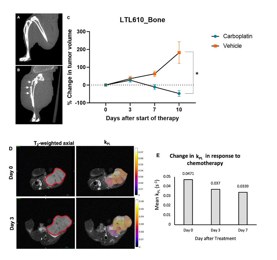
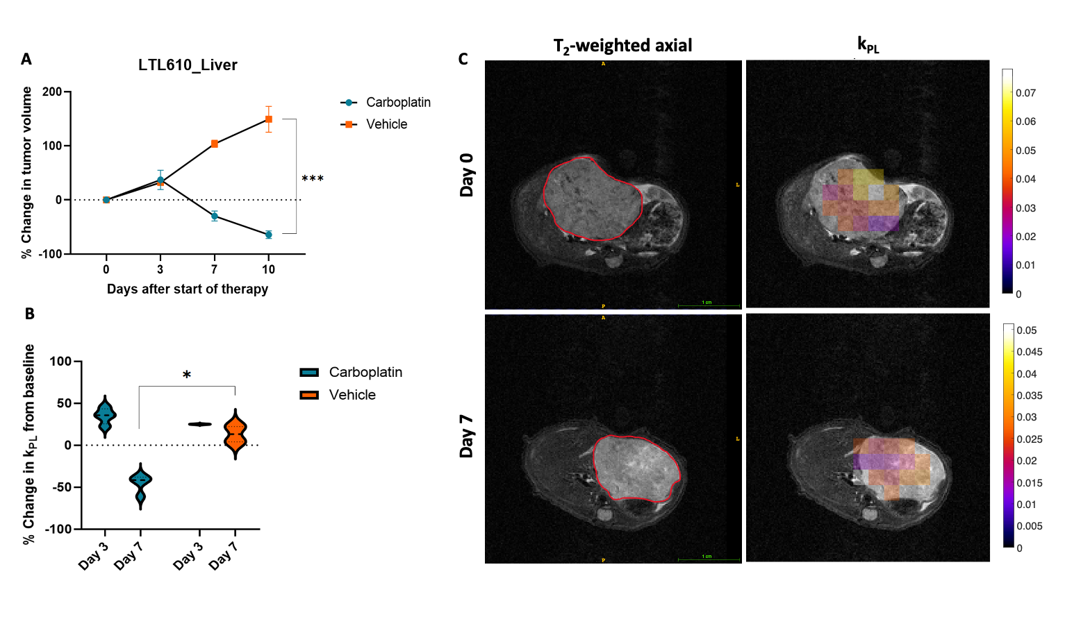
We have successfully established metastatic models of SCNC PDXs in the bone and liver with a take rate of 100% for LTL610. The doubling time of the tumors in both sites were ~5 days. Tibial outgrowth of LTL610 tumors were characterized for the first time which is manifested as osteolytic response in the left tumor-bearing tibia as evidenced by the u-CT scans relative to the normal contralateral limb (Figure 1A&B). LTL610 tumors (n=3) in the tibia demonstrated a significant decrease (p<0.05) in volume by day 7 after the first cycle of carboplatin in comparison to the placebo control animals (n=3), where the tumor volume continued to increase with time (figure 1C). Representative T2-weighted axial images and kPL map overlaid on the tumor of LTL610 PDX in tibia at baseline and three days after carboplatin treatment are shown in figure 1D. Figure 1E shows a decrease in kPL value from 0.0471s-1 to 0.0370s-1 three days after chemotherapy. In the liver, there was an initial increase in both tumor volume and kPL, three days after the first cycle of carboplatin in both the treated and control mice. However, the tumor volume of the mice decreased by day 7 in the treated group (n=4) relative to the control group (n=3) (Figure 2A). A concordant significant decrease (p<0.05) in percent change of kPL was observed on day 7 after start of therapy in carboplatin-treated animals (Figure 2B), while the mice in the control group had no change. Representative T2-weighted axial images of LTL610 PDX in liver at baseline and day 7 after carboplatin treatment are shown in figure 2C.Conclusion
These studies demonstrate that decreases in kPL, which were congruent with changes in tumor volume can be used to non-invasively assess tumor response to carboplatin in liver and bone metastases. Interestingly, the same dose of the drug had a delayed therapeutic response in the liver vs bone microenvironments. These observations reinforce the differences conferred upon tumor growth and treatment efficacy due to the microenvironment. Additionally, the kPL changes denote a potentially early biomarker of response in the bone which is generally not amenable to RECIST evaluation. This is an ongoing study and in addition to imaging more mice, future work includes the use of more SCNC PDXs along with metabolic characterization using stable isotope-resolved NMR metabolomics and immunohistochemistry.Acknowledgements
- Preclinical MR Imaging and Spectroscopy Core at UCSF
- This study was funded by the following grants: NIH P41EB013598, UCSF Prostate Cancer Pilot Award, DoD PCRP PC160630 (Idea Development Award), NIH R01 CA215694, NIH U24 CA0163815.
References
1. Crona, D. J. & Whang, Y. E. Androgen Receptor-Dependent and -Independent Mechanisms Involved in Prostate Cancer Therapy Resistance. Cancers (Basel) 9, (2017).
2. Aggarwal, R., Zhang, T., Small, E. J. & Armstrong, A. J. Neuroendocrine prostate cancer: subtypes, biology, and clinical outcomes. J Natl Compr Canc Netw 12, 719–726 (2014).
3. Singh, A. et al. Liver Metastases in Prostate Carcinoma Represent a Relatively Aggressive Subtype Refractory to Hormonal Therapy and Short-Duration Response to Docetaxel Monotherapy. World J. Oncol. 6, 265–269 (2015).
4. Drake, C. G. Visceral Metastases and Prostate Cancer Treatment: ‘Die Hard,’ ‘Tough Neighborhoods,’ or ‘Evil Humors’? 14 (2016).
5. Lin, D. et al. High Fidelity Patient-Derived Xenografts for Accelerating Prostate Cancer Discovery and Drug Development. Cancer Res 74, 1272–1283 (2014).
6. Agarwal, S., Peehl, D. M. & Sriram, R. Intratibial implantation of tumor cells. (2021).
7. Agarwal, S., Bok, R., Peehl, D. M. & Sriram, R. Intrahepatic implantation of tumor cells. (2021).
8. Ardenkjaer-Larsen, J. H., Bowen, S., Petersen, J. R., Rybalko, O., Vinding, M. S., Ullisch. M., Nielsen, N. C. Cryogen-free dissolution dynamic nuclear polarization polarizer operating at 3.35 T, 6.70 T, and 10.1 T. Magn Reson Med 81, 2184-2194 (2019).
9. Sahin, S. I., Ji, X., Agarwal, S., Sinha, A., Mali, I., Gordon, J. W. et al. Metabolite-Specific Echo Planar Imaging for Preclinical Studies with Hyperpolarized 13C-Pyruvate MRI. Tomography 9, 736-749 (2023).
10. Larson, P. E. Z. et al. Investigation of analysis methods for hyperpolarized 13C-pyruvate metabolic MRI in prostate cancer patients. NMR Biomed 31, e3997 (2018).
Figures