3515
Development of a Whole Abdominopelvic Variable Resolution Hyperpolarized 13C MRI Approach for Advanced Prostate Cancer Clinical Research1Department of Radiology and Biomedical Imaging, University of California San Francisco, San Francisco, CA, United States, 2Bioengineering Joint PhD Program, UC Berkeley-UCSF, San Francisco, CA, United States, 3Helen Diller Family Comprehensive Cancer Center, University of California San Francisco, San Francisco, CA, United States
Synopsis
Keywords: Prostate, Prostate, Cancer, Hyperpolarized MR
Motivation: Monitoring the progression or response of advanced prostate metastases is a current clinical unmet need that is not reliably delineated with current CT and PET.
Goal(s): Here, we developed a high-resolution whole abdominopelvic [1-13C]pyruvate HP MRI approach for the metabolic biomarker characterization of metastases in prostate cancer patients.
Approach: A variable-resolution imaging approach was used to provide high-resolution [1-13C]pyruvate, robust spatiotemporal denoising and B1+ variation correction methods were used to quantify the rate-constant for the conversion of [1-13C]pyruvate to lactate, kPL.
Results: Improved conspicuity of [1-13C]pyruvate distribution and kPL conversion maps of metastatic lesions were achieved with the new approach.
Impact: The improvement in [1-13C]pyruvate resolution and clear delineation of highly metabolically active metastatic lesions in kPL maps demonstrated the potential of [1-13C]pyruvate HP MRI in advanced prostate cancer.
Introduction:
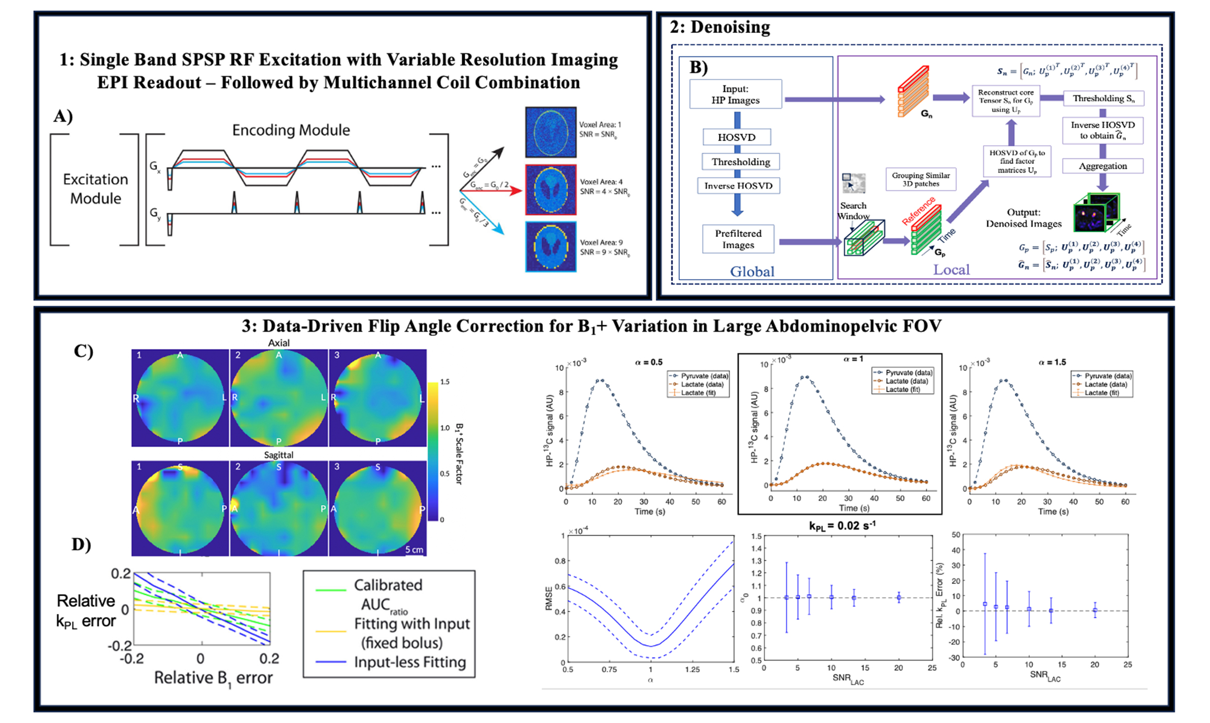
Hyperpolarized (HP) 13C MRI is a safe, noninvasive, and quantitative imaging technique that visualizes dynamic metabolic processes and can be used to detect and monitor treatment response of advanced prostate cancer metastases1. In an approximately 2-minute addition to a MRI exam, HP [1-13C]pyruvate MRI can detect metabolic reprogramming through voxel-wise quantification of biomarkers, such as the first-order enzymatic conversion rates of pyruvate to lactate (kPL). Changes in kPL reflect oncogenomic alterations that occur with the progression or response of advanced prostate cancer to targeted therapies—such as PI3K, MYC inhibitors—that CT and PET with FDG or PSMA cannot reliably provide2. This project was designed to develop a robust, efficient and variable-resolution 13C EPI-based approach of advanced prostate cancer metastases which includes spatiotemporal denoising to improve image quality and quantification of dynamic [1-13C]lactate as well as voxel-wise data-constrained flip angle correction to improve kinetic modeling given the approximately ±50% B1+ variations encountered in large FOV abdominopelvic imaging6,7,8 (Figure 1).Methods:
Sequences: The HP-13C images were acquired using a variable-resolution metabolite-selective imaging approach using a single-band SPSP RF pulse and a symmetric EPI readout to independently excite [1-13C]pyruvate and [1-13C]lactate. Each metabolite’s readout waveform was independently scaled to produce 5 x 5 mm2 pyruvate and 10 x 10 mm2 lactate resolution3 (Figure 1A). Additional sequence parameters were TR/TE = 150ms/22.3ms with ramp sampling enabled, 3s temporal resolution, slice thickness = 16 mm, and constant flip angles of Pyr 15°, Lac 30°. A lower resolution [1-13C]pyruvate EPI acquisition was also acquired for comparison at a coarser 10 x 10 mm2 resolution, TR/TE = 110ms/25.3ms, 3s temporal resolution, slice thickness = 20 mm, and constant flip angles of Pyr 15°, Lac 30°.Patient Study: A representative 4+4 advanced prostate cancer patient with several metastases was studied using HP 13C and 1H conventional MRI on a clinical 3T MRI following injection of a sterile dose of 250 mM HP [1-13C]pyruvate (FDA IND approved). A flexible vest 13C QTAR coil for RF transmit and an 8-channel array for receive (Clinical MR Solutions) enabled coverage of the whole abdomen or pelvis to assess the metastases. This study was approved by the IRB at UCSF and the 1H MRI images were assessed by research radiologists at UCSF.
Data Processing: The EPI datasets were denoised using spatiotemporal GL-HOSVD denoising to increase spatial-fidelity and improve SNR of the [1-13C]lactate dynamic data4 (Figure 1B). kPL was calculated using an inputless two-site exchange model6. The mean total SNR (area under the curve SNR, AUCSNR) was quantified, and voxel-wise kinetic modeling was performed on denoised data to original SNRAUC < 10 for pyruvate and original SNRAUC < 5 lactate, along with fitting (<30% relative error) criteria. For B1+ correction, during fitting the flip angles were varied ± 50% of nominal flip angle. A B1+ scale-factor (α) was introduced to apply this flip angle variations. For each voxel, the optimal scale factor (α0) was determined by minimizing the RMSE of the model fitting. Subsequently, the nominal flip angle was scaled by α0 in fitting to determine the predicted kPL7,8 (Figure 1E and 1F).
Results:
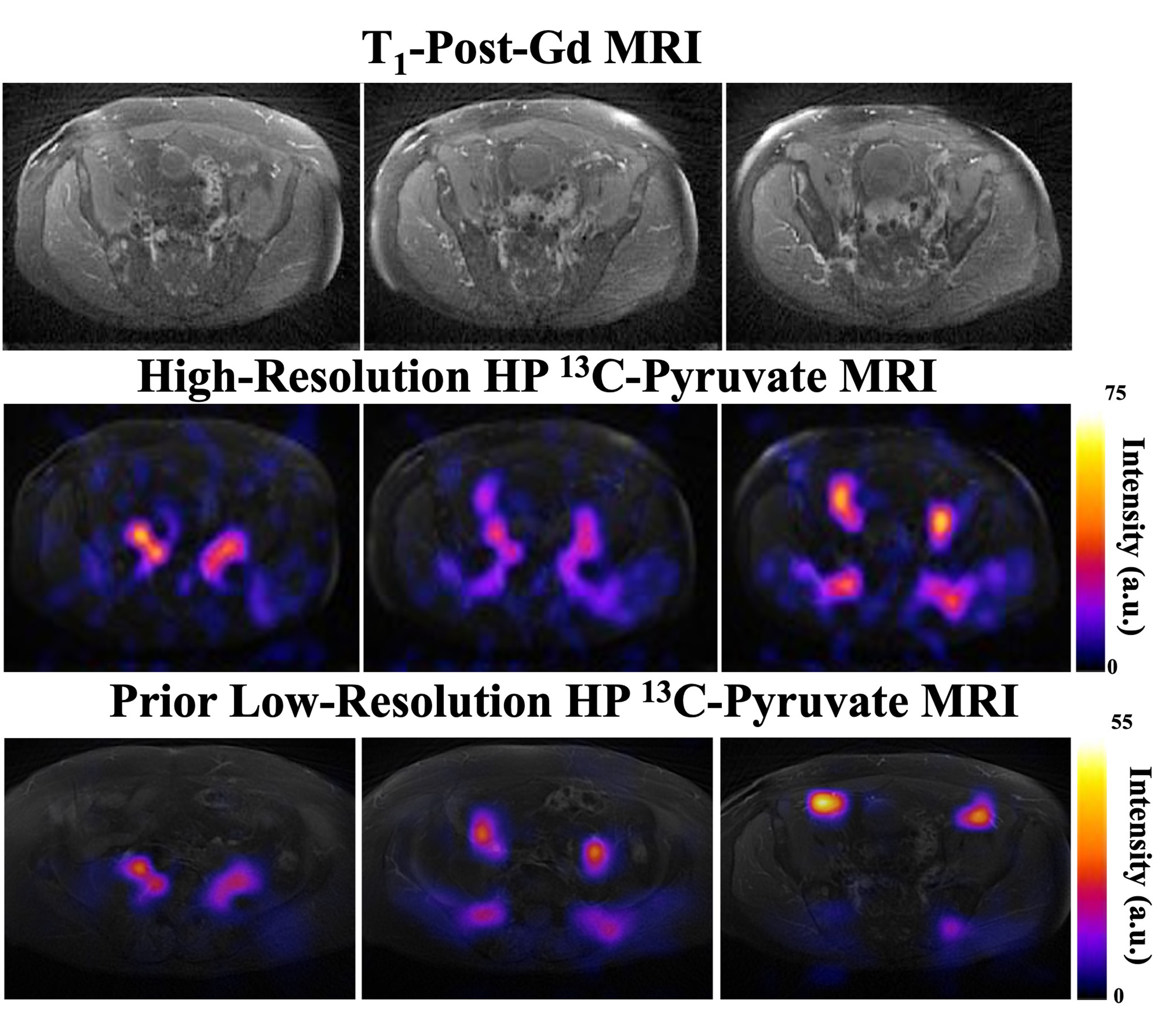
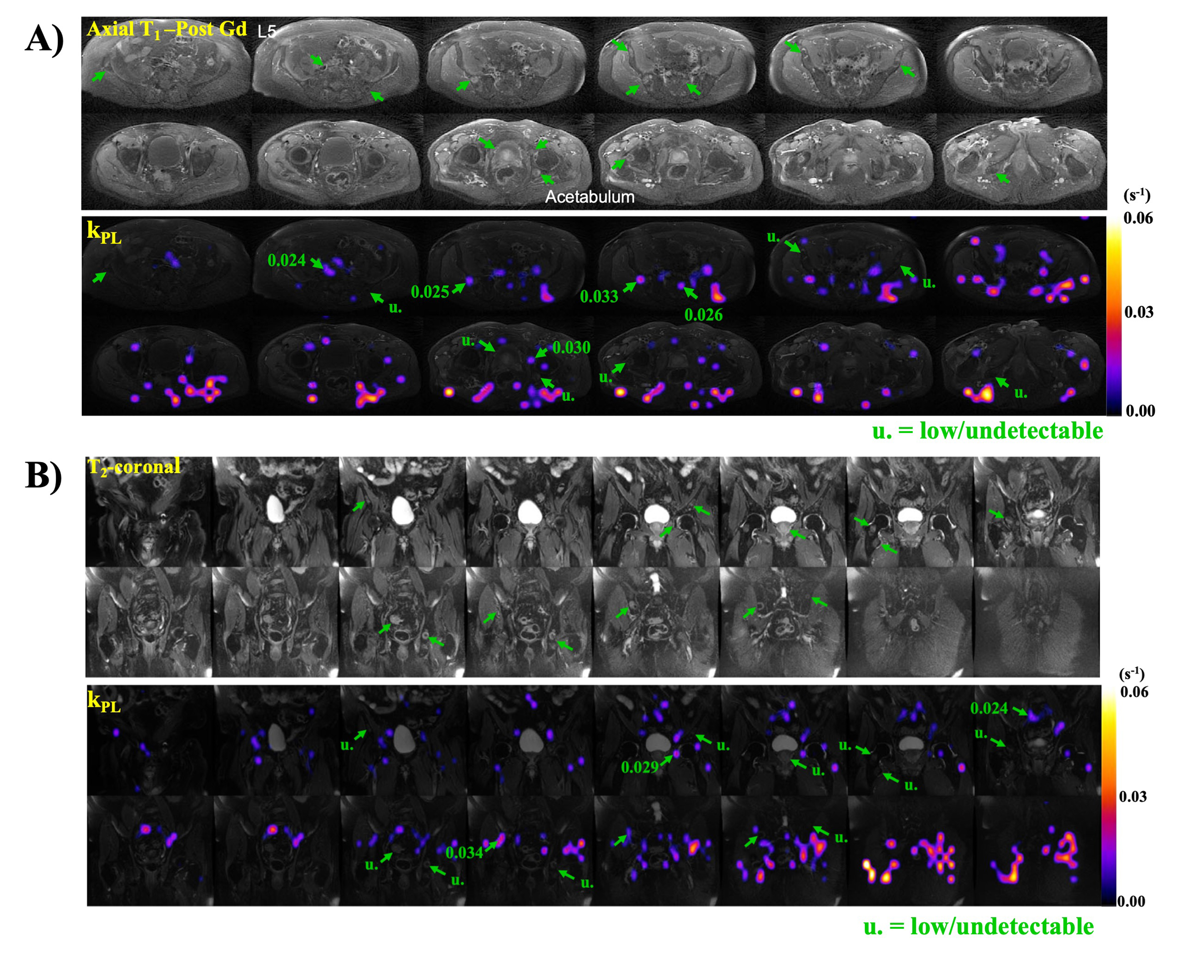
Figure 2 shows the comparison of higher- and lower resolution acquisitions of [1-13C]pyruvate AUC (area under the curve summed signal across all 20 timepoints), images in the prostate overlaid on 1H T1-post Gd anatomical MRI. Normal metabolite distributions can be seen in the [1-13C]pyruvate images. This figure demonstrates improved conspicuity of pyruvate distributions throughout the abdomen and pelvis when comparing anatomical regions in each respective HP acquisition. Figure 3 shows regions of high kPL correlated with the metastatic lesions in two orientations, axial and coronal (Figure 3A and Figure 3B). Additionally, arrows point to some regions in which some metastatic lesions had undetectable kPL.Discussion and Conclusion:
This study investigated an improved HP-13C MR imaging approach to evaluate the metabolism of metastatic lesions in patients with prostate cancer. In the representative post-therapy patient data shown, many of the metastatic lesions demonstrated low to undetectable kPL, indicating successful treatment. However, several lesions showed moderate to high pyruvate-to-lactate conversion reflecting metabolically active tumor. This study supports the potential of high resolution [1-13C]pyruvate to find metastases that are highly metabolically active, and demonstrated the potential of kPL as a quantitative biomarker for monitoring early response of systemic drug treatment of advanced metastatic prostate cancer.Acknowledgements
This work was supported by NIH grants R01CA256740, R01CA238379, & P41EB013598 and a UCSF Resource Allocation Program Grant. We would also like to acknowledge Mary Frost, Kimberly Okamoto, Duy Dang and Evelyn Escobar for their assistance with the patient studies.References
- Wang ZJ, et al. Hyperpolarized 13C MRI: State of the Art and Future Directions. Radiology 2019; 291:273–284.
- Chen HY, Aggarwal R, Bok RA, Ohliger MA, Zhu Z, Lee P, Gordon JW, van Criekinge M, Carvajal L, Slater JB, Larson PEZ, Small EJ, Kurhanewicz J, Vigneron DB. Hyperpolarized 13C-pyruvate MRI detects real-time metabolic flux in prostate cancer metastases to bone and liver: a clinical feasibility study. Prostate Cancer Prostatic Dis. 2020 Jun;23(2):269-276. doi: 10.1038/s41391-019-0180-z. Epub 2019 Nov 4. PMID: 31685983; PMCID: PMC7196510.
- Gordon JW, Autry AW, Tang S, Graham JY, Bok RA, Zhu X, Villanueva-Meyer JE, Li Y, Ohilger MA, Abraham MR, Xu D, Vigneron DB, Larson PEZ. A variable resolution approach for improved acquisition of hyperpolarized 13C metabolic MRI. Magn Reson Med. 2020 Dec;84(6):2943-2952. doi: 10.1002/mrm.28421. Epub 2020 Jul 22. PMID: 32697867; PMCID: PMC7719570.
- Nickles TM, et al. Hyperpolarized 13C Metabolic Imaging of the Human Abdomen with Spatiotemporal Denoising. ISMRM 2023, Toronto. Abstract ID: 4096.
- Kim Y, et al. Denoising of hyperpolarized 13C MR images of the human brain using patch-based higher-order singular value decomposition. Magn Reson Med. 2021; 86: 2497-25.
- Larson PEZ, Chen H, Gordon JW, et al. Investigation of analysis methods for hyperpolarized 13C‐pyruvate metabolic MRI in prostate cancer patients. NMR in Biomedicine. 2018;31(11). doi:10.1002/nbm.3997.
- Lee PM, Chen H, Gordon JW, et al. Whole Abdomen Metabolic Imaging of Healthy Volunteers Using Hyperpolarized [1‐13C]pyruvate MRI. Magnetic Resonance Imaging. 2022;56(6):1792-1806. doi:10.1002/jmri.28196.
- Nickles TM, et al. Data-constrained Determination of Applied Flip Angles to Improve Hyperpolarized 13C MR Kinetic Modeling in the Presence of Large B1 Variations Encountered in Abdominal Imaging. ENC 2023, P145.
Figures