3513
Whole tumor amide proton transfer–weighted imaging histogram analysis to predict prostate cancer bone metastases: a preliminary study1Shaanxi Provincial People's Hospital, xi'an, China, 2Philips Healthcare, xi'an, China, 3Philips Healthcare, beijing, China
Synopsis
Keywords: Prostate, Prostate
Motivation: Accurate prediction of prostate cancer(PCa) bone metastases can be challenged by radiologists. The utility of APTw histogram methods for evaluating bone metastases involvement in PCa is still unclear.
Goal(s): The purpose of this study was to evaluate APTw-derived whole-tumor histogram analysis parameters in predicting PCa bone metastases.
Approach: Diagnostic performance was evaluated using ROC analysis and the AUC comparisons were conducted using the DeLong method.
Results: Our preliminary research showed that whole-tumor histogram analysis of APTw images combined with clinical factors (tPSA, cT stage) showed good diagnosis efficiency in predicting PCa bone metastases while using 68Ga-PSMA PET/CT as the reference standard.
Impact: Our preliminary research showed that whole-tumor histogram analysis of APTw images combined with clinical factors(tPSA, T stage) showed good diagnosis efficiency in predicting prostate cancer bone metastases while using 68Ga-PSMA PET/CT as the reference standard.
Introduction
A significant portion, approximately 12%, of prostate cancer (PCa) patients exhibit bone metastases at diagnosis, which severely diminishes life quality and results in a 5-year survival rate of a mere 6%. ^68Ga-PSMA PET/CT has proven effective in diagnosing bone metastases in PCa patients. However, its widespread clinical application is curtailed by the prohibitively high costs associated with the procedure. Recently, APTw imaging has gained traction for its precise detection capabilities in metastatic lesions, high-grade cancers, and other malignancies. Histogram analysis, a first-order statistical method, evaluates individual pixel values, offering a detailed perspective on tumor heterogeneity that surpasses the conventional region of interest (ROI) mean values. Yet, its application in evaluating bone metastases in PCa remains unclear. Consequently, this study investigates APTw-derived histogram analysis parameters as predictors for bone metastases in PCa.Materials and methods
In this retrospective study, preoperative MRI scans, inclusive of APTw imaging of 65 patients, were examined using a 3.0T scanner (Ingenia CX 3.0T, Philips Healthcare). Tumors were segmented by two independent radiologists into the volume of interest (VOI), which were then applied to the corresponding APTw images to generate APTw VOIs. Histogram parameters of primary tumors were acquired automatically via whole-tumor volume histogram analysis. The intraclass correlation coefficient (ICC) was performed to evaluate the inter-observer consistency of histogram parameters. Univariate and multivariate logistic regression analyses were utilized to discern the independent risk factors significantly associated with bone metastases. Diagnostic performance was evaluated using receiver operating characteristic (ROC) curve analysis, and the area under the curve (AUC) comparisons were conducted using the DeLong method.Results
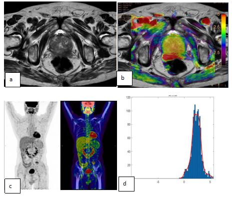
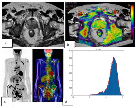
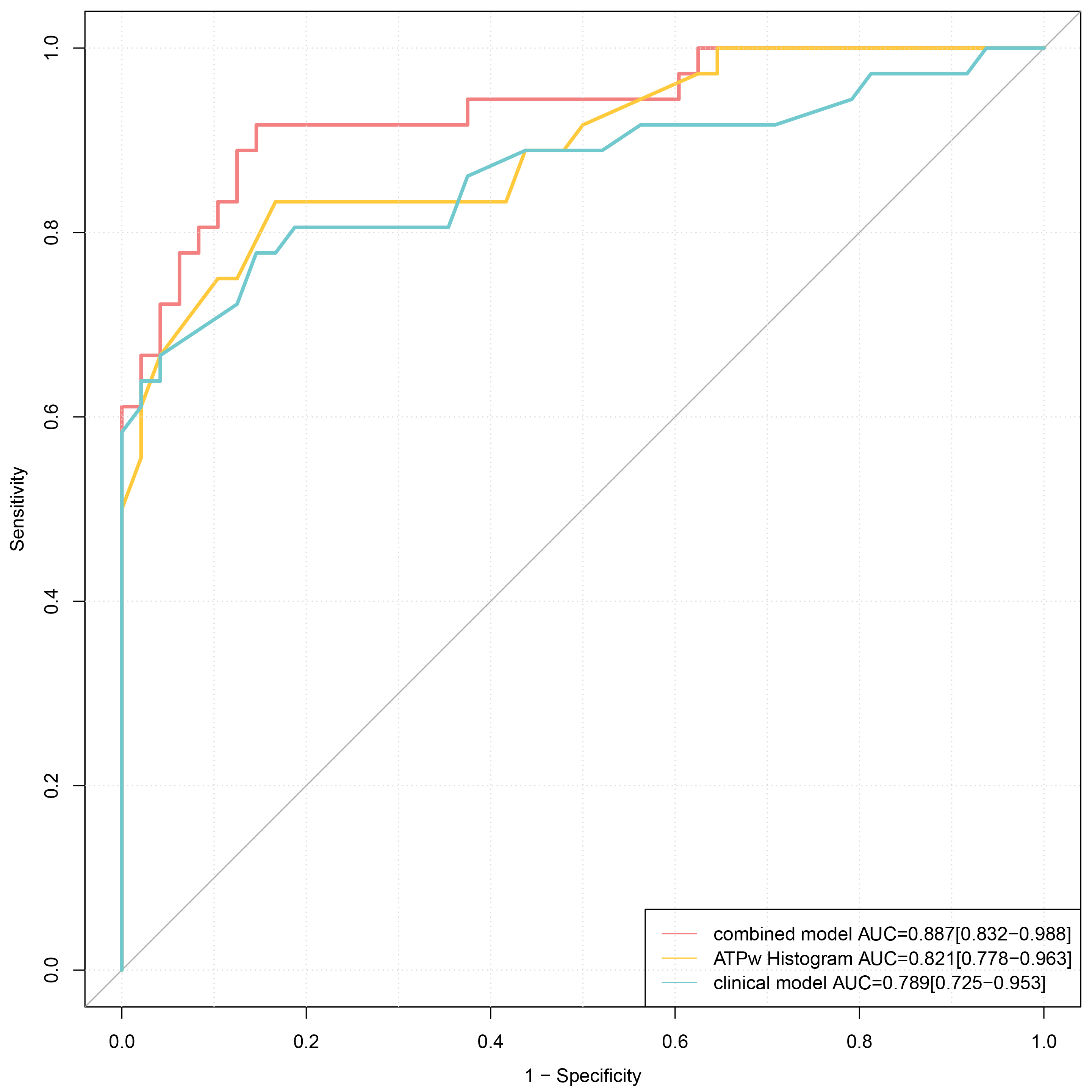
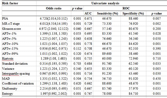
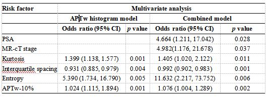
The ICC of the histogram parameters assessment of the two radiologists was pretty good (ICC value > 0.89). Figure 1 and 2 showed two representative examples of PCa without/with bone metastasis. Univariate analysis identified significant associations between bone metastases and variables such as total PSA, clinical T stage, Gleason score, APTw percentiles, standard deviation, variance, interquartile range, mean absolute deviation, coefficient of variation, uniformity, entropy, and kurtosis (all p<0.05). The multivariate analysis highlighted MR-T stage, total PSA, interquartile range, entropy, and kurtosis as independent predictors of bone metastasis (Table 1 and Table 2). AUC values for clinical model assessment, the APTw histogram model, and the combined model were 0.789, 0.821, and 0.887, respectively, with the combined model significantly outperforming both the APTw histogram and clinical models (Figure 3).Discussion
The study probes the efficacy of APTw histogram analyses in differentiating between metastatic and non-metastatic bones in PCa patients, anchored by ^68Ga-PSMA PET/CT as the gold standard. It unearthed independent risk factors such as MR-T stage, total PSA, interquartile range, entropy, and kurtosis after multivariable analysis. The combined model encompassing APTw histogram parameters and clinical variables yielded the most accurate predictions for PCa bone metastases. APT imaging may serve as a cost-effective, readily accessible, and expedited alternative to identify bone metastases in metastatic PCa patients, demonstrating commendable diagnostic precision in a moderately large cohort and potentially enhancing diagnostic and therapeutic approaches, especially for patients at elevated risk for bone metastases.Conclusions
Our preliminary research corroborates the potential of whole-tumor histogram analysis of APTw images, when combined with clinical factors, to efficiently predict bone metastases in prostate cancer. This study broadens the applicability of APTw imaging, potentially paving the way for more tailored medical treatments that could enhance the survival prospects for PCa patients.Acknowledgements
We are grateful to all the participants for their cooperation and patience.References
1 .Chafer CL, Weinberg RA. A perspective on cancer cell metastasis. Science. 2011;331:1559–1564.
2.Svensson E, Christiansen CF, Ulrichsen SP,et al. Survival after bone metastasis by primary cancer type: a Danish population-based cohort study. BMJ Open.2017;7:e016022.
3. Nørgaard M, Jensen A, Jacobsen JB, et al. Skeletal related events, bone metastasis and survival of prostate cancer: a population based cohort study in Denmark (1999 to 2007). J Urol. 2010;184:162–167.
4.Fendler WP, Eiber M, Beheshti M, et al. 68 Ga-PSMA PET/CT: Joint EANM and SNMMI procedure guideline for prostate cancer imaging: version 1.0. Eur J Nucl Med Mol Imaging. 2017; 44: 1014–1024.
5.Fu F, Meng N, Huang Z, et al. Identifcation of histological features of endometrioid adenocarcinoma based on amide proton transfer-weighted imaging and multimodel difusion-weighted imaging. Quant Imaging Med Surg.2022;12:1311.
6. Chen W, Liu G, Chen J, et al. Whole-tumor amide proton transfer-weighted imaging histogram analysis to predict pathological extramural venous invasion in rectal adenocarcinoma: a preliminary study. Eur Radiol. 2023; 33(7): 5159-5171.
7.Zhao L, Liang M, Yang Y, et al.Histogram models based on intravoxel incoherent motion difusion-weighted imaging to predict nodal staging of rectal cancer. Eur J Radiol. 2021; 142:1098698.
8.Kamimura K, Nakajo M, Yoneyama T, et al. Amide proton transfer imaging of tumors: theory, clinical applications, pitfalls, and future directions. Jpn J Radiol. 2019;37:109–116.
9. Nishie A, Asayama Y, Ishigami K, et al. Amide proton transfer imaging to predict tumor response to neoadjuvant chemotherapy in locally advanced rectal cancer. J Gastroenterol Hepatol. 2019; 34:140–146.
Figures