3505
Improved correction of B0 inhomogeneity-induced distortions in prostate diffusion images1Department of Radiology, University of California San Diego, La Jolla, CA, United States, 2Department of Radiation Medicine and Applied Sciences, University of California San Diego, La Jolla, CA, United States, 3Department of Bioengineering, University of California San Diego, La Jolla, CA, United States, 4Department of Neurosciences, University of California San Diego, La Jolla, CA, United States, 5Halıcıoğlu Data Science Institute, University of California San Diego, La Jolla, CA, United States
Synopsis
Keywords: Prostate, Diffusion/other diffusion imaging techniques, B0-inhomogeneity distortion correction
Motivation: The Jacobian intensity correction (JIC) of conventional EPI-distortion correction methods can conceal severe under-correction and even create false anatomical structure.
Goal(s): Demonstrate the shortcomings of JIC methods and present an alternative distortion-correction technique that obviates the need for a JIC.
Approach: Acquiring DWI images with opposite phase-encoding polarity at multiple b-values and normalizing prior to estimating tissue displacement eliminates the need to account for intensity scaling with the JIC. This approach, referred to as mRPG, was compared against conventional distortion correction methods.
Results: mRPG significantly improved the estimation and removal of spatial distortions compared to conventional methods.
Impact: Jacobian intensity correction (JIC) can generate misleading improvement of EPI distortion and create false structure in prostate diffusion images. Eliminating the JIC, by acquiring opposed-phase images at multiple b-values and normalizing prior to correction, results in improved EPI distortion correction.
Introduction
Echo-planar imaging (EPI) methods for diffusion-weighted imaging (DWI) are prone to distortions arising from B0-field inhomogeneity. These distortions appear as spatial displacement in the phase-encoding direction and signal-intensity scaling proportional to the degree of tissue compression or expansion.A common approach to correct for such distortion involves the acquisition of two images, typically without diffusion weighting (b=0s/mm2) and with opposite phase-encoding polarity. These images will necessarily show equal and opposite spatial displacements, with tissue compression in one image and expansion in the other. By estimating the displacements and associated signal-intensity changes between opposite-polarity images, distortions can be undone. Techniques utilizing this approach include the Reversed Polarity Gradient (RPG) method1,2 and FSL-topup3,4, which simultaneously estimate the spatial and signal-intensity aspects of distortion, with intensities scaled by the Jacobian of the estimated tissue displacement.
In this study, we demonstrate that Jacobian intensity correction (JIC) can generate misleading improvement of EPI distortion and even create false anatomical structure. We also propose an alternative distortion correction method that avoids these issues by eliminating the intensity scaling factor prior to estimating spatial displacements, specifically by normalizing the opposite-polarity images across multiple b-values.
Methods
Patients were considered for this study if they underwent a prostate MRI exam with additional DWI and did not have implanted devices that caused severe EPI distortion.MRI acquisition and pre-processing
Imaging was performed on 3T scanners (SIGNA Premier; GE Healthcare), using 32-channel body coils. Acquisition parameters are summarized in Table 1. Two multi-b-value DWI volumes were acquired per patient, with opposite phase-encoding polarity along the anterior-posterior axis (arbitrarily referred to as “forward” and “reverse”).
DWI samples acquired at each b-value were averaged. ADC maps were computed from the forward- and reverse-polarity volumes by fitting the signal from b-values<1000s/mm2 with a monoexponential decay model.
Proposed distortion correction method
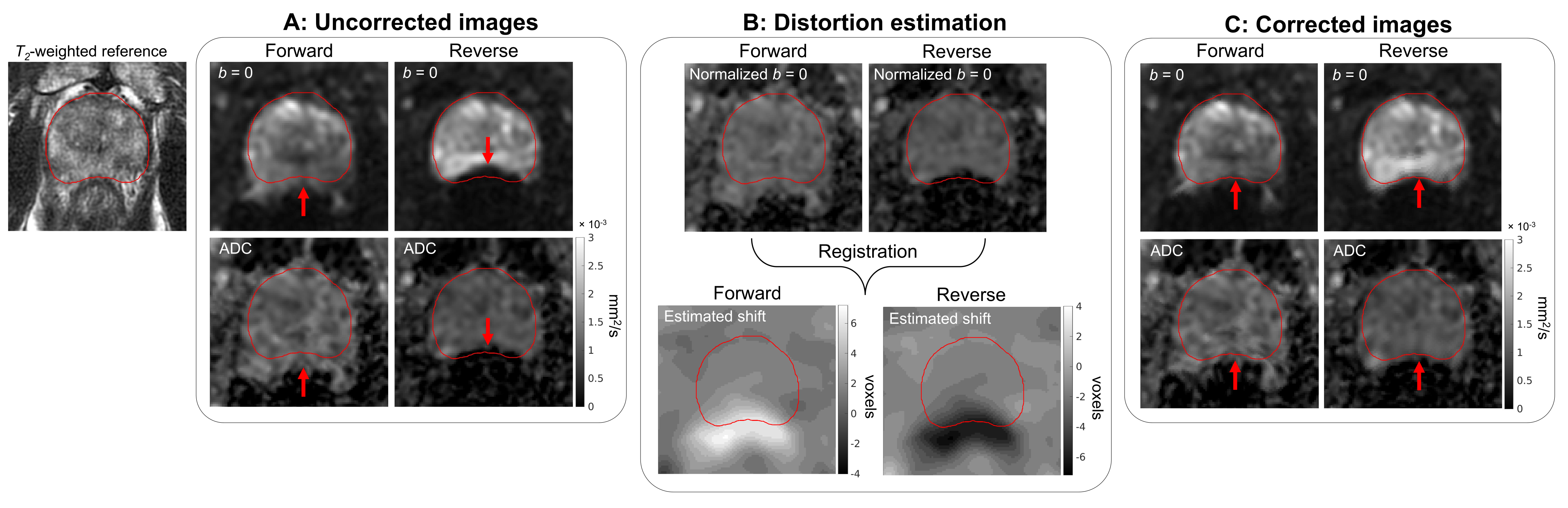
Forward and reverse DWI volumes were first normalized by their sum across all b-values to eliminate intensity changes arising from tissue compression or expansion (see Figure 1B). This obviated the need to fit a Jacobian term when registering forward and reverse images, simplifying estimation of the spatial transformation. In this study, the transformation was estimated by shifting the opposite-polarity volumes along the phase-encoding direction and computing the shift for each voxel that minimized the difference between forward and reverse images. This approach amounts to a multi-b-value adaptation of RPG, so we denote it as multi-b RPG (mRPG).
Evaluation of distortion correction methods
Spatial shift maps were estimated from the forward- and reverse-polarity volumes using RPG, topup, and mRPG. RPG and topup used b=0s/mm2 volumes for the estimation as detailed previously1,4. Spatial shifts estimated from each method were then applied to correct the b=0s/mm2 images and ADC maps. For RPG and topup, a second correction incorporating the JIC was applied to b=0s/mm2 images.
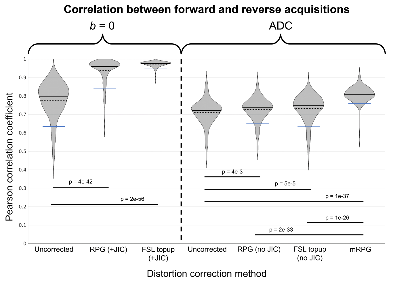
Distortion was quantified by the Pearson correlation between forward and reverse volumes. The distribution of correlation coefficients was examined for b=0s/mm2 images and ADC maps, before and after correction by each method, and between the three methods. Wilcoxon signed-rank tests (α=0.05) were used to assess whether correlation increased significantly after distortion correction, and with mRPG vs RPG and topup.
Results
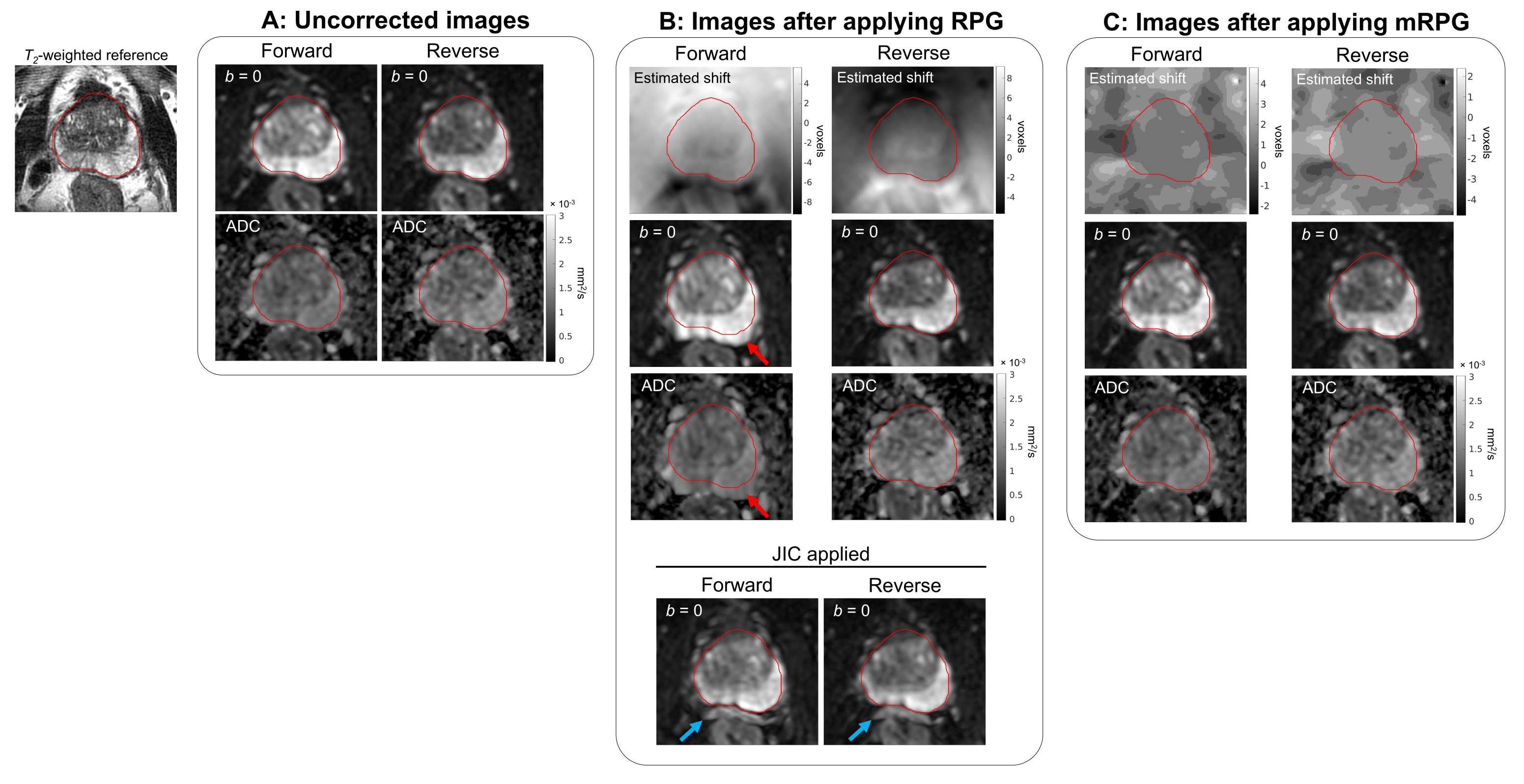
This study included 163 patients. Figure 1 shows an example application of mRPG. Figure 2 shows the distribution of correlation coefficients between opposite-polarity volumes. Point estimates for these distributions are listed in Table 2. With JIC, RPG and topup substantially increased the correlation of b=0s/mm2 images. Without JIC, however, they produced only slight increases in the correlation of ADC maps, compared to the much larger increase from mRPG. Figure 3 shows an example of a false correction generated with the JIC, vs an appropriate correction from mRPG.Discussion
Simultaneous estimation of spatial distortion and intensity scaling between opposite-polarity images is challenging and prone to overfitting, resulting in overemphasized intensity changes that compensate for underestimated tissue displacement. Such overfitting underlies the disparity shown in Figure 2, wherein RPG and topup yield large improvements on b=0s/mm2 images after JIC, which are not reflected on corresponding ADC maps.mRPG avoids overfitting by normalizing opposite-polarity images across multiple b-values. Normalization removes the intensity scaling (JIC) factor prior to estimating tissue displacement, resulting in significantly improved estimation and removal of spatial distortions.
Although mRPG requires additional scan time to acquire multiple b-values in both directions, it preserves anatomical resolution since any tissue that is compressed in one direction is expanded in the other. Furthermore, it is not necessary to acquire as many b-values as used here. Companion abstract #4876 demonstrates that as few as two b-values can be used effectively.
Acknowledgements
This work was supported, in part, by the National Institutes of Health (NIH/NIBIB K08 EB026503), the Department of Defense (DOD/CDMRP PC220278), the American Society for Radiation Oncology, and the Prostate Cancer Foundation.References
1. Holland D, Kuperman JM, Dale AM. Efficient correction of inhomogeneous static magnetic field-induced distortion in Echo Planar Imaging. NeuroImage. 2010;50(1):175–183.
2. Rakow-Penner RA, White NS, Margolis DJA, et al. Prostate diffusion imaging with distortion correction. Magnetic Resonance Imaging. 2015;33(9):1178–1181.
3. Andersson JLR, Skare S, Ashburner J. How to correct susceptibility distortions in spin-echo echo-planar images: application to diffusion tensor imaging. NeuroImage. 2003;20(2):870–888.
4. Smith SM, Jenkinson M, Woolrich MW, et al. Advances in functional and structural MR image analysis and implementation as FSL. NeuroImage. 2004;23:S208–S219.
Figures