3499
Point spread function(PSF) encoding EPI versus BLADE DWI in brain tumor diagnosis1Center for Biomedical Imaging Research, Department of Biomedical Engineering, School of Medicine, Tsinghua University, Beijing, China, 2Department of Neurosurgery,Yuquan Hospital, School of Clinical Medicine, Tsinghua University, Beijing, China, 3Siemens Shenzhen Magnetic Resonance Ltd., Shenzhen, China, 4MR Research Collaboration Team, Siemens Healthineers Ltd., Beijing, China, 5Department of Mathematics and Statistics, Whiting School of Engineering, Johns Hopkins University, Baltimore, MD, United States
Synopsis
Keywords: Diffusion Acquisition, Diffusion Tensor Imaging
Motivation: High-resolution DWI plays a crucial role in brain tumor diagnosis. Previous studies have introduced two high-resolution distortion-free DWI techniques: PSF and BLADE. However, no one has yet compared the two in brain MR imaging.
Goal(s): To compare the image quality of PSF and BLADE in high-resolution DWI.
Approach: In this study, scan parameters were adjusted to achieve the optimized image quality for PSF and BLADE. Subsequently, scans were performed on patients, and the final image quality was compared.
Results: With scanning times being similar, PSF exhibits a superior SNR compared to BLADE while its performance is suboptimal at the boundaries of brain tissues.
Impact: A preliminary comparison of the image quality between PSF and BLADE has been conducted, paving the way for more in-depth clinical research in areas such as diagnostic accuracy and imaging quality control.
Introduction
DWI is a critical tool for diagnosing brain tumors. High-resolution DWI enhances our ability to detect smaller tumors and early lesions while improving the precision of tumor localization. However, commonly used EPI DWI often suffers from severe susceptibility-induced geometric distortion due to its limited encoding bandwidth along the phase-encoding (PE) direction, which hampers image quality. To address this issue, recent advancements in MRI technology have introduced the point spread function (PSF) encoding EPI DWI technique1, known for its distortion-free imaging capabilities. By employing tilted-CAIPI reconstruction2, PSF DWI can achieve a high acceleration factor exceeding 20. Another promising development is the turbo gradient and spin echo (TGSE) BLADE DWI technique3,4,5,6 in which the TGSE readout can effectively mitigates magnetic susceptibility-induced artifacts and distortions.In this study, we aim to compare the image quality of highly accelerated PSF encoding DWI and TGSE-BLADE techniques, focusing on their performance in visualizing brain lesions and normal brain structures.
Method
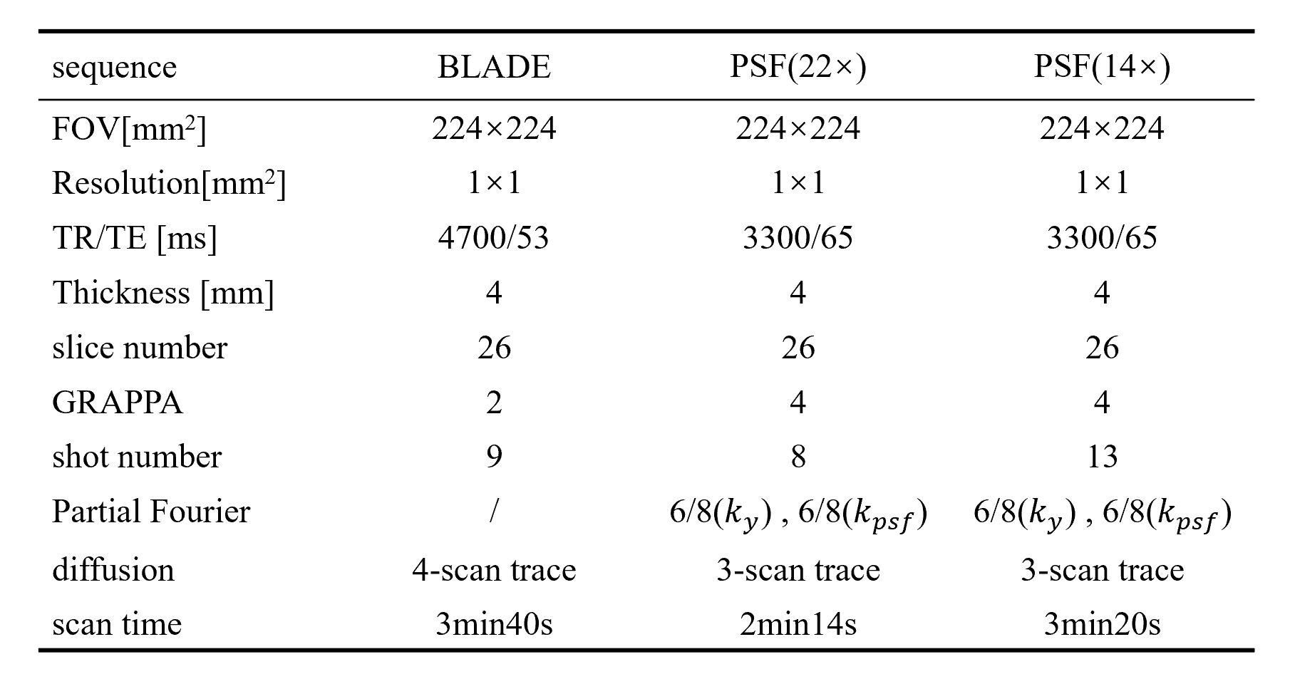
This study included a total of six patients. The study was approved by the local Ethical Standards Committee, and written informed consents were obtained from both subjects. All data were collected using a 64-channel Head/Neck coil on a Siemens Prisma 3T MR system(Siemens Healthineers, Erlangen, Germany). The experiment employed the prototyping PSF sequence and the TGSE-BLADE sequence, following the acquisition protocol shown in Table 1. High-resolution DWI data with a slice thickness of 4mm and no inter-slice gap were acquired at an in-plane resolution of 1mm×1mm, along with other anatomical scans. Experimental parameters were adjusted to achieve the best possible image quality for PSF and BLADE individually. 8-shot PSF images were acquired which can be robustly reconstructed in healthy volunteers, while 13-shot PSF images which had a similar scan time with BLADE were also obtained for comparison purposes.Following data acquisition, BLADE images were reconstructed online, while PSF images were reconstructed offline using Matlab R2023b. To reduce the reconstruction time, the PSF data underwent channel compression to 8 channels using the GCC method7 before subsequent reconstruction steps. Both BLADE and PSF images underwent intensity correction , and no denoising was applied to the original images. After reconstruction, DWI trace-weighted images of the two sequences at the locations with brain lesions in the patients and images at different locations within one patient's brain were compared.
Results and Discussion
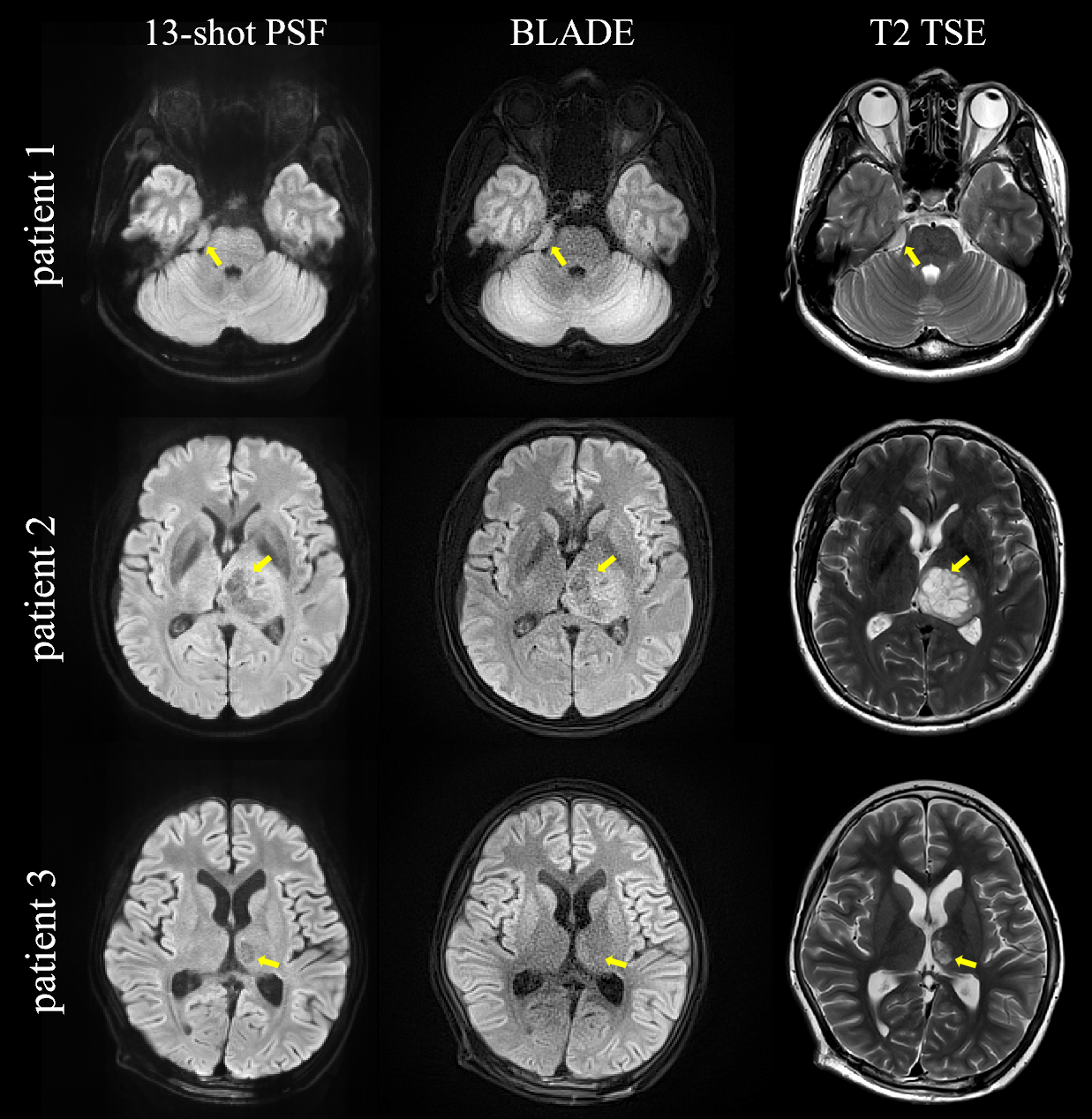
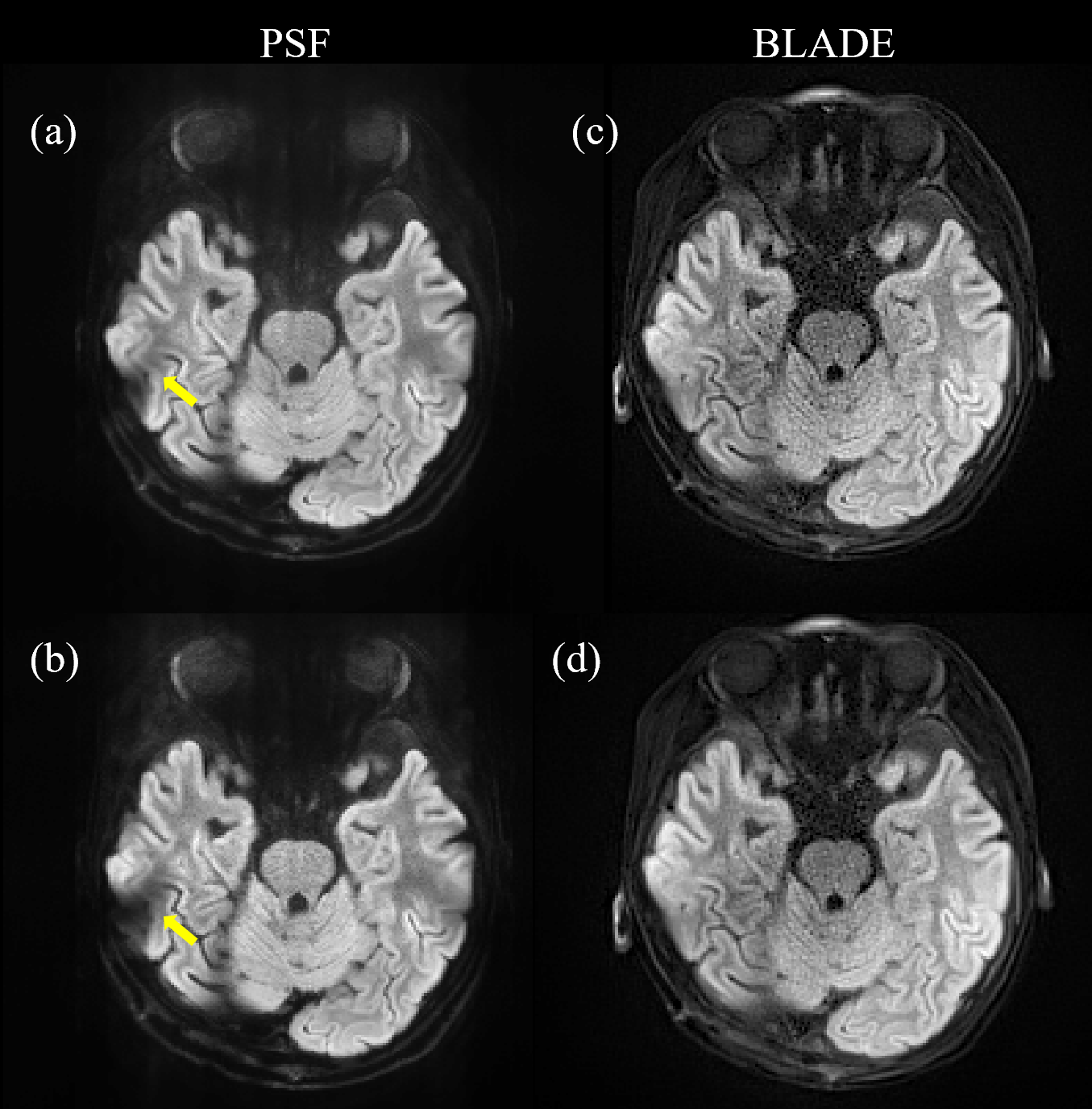
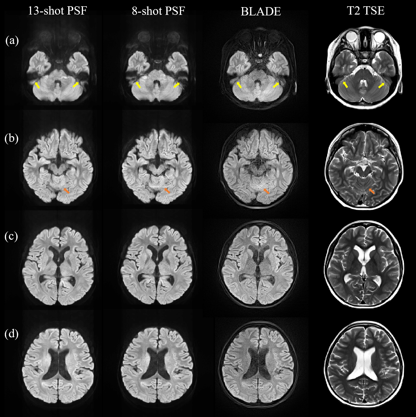
Figures 1~3 present comparisons of PSF and BLADE image in the patients. From the images, it can be observed that both PSF and BLADE exhibit minimal distortion and similar sharpness compared to the T2 TSE images. However, BLADE exhibits lower SNR than PSF, especially in the regions around the brainstem and corpus callosum. In Figure 1, a glioma in the brainstem midbrain area of patient 3 is clearly visible in PSF, with distinct lesion contours and internal details, whereas no lesion is evident in BLADE due to limited SNR. Figure 2 shows the comparison between BLADE (after total variation(TV) denoising) and PSF (without denoising).However, in the location beneath the temporal lobe of the cranial base, the reconstruction quality of PSF is poorer than BLADE. Furthermore, the distortion in the 22-fold accelerated 8-shot PSF images is more pronounced compared to the 14-fold accelerated 13-shot PSF images. This is due to the assumption in the PSF reconstruction that the B0 field's inhomogeneity is smooth2. In locations where B0 inhomogeneity changes significantly, particularly air-bone-tissue interfaces, the reconstruction with tilted-GRAPPA kernels is less effective, and the higher the acceleration factor in PSF, the more severe the impact. The yellow arrows in Figure 2(a), (b), and Figure 3(a) illustrate these reconstruction artifacts. Therefore, it is recommended to use different sampling methods for different lesion locations.
Conclusion
Both PSF and BLADE provide distortion-free images. PSF holds an advantage over BLADE in terms of SNR, but it performs less effectively near air-bone-tissue interfaces. Increasing the number of PSF shots at the cost of adding some scanning time can reduce reconstruction artifacts and enhance clinical diagnosis.Acknowledgements
NoReferences
[1] Robson MD, Gore JC, Constable RT. (1997). Measurement of the point spread function in MRI using constant time imaging. Magn Reson Med. 38, 733–740.
[2] Dong, Z., Wang, F., Reese, T. G., Manhard, M. K., Bilgic, B., Wald, L. L., Guo, H., & Setsompop, K. (2019). Tilted-CAIPI for highly accelerated distortion-free EPI with point spread function (PSF) encoding. Magnetic Resonance in Medicine, 81(1), 377–392.
[3] Hu, H. H., McAllister, A. S., Jin, N., Lubeley, L. J., Selvaraj, B., Smith, M., Krishnamurthy, R., & Zhou, K. (2019). Comparison of 2D BLADE Turbo Gradient- and Spin-Echo and 2D Spin-Echo Echo-Planar Diffusion-Weighted Brain MRI at 3 T: Preliminary Experience in Children. Academic Radiology, 26(12), 1597–1604.
[4] Lin, M., Lin, N., Sheng, Y., Sha, Y., Zhang, Z., & Zhou, K. (2022). Detection of cholesteatoma: 2D BLADE turbo gradient- and spin-echo imaging versus readout-segmented echo-planar diffusion-weighted imaging. European Archives of Oto-Rhino-Laryngology, 279(11), 5223–5229.
[5] Kun Zhou, Wei Liu, & Shi Cheng. (2018). Non-CPMG PROPELLER diffusion imaging: comparison of phase insensitive preparation with split acquisition. Proc. Intl. Soc. Mag. Reson. Med.26.
[6] Kun Zhou & Wei Liu. (2016). Multi-Blade Acquisition of Split Turbo Spin Echoes: A Robust and Fast Diffusion Imaging Technique. Proc. Intl. Soc. Mag. Reson. Med.24.
[7] Zhang T, Pauly JM, Vasanawala SS, Lustig M. Coil compression for accelerated imaging with Cartesian sampling. Magn Reson Med. 2013 Feb;69(2):571-82.
Figures