3488
Time-dependent diffusion MRI could distinguish between functioning and non-functioning pituitary adenoma1Department of Advanced Radiological Imaging, Kagoshima University Graduate School of Medical and Dental Sciences, Kagoshima, Japan, 2Radiology, Kagoshima University Graduate School of Medical and Dental Sciences, Kagoshima, Japan, 3Neurosurgery, Kagoshima University Graduate School of Medical and Dental Sciences, Kagoshima, Japan, 4Radiological Technology, Kagoshima University Graduate School of Medical and Dental Sciences, Kagoshima, Japan, 5Siemens Healthcare K.K., Tokyo, Japan, 6Siemens Healthcare GmbH, Erlangen, Germany, 7Advanced Radiological Imaging, Kagoshima University Graduate School of Medical and Dental Sciences, Kagoshima, Japan
Synopsis
Keywords: Microstructure, Diffusion/other diffusion imaging techniques
Motivation: Differentiation between functioning and non-functioning pituitary adenoma (PA) is clinically relevant. Feasibility of their microstructural differentiation is undetermined.
Goal(s): To investigate whether the time-dependent diffusion MRI can distinguish functioning from non-functioning PA.
Approach: Twenty-four patients with functioning and 30 with non-functioning PA were examined. Pituitary DWI was performed using inner FOV EPI based on 2D-selective radiofrequency excitations with oscillating (diffusion time=7.1ms) and pulsed (36.3ms) gradient preparations. ADC change (cADC) and relative ADC change (rcADC) between two diffusion-times were calculated.
Results: cADC was significantly higher in functioning than in non-functioning PAs (P=0.012), showing that time-dependent diffusion MRI can distinguish them.
Impact: Our results clarified that there are microstructural differences between functional and non-functional pituitary adenomas. Time-dependent diffusion MRI of the pituitary gland can detect their microstructural characteristics, providing clues as to their imaging differentiation.
INTRODUCTION
Pretreatment differentiation between functioning and non-functioning pituitary adenoma (PA) is clinically relevant. Microstructural characteristics of functioning versus non-functioning PA is unknown, and to date there is no report regarding the ability of diffusion MRI to differentiate functioning and non-functioning PAs. Complementary to conventional pulsed gradient spin-echo sequences (PGSE), oscillating gradient spin-echo (OGSE) sequences allow for DWI with a short diffusion time1, enabling time-dependent diffusion analysis which could provide specific information regarding restricted diffusion. Our purpose was to investigate whether the time-dependent diffusion MRI can distinguish functioning from non-functioning PA.METHODS
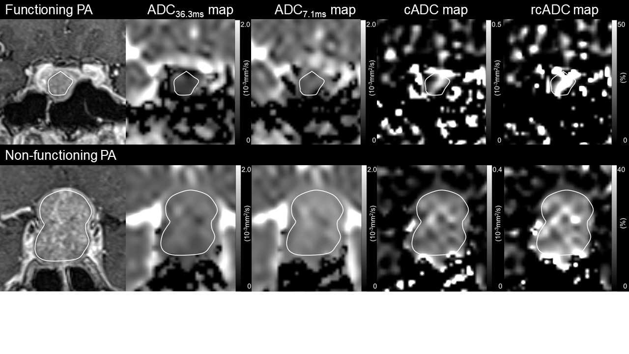
A retrospective study was performed including 24 patients (mean age, 45 ± 17 years) with functioning PA (12 growth hormone [GH]-producing, 9 prolactin [PRL]-producing, 2 thyroid stimulating hormone [TSH]-producing, 1 adrenocorticotropic hormone [ACTH]-producing) and 30 patients with non-functioning PA (59 ± 17 years). All patients underwent preoperative MR imaging using a 3T system (MAGNETOM Prisma; Siemens Healthcare, Erlangen, Germany) with a 20-channel head/neck coil. DWI of the pituitary gland was performed using a research sequence with inner FOV EPI sequences based on 2D-selective radiofrequency excitations with OGSE preparation using sine-modulated trapezoidal waveforms (effective diffusion time = 7.1 ms) and PGSE (36.3 ms) preparation with b values of 0 and 1,000 s/mm2. The sequences shared the following parameters: TR, 4,000 ms; TE, 103 ms; FOV, 180 × 60 mm2; matrix size, 72 × 24; slice thickness, 3 mm; slice gap, 0.3 mm; and acquisition time, 1 min and 20 sec. In addition to maps of ADC obtained with OGSE (ADC7.1ms) and PGSE (ADC36.3ms), maps of the change in ADC values between OGSE and PGSE sequences were generated: cADC = ADC7.1ms – ADC36.3ms. Moreover, maps of the relative ADC change between OGSE and PGSE sequences were generated: rcADC = (ADC7.1ms – ADC36.3ms)/ADC36.3ms × 100 (%). ROI was drawn in each adenoma by a neuroradiologist (Fig. 1). The ROI mean values of the diffusion parameters were compared between functioning and non-functioning pituitary adenoma using the Mann-Whitney U test. Furthermore, we compared the diffusion parameters between non-functioning PA and each hormone-producing PA. The diagnostic performances of the parameters were evaluated using ROC curve analysis.RESULTS
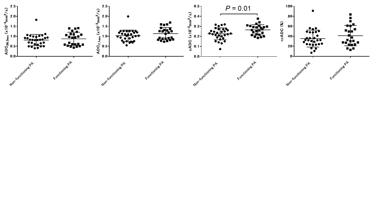
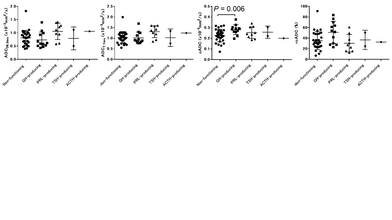
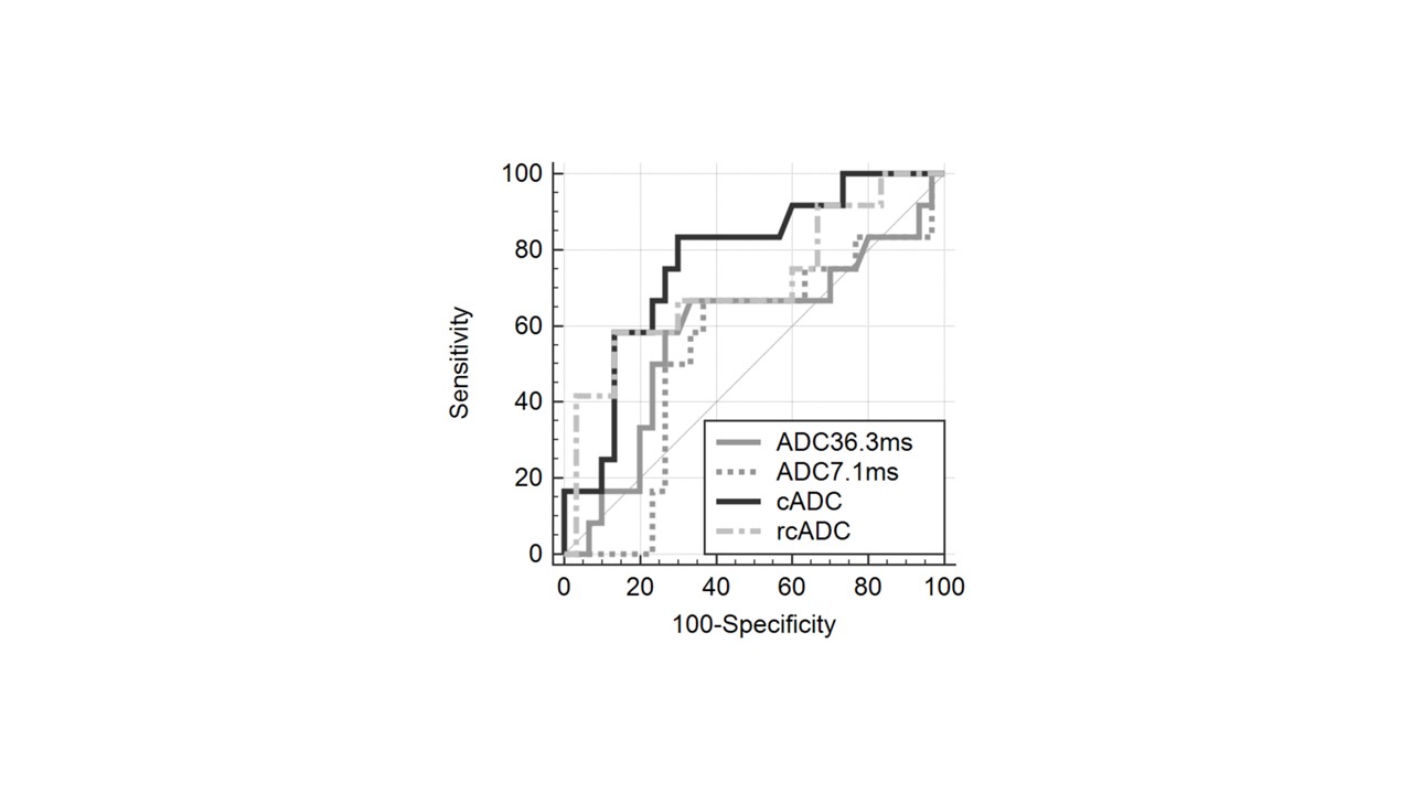
No significant difference was noted between functioning and non-functioning PA in ADC36.3ms, ADC7.1ms, and rcADC, whereas cADC was significantly higher in functioning PAs than in non-functioning PAs (P = 0.012) (Figure 2). The ROC curve analysis showed significance for cADC (AUC = 0.677, P = 0.017) for identifying functioning PA from non-functioning PA. Moreover, cADC was significantly higher in GH-producing PAs than in non-functioning PAs (P = 0.006) (Figure 3). The ROC curve analysis showed significance for cADC (AUC = 0.771, P < 0.001) and rcADC (AUC = 0.708, P = 0.034) for identifying GH-producing PA from non-functioning PA. The ROC curves for the diffusion parameters to distinguish GH-producing PA from non-functioning PA are shown in Figure 4.DISCUSSION
Our results suggest that cADC is a useful imaging marker for distinguishing functioning PAs, especially GH-producing PAs, from non-functioning PAs, while ADC nor rcADC are not. The underlying mechanism for the stronger ADC diffusion time dependence in functioning PA than in non-functioning PA is unknown. Non-functioning PAs consist of a wide spectrum of morphologic lesions, and more than 60% were classified as gonadotropin-producing PAs2. The tumors consist of polygonal cells with prominent sinusoidal, trabecular, or tubular architecture along the blood vessels, which often show markedly elongated cells arranged in a pseudorosette pattern3. This adenoma has rich extracellular matrices. On the other hand, half of our functioning PAs were GH-producing PAs. Those tumors consist of round cells with diffuse pattern3. Therefore, the extracellular space is small. Theoretically, narrower extracellular space could explain stronger diffusion time-dependence of ADC in functioning PAs. Further studies are needed to elucidate the pathological factors that account for our findings.CONCLUSION
The cADC derived from the time-dependent diffusion MRI could distinguish functioning PAs, especially GH-producing PAs, from non-functioning PAs.Acknowledgements
No acknowledgement found.References
1. Schachter M, Does MD, Anderson AW, et al. Measurements of restricted diffusion using an oscillating gradient spin-echo sequence. J Magn Reson. 2000;147:232–237.
2. Yamada S, Ohyama K, Taguchi M, et al. A study of the correlation between morphological findings and biological activities in clinically nonfunctioning pituitary adenomas. Neurosurgery. 2007;61:580-4.
3. Osamura RY, Lopes MBS, Grossman A, et al. Tumors of the pituitary gland. In WHO Classification of Tumors of Endocrine Organs, 4th ed.; Ricardo, V.L., Osamura, R.Y., Gunter, K., Juan, R., Eds.; International Agency for Research on Cancer: Lyon, France, 2017;11–64.
Figures