3487
Brain glymphatic system impairment in multiple sclerosis patients based on diffusion tensor imaging1The Second Clinical College, Lanzhou University, Lan Zhou, China, 2The Second Hospital of Lanzhou University, Lan Zhou, China, 3Department of Clinical and Technical Support, Philips Healthcare, Xi’an, China
Synopsis
Keywords: DWI/DTI/DKI, Brain, glymphatic system, along perivascular space, clinical fatigue
Motivation: Glymphatic abnormalities have been reported in several neurodegenerative disorders. However, glymphatic function has not been thoroughly investigated in multiple sclerosis, especially its relationship with clinical fatigue.
Goal(s): We aimed to investigate glymphatic system function in multiple sclerosis and to evaluate its association with clinical fatigue.
Approach: We prospectively enrolled 36 multiple sclerosis patients and 31 healthy controls. All subjects underwent diffusion tensor imaging, and the along perivascular space (ALPS) indexes were calculated. Correlations between ALPS indexes and clinical fatigue parameters were analyzed.
Results: The brain glymphatic system is impaired in multiple sclerosis. Impaired glymphatic function was associated with multiple sclerosis-related fatigue.
Impact: Non-invasive diffusion-based imaging approach could be used as a proxy for displaying an impaired glymphatic system in multiple sclerosis patients, suggesting that glymphatic impairment may be a pathological mechanism underpinning clinical fatigue.
Summary of Main Findings
Our results suggest that the brain glymphatic system is impaired in multiple sclerosis. Impaired glymphatic function was associated with multiple sclerosis-related fatigue. Those findings confirmed that glymphatic system may be involved in the pathogenesis of fatigue in multiple sclerosis patients.Introduction
Multiple sclerosis (MS) is a chronic central nervous system (CNS) inflammatory disease of autoimmune etiology [1]. Primary fatigue is a common symptom in MS patients, which may depend on demyelination, inflammation, or axonal loss [2]. Mitochondrial dysfunction, cytotoxic factors such as reactive oxygen species (ROS) production, leucocyte infiltrates, neuronal acidosis and accumulation of iron fuel the neuronal loss [3, 4]. Therefore, given the causative role of toxic waste products, it is essential for the brain to have a waste clearing system, which was defined as the glymphatic system. Previous studies have described the glymphatic abnormalities in several neurodegenerative conditions, including Alzheimer’s and Parkinson’s disease. However, glymphatic function has not been thoroughly investigated in multiple sclerosis, particularly in relation to clinical fatigue.Material and Methods
Thirty-six patients diagnosed with clinically definite Relapsing Remitting MS (RRMS) and 31 age- and sex-matched healthy controls (HCs) were prospectively enrolled from the Lanzhou University Second Hospital. All patients were assessing using the Fatigue Severity Scale (FSS) and the modified fatigue impact scale (MFIS), and those who had a mean FSS score of≥4 was considered to have noticeable fatigue [5]. Accordingly, the patients were divided into MS presenting fatigue (MS-F) and MS not presenting fatigue (MS-NF) groups. All patients underwent MR imaging on a 3.0 T scanner (Ingenia CX, Philips Healthcare, The Netherlands) with a 32-channel phased-array head coil. More details about scanning parameters were listed in Table 1. The along perivascular space (ALPS) index was calculated using FSL (https://fsl.fmrib.ox.ac.uk/fsl/) with the following formula: ALPS index = mean (Dxproj, Dxassoc)/mean (Dyproj, Dzassoc). One-way ANOVA analysis was carried out to determine the differences among three groups (MS-F, MS-NF and HCs). Between-group comparisons of demographic, clinical and MRI variables were performed using t-test and Chi-square, and correlation analysis was measured by the Pearson correlation. All analyses were conducted using IBM SPSS Statistics software (version 26.0, Armonk, NY, USA).Results
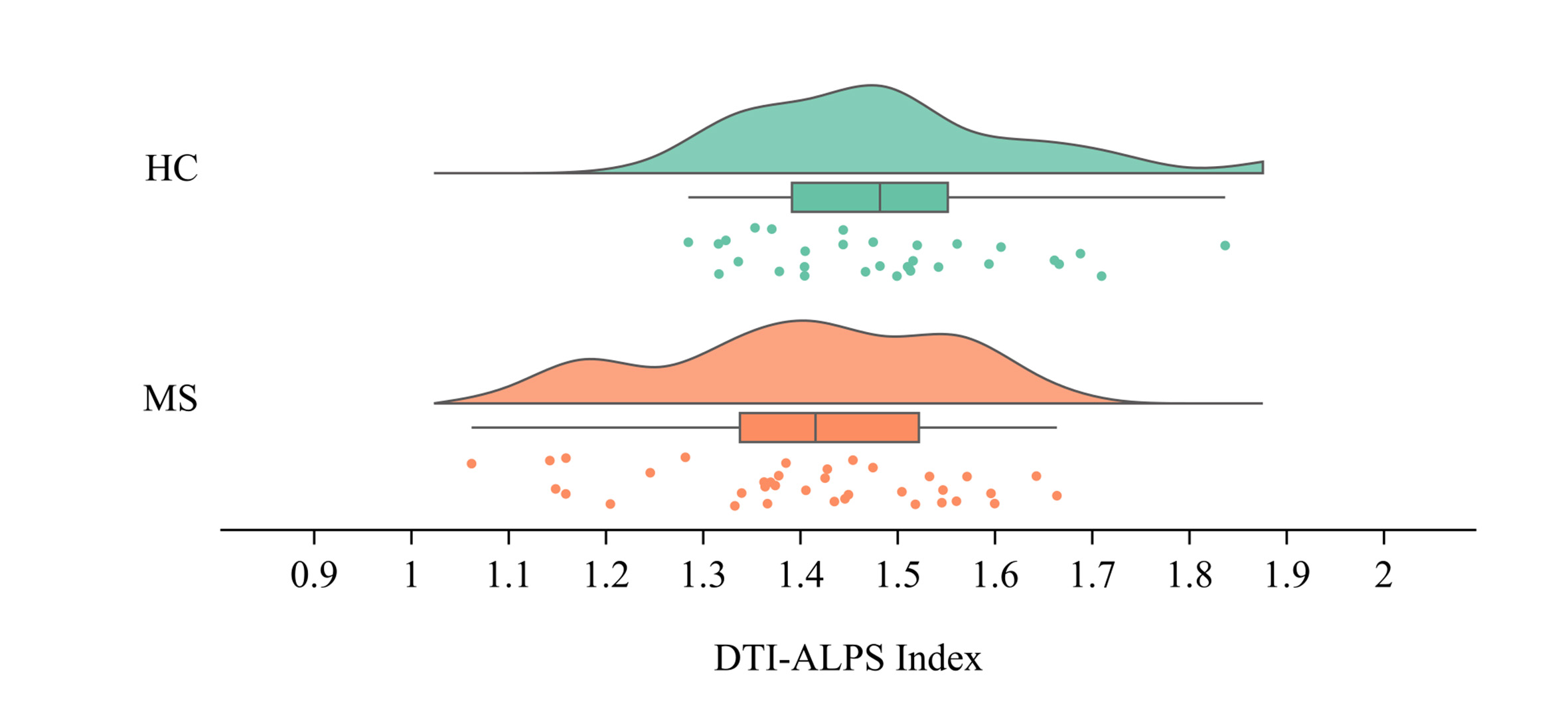
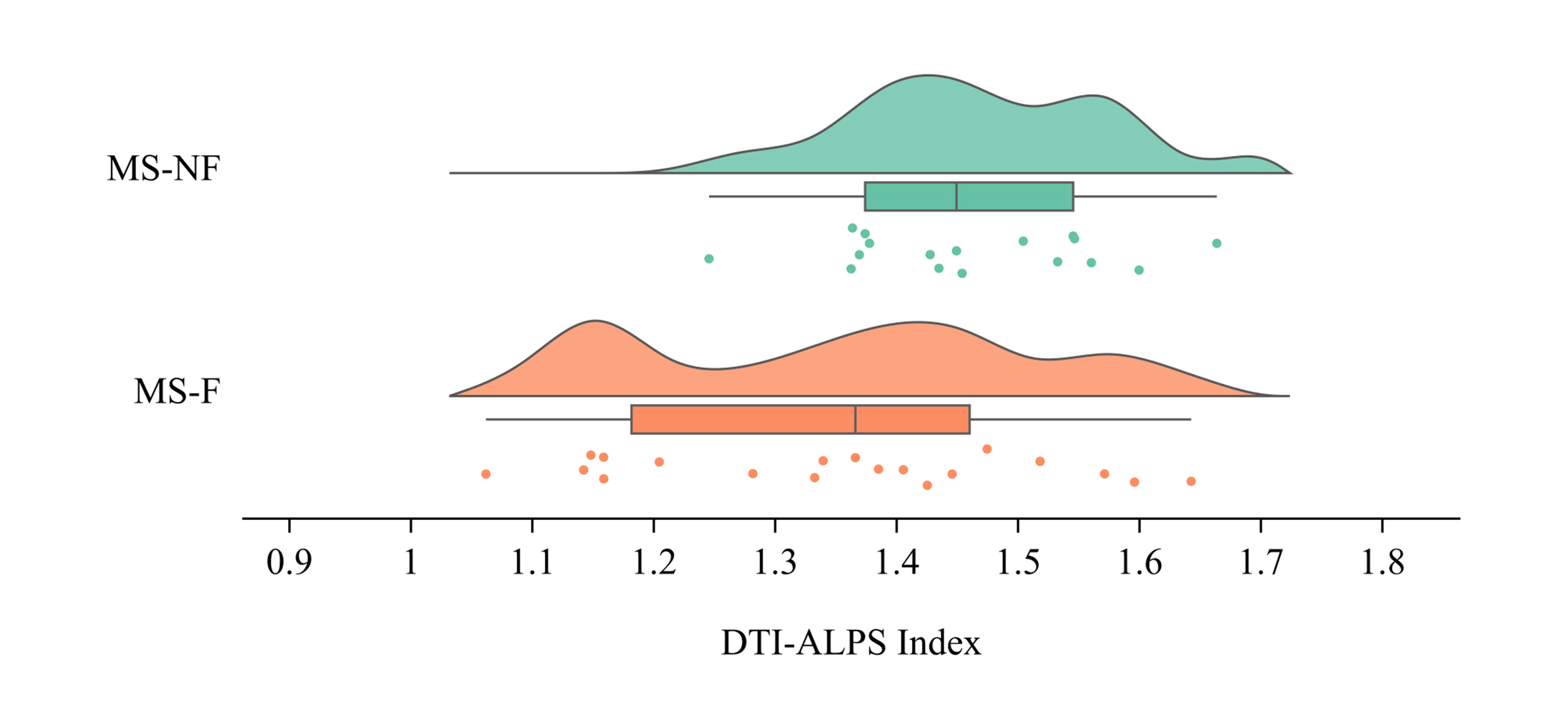
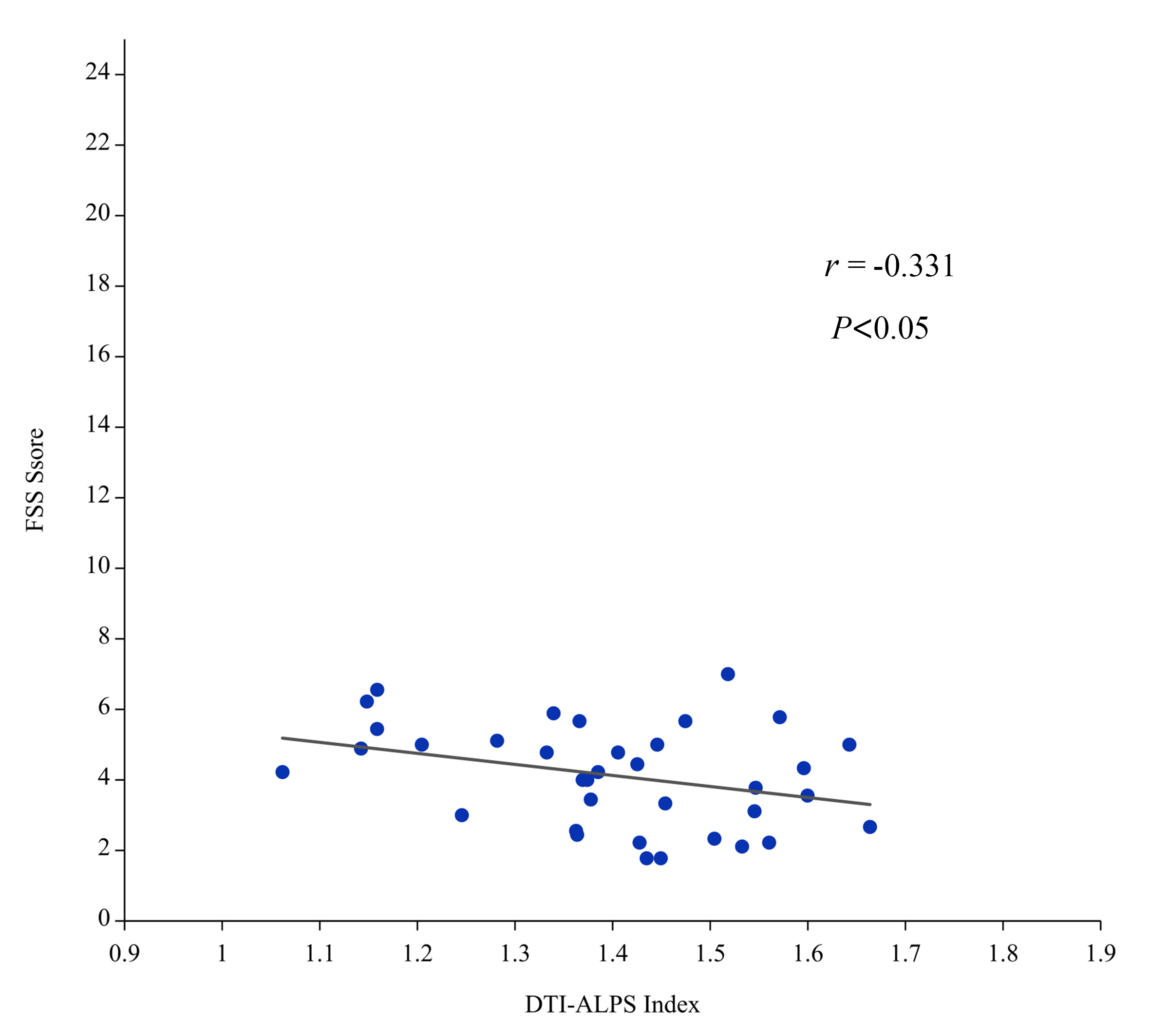
Demographic, clinical and MRI variables from study subjects were summarized in Table 2. There were 19 MS-F, 17 MS-NF, and 31 HCs. The sex and age showed not significantly different between HC and MS groups as well as MS-F and MS-NF groups (P>0.05). Compared with both the HC and MS-NF groups, MS-F patients had significantly higher FSS scores (both, P < 0.001). In addition, MS patients showed lower diffusion ALPS-index than healthy controls (P=0.017) (Figure 1). As is shown in Table 2 and Figure 2, MS-F groups had lower diffusion ALPS index versus MS-NF patients. In MS patients, lower diffusion ALPS index was negatively associated with more severe clinical fatigue (r =−0.331, P = 0.048) (Figure 3).Discussion and Conclusion
Our study investigated the brain glymphatic system function in MS patients using a non-invasive MRI index of diffusivity along the perivascular space: DTI-ALPS. The study suggested that MS patients displayed a glymphatic system impairment compared with HCs, and glymphatic system was more severely affected in MS patients presenting clinical fatigue. Our studies found a negative correlation between clinical fatigue and glymphatic system impairment, highlighting a particular role of the diffusivity along the perivascular space in contributing to the clinical fatigue in MS patients. The glymphatic system crosses the perivascular space, and when perivascular space expands, the flow rate of the glymphatic fluid slows down, contributing to an accumulation of inflammatory molecules and toxic substances, thereby fueling the demyelination process of white matter and gray matter damage.In conclusion, our study confirmed the hypothesis that glymphatic system impairment possibly contributes to demyelination and neuronal loss processes and causes clinical fatigue. Particularly, we verified a significant correlation between the diffusivity along the perivascular space and clinical fatigue measured by FSS, supporting the hypothesis of the glymphatic system involvement in the pathogenesis of fatigue in MS patients. However, notably, further investigation with larger sample sizes is required to verify the interplay with MRI markers of disease pathology with more certainty.Acknowledgements
Z.W. thanks J.Z. for guidance and advice during the writing process of the article. The authors are grateful to K.A. of Philips Healthcare China for their help with the MRI technique used in this article.References
[1] Marcus R. What Is Multiple Sclerosis? . JAMA. 2022;328(20):2078. doi:10.1001/jama.2022.14236.
[2] Manjaly ZM, Harrison NA, Critchley HD, et al. Pathophysiological and cognitive mechanisms of fatigue in multiple sclerosis. J Neurol Neurosurg Psychiatry. 2019;90(6):642-651. doi:10.1136/jnnp-2018-320050.
[3] Magliozzi R, Howell O, Vora A, et al. Meningeal B-cell follicles in secondary progressive multiple sclerosis associate with early onset of disease and severe cortical pathology. Brain. 2007;130(Pt 4):1089-1104. doi:10.1093/brain/awm038.
[4] Ciccarelli O, Barkhof F, Bodini B, et al. Pathogenesis of multiple sclerosis: insights from molecular and metabolic imaging. Lancet Neurol. 2014;13(8):807-822. doi:10.1016/S1474-4422(14)70101-2.
[5] Herlofson K, Larsen JP. Measuring fatigue in patients with Parkinson's disease - the Fatigue Severity Scale. Eur J Neurol. 2002;9(6):595-600. doi:10.1046/j.1468-1331.2002.00444. x.
Figures
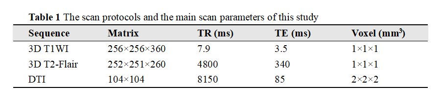
* Abbreviation: 3D T1WI: 3D T1-weighted imaging, 3D T2-Flair: 3D T2 fluid-attenuated inversion recovery (FLAIR), DTI: diffusion tensor imaging.
** Slice thickness: 1 mm, slice gap: 0 mm.
***For DTI, b-value=1000s/mm2.
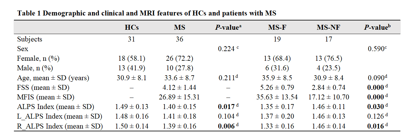
*HCs, Healthy controls; MS, Multiple sclerosis; MS-F, MS presenting fatigue; MS-NF, MS not presenting fatigue; FSS, Fatigue Severity Scale; MSIF, Modified Fatigue Impact Scale; ALPS, along perivascular space.
**a Multiple sclerosis versus healthy controls.
b MS presenting fatigue versus MS not presenting fatigue.
c Chi-square test.
d Student’s t-test.