3481
Assessment of the repeatability and stability of NODDI diffusion modelling using phantom and in vivo acquisitions.1Department of Computer Science, University of Pisa, Pisa, Italy, 2INFN, Division of Bologna, Bologna, Italy, 3Oxford Centre for Clinical Magnetic Resonance Research, University of Oxford, Oxford, United Kingdom, 4Department of Physiology, Anatomy, and Genetics, University of Oxford, Oxford, United Kingdom, 5Department of Oncology, University of Oxford, Oxford, United Kingdom, 6Department of Radiology, Cambridge University Hospitals, Cambridge, United Kingdom, 7Nuffield Department of Medicine, University of Oxford, Oxford, United Kingdom, 8Department of Physics and Astronomy, University of Bologna, Bologna, Italy, 9Department of Radiology, Oxford University Hospitals, Oxford, United Kingdom
Synopsis
Keywords: Diffusion Modeling, Microstructure, NODDI model, time stability and consistency, single centre
Motivation: The NODDI diffusion-MRI model shows promising results in characterising brain microstructure and capturing neurological disease-related changes. However, the NODDI model lacks validation, limiting its clinical application.
Goal(s): The goal is to validate the diffusion MRI NODDI model, assessing its consistency over time and addressing the need for robust methods in clinical research.
Approach: By scanning several times phantoms simulating brain-restricted diffusion and healthy volunteers with the same acquisition protocol, we meticulously assess NODDI's stability over time and in the presence of magnetic gradient coil heating.
Results: The study confirms the NODDI model's exceptional consistency and stability, establishing its credibility for future clinical applications.
Impact: The study confirms the reliability and stability of the NODDI model in assessing brain microstructure over time. This has significant implications for monitoring neurological disease progression and may lead to standardised MRI calibration protocols for collaborative research and clinical applications.
Introduction
The structure and function of the brain are key to ongoing health and well-being. It is known that when the brain is affected by pathologies such as Multiple Sclerosis (a condition whereby the brain is attacked by the immune system), changes in structure and tissue volume are a hallmark of disease progression1. Neurite orientation dispersion and density imaging (NODDI) is a diffusion model for estimating the microstructural complexity of dendrites and axons in vivo2, providing more specific markers of brain tissue microstructure than indices from standard diffusion tensor imaging (DTI), such as fractional anisotropy (FA)3. Despite the strengths of NODDI to assess changes in brain microstructure, there is a lack of research examining the consistency and repeatability over time of NODDI results, and only a limited number of studies have evaluated the reliability of these metrics4. Thus, it is crucial to thoroughly evaluate the typical fluctuations and consistency over time of the most used diffusion metrics5,6. This work aims to evaluate the consistency and accuracy of the NODDI fit parameters, such as the β-fraction, the Orientation Dispersion Index (ODI) and Intra- and Extra- neurite volume fractions of the NODDI model.Methods
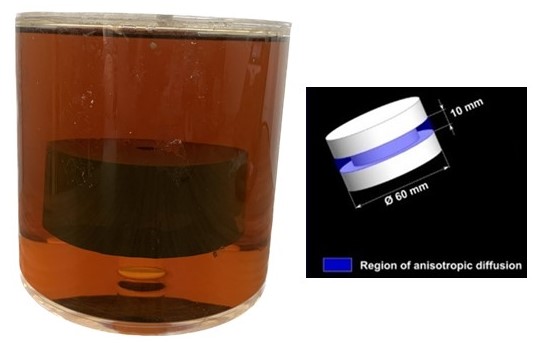
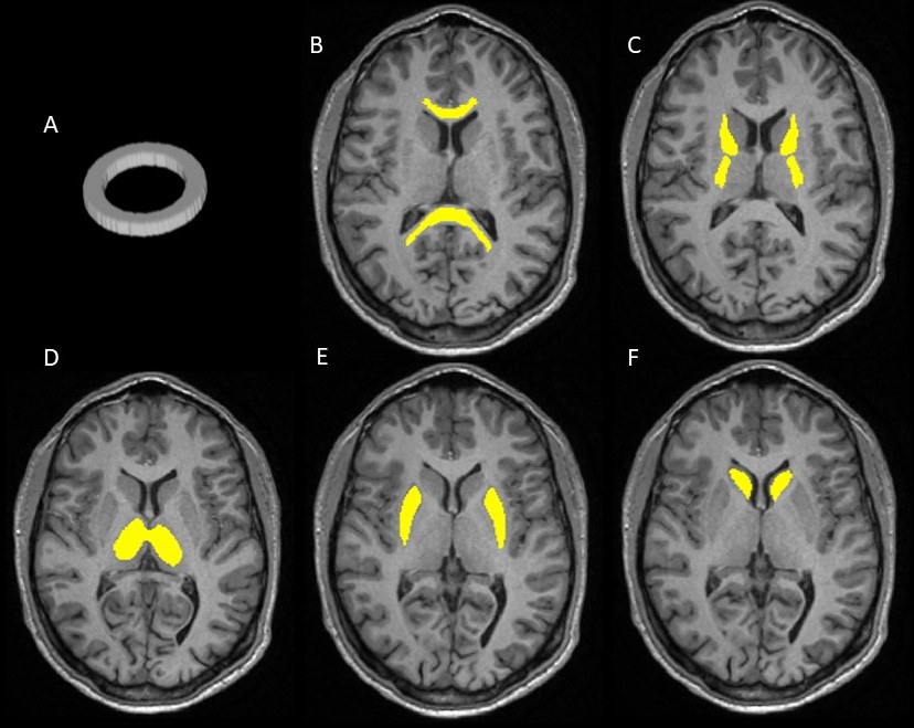
All acquisitions were performed using a 3T (GE-HealthCare, WI, USA) scanner and 21-channel head and neck coil. A phantom7, shown in Figure 1, was scanned four times on different days over a period of one month. Additionally, it underwent eight consecutive scans on the same day. The acquired data was pre-processed using the FMRIB Software Library (FSL), including topup and eddy, and then fit to the NODDI model. The mean and standard deviation for each metric were extracted from the fibre ring using a binary mask, shown in Figure 2. Along with the phantoms, a group of four healthy volunteers – three males and one female, age between 24 and 30, mean age 28.5 – was scanned twice on the same day with a ten-minute interval between the two scans. The in vivo results were extracted from the ROIs in Figure 2. The acquisition protocol consisted of 90 diffusion directions: 30 with a b-value of 1000 s/mm2 and 60 with a b-value of 2600 s/mm2, with 9 b=0 images randomly dispersed, Repetition Time (TR) = 6000ms, Echo Time (TE) = 70ms, Field of View (FOV) = 240x240 mm2, Slice thickness = 2.5 mm with 32 slices for the phantom and 66 for the brain, ASSET factor of 2, bandwidth = 250 kHz and spectral fat saturation with non-spectral selective excitation. For in vivo scans, simultaneous multi-slice (factor 2) was used. The sequence was adapted to output the integrated reverse polarity phase encode acquisition. Finally, the coefficient of variation (CV) was computed to assess the stability of the phantom results over time. For the brain results, instead, the repeatability coefficient (RC) was computed.Results and Discussion
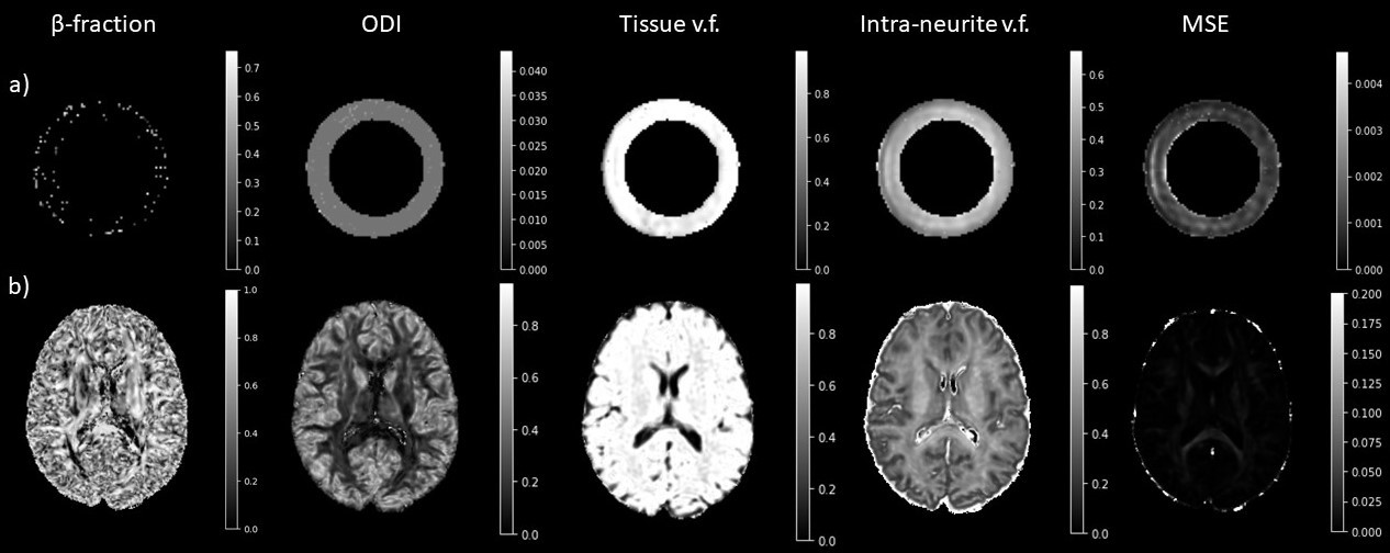
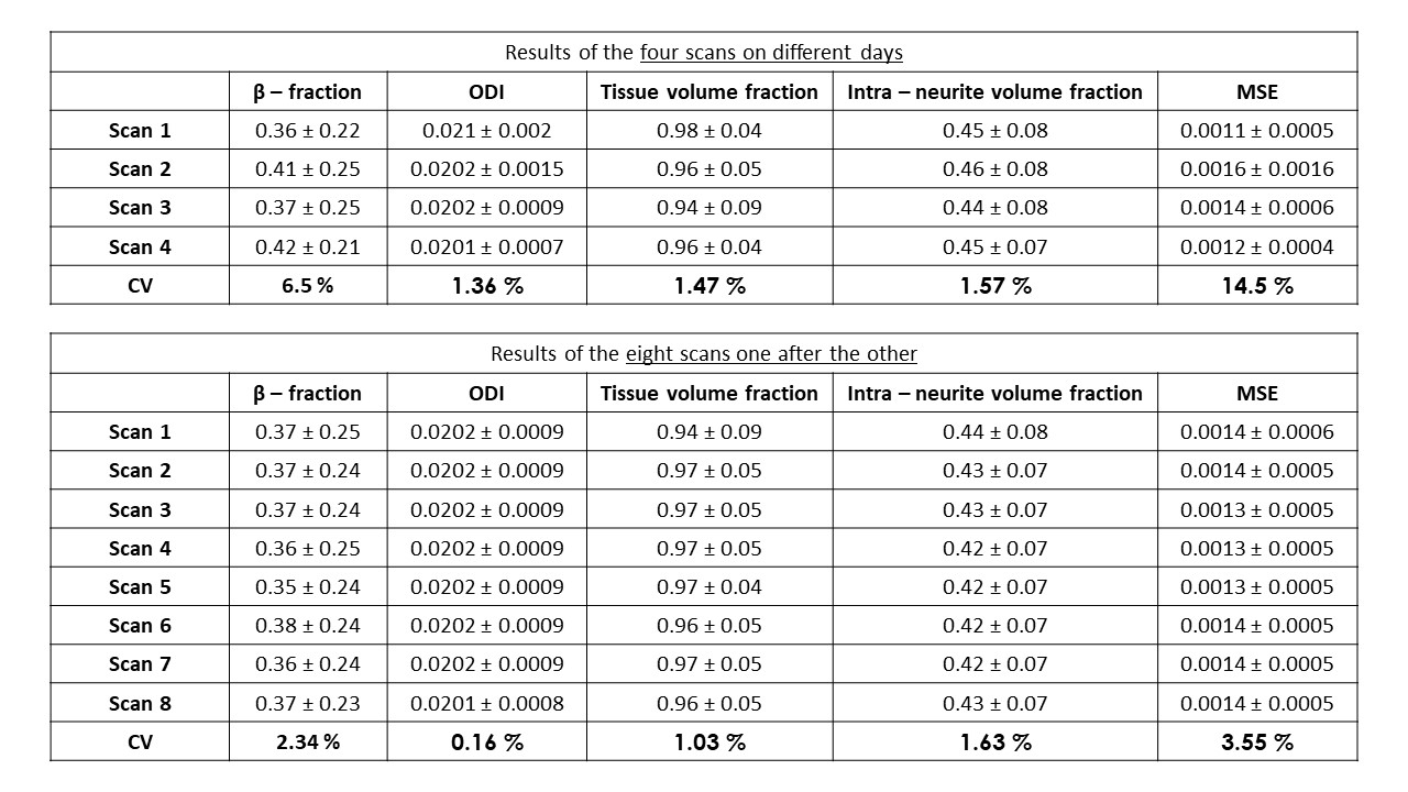
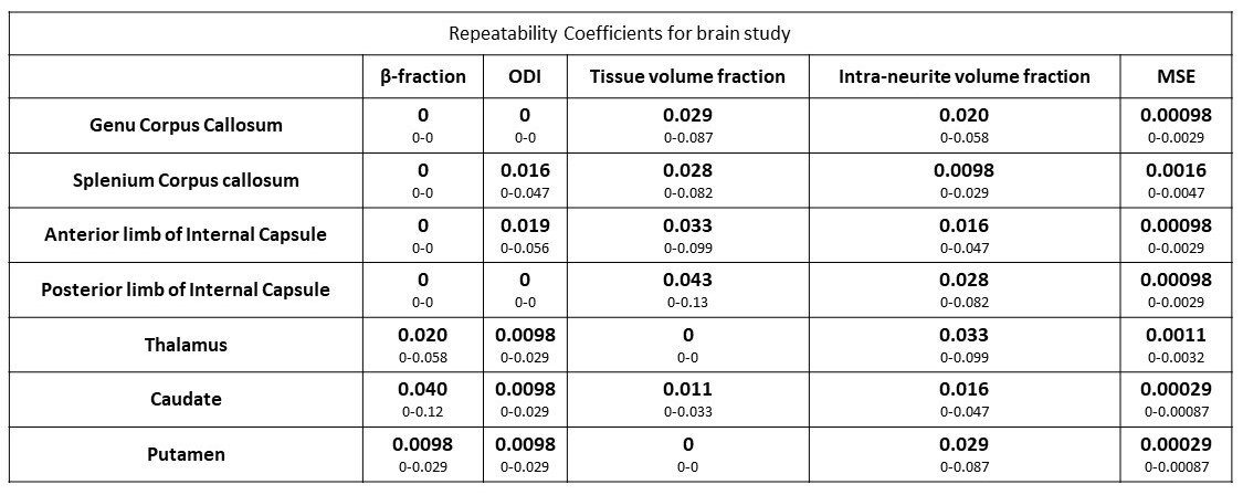
In Figure 3a all the obtained quantitative maps for the phantom study are displayed, showing uniform distribution of all the indices along the fibre ring, except for the β-fraction. The very low values obtained for the CVs, around 1.5%, in the phantom study, presented in Table 1, demonstrate the consistency over time of the NODDI metrics, on and between scanning days. Moreover, the values obtained for each NODDI metric are consistent with phantom manufacturing. Figure 3b presents all the quantitative maps obtained in the in vivo study. The results of the brain study, presented in Table 2, demonstrate how the NODDI model is consistent over time, with low RCs, close to zero, for all the ROIs and each NODDI metric examined. The obtained results confirm the stability of the NODDI model and its suitability for longitudinal studies. The consistency of the measurements shows how NODDI can be applied in different research and clinical scenarios, where stability is critical for identifying changes in brain microstructure over time and making well-informed decisions about patient care.Conclusions
The consistency of the NODDI results demonstrates the reliability of the model and serves as a foundation for detecting minor changes in brain microstructure over time, allowing for the monitoring of the progression of neurological diseases. The next stage in the research involves a multi-centre study to compare the outcomes of different MRI scanners, which may lead to different results. If successful, this study may lead to the integration of the phantom into a calibration protocol which would ensure consistent results across different MRI scanners, opening new possibilities for collaborative research and clinical applications.Acknowledgements
No acknowledgement found.References
- Spano, B. et al. Disruption of neurite morphology parallels MS progression. Neurology - Neuroimmunology Neuroinflammation 5, e502 (2018).
- Zhang, H., Schneider, T., Wheeler-Kingshott, C. A. & Alexander, D. C. NODDI: Practical in vivo neurite orientation dispersion and density imaging of the human brain. Neuroimage 61, 1000–1016 (2012).
- Tariq, M., Schneider, T., Alexander, D. C., Gandini Wheeler-Kingshott, C. A. & Zhang, H. Bingham–NODDI: Mapping anisotropic orientation dispersion of neurites using diffusion MRI. Neuroimage 133, 207–223 (2016).
- Huang, L. et al. Reproducibility of Structural, Resting-State BOLD and DTI Data between Identical Scanners. PLoS One 7, e47684 (2012).
- Hakulinen, U. et al. Repeatability and variation of region-of-interest methods using quantitative diffusion tensor MR imaging of the brain. BMC Med Imaging 12, 30 (2012).
- Zhou, X. et al. Scan-rescan repeatability and cross-scanner comparability of DTI metrics in healthy subjects in the SPRINT-MS multicenter trial. Magn Reson Imaging 53, 105–111 (2018).
- Laun, F. B., Huff, S. & Stieltjes, B. On the effects of dephasing due to local gradients in diffusion tensor imaging experiments: relevance for diffusion tensor imaging fiber phantoms. Magn Reson Imaging 27, 541–548 (2009).
Figures