3480
Multi-echo NODDI with released intrinsic diffusivity: Initial insights for rat brain tissue1Institute of Neuroscience and Medicine - 4, Medical Imaging Physics, Forschungszentrum Jülich, Juelich, Germany, 2Institute of Biomedical Engineering and Nanomedicine, National Health Research Institutes, Miaoli, Taiwan, 3Institute of Medical Device and Imaging, National Taiwan University College of Medicine, Taipei, Taiwan, 4Department of Neurology, Faculty of Medicine, RWTH Aachen University, Aachen, Germany, 5JARA – BRAIN – Translational Medicine, RWTH Aachen University, Aachen, Germany, 6Institute of Neuroscience and Medicine - 11, Forschungszentrum Jülich, Juelich, Germany
Synopsis
Keywords: Diffusion Modeling, Microstructure, relaxometry; multi-dimensional; stroke; ischaemia; pre-clinical
Motivation: The applicability of NODDI and its spinoffs, e.g. multi-echo (MTE) NODDI, is limited to those tissue conditions where the intrinsic diffusivity is known a priori.
Goal(s): We propose an estimation approach for MTE-NODDI parameters in which the intrinsic diffusivity is released whilst ensuring fitting stability by adding an l2-norm regularisation term to the cost function.
Approach: The regularisation parameter was optimised via the generalised cross-validation approach. The MTE-NODDI with released intrinsic diffusivity was tested in a healthy rat at 3 T.
Results: The estimation of MTE-NODDI with released intrinsic diffusivity is well-conditioned if an l2-norm regularisation term is used.
Impact: The proposed estimation approach for MTE-NODDI parameters with released intrinsic diffusivity was shown to be stable. Thus, a new range of tissue conditions in which the intrinsic diffusivity is known to be affected, e.g. stroke, can be accurately characterised.
Introduction
Neurite orientation dispersion and density imaging (NODDI) is widely used for the investigation of tissue microstructure1,2 in diffusion MRI. However, two main limitations restrict its applicability in some tissue pathologies. Firstly, the intrinsic diffusivity, $$$d_0$$$, must be fixed to a priori values in order to stabilise the fitting1,3,4. Secondly, the compartment-specific volume fractions suffer from echo-time (TE) dependence due to differences in the compartment-specific transverse relaxation times5,6,7 (T2). To overcome the latter, Gong et al.7 proposed the multi-TE NODDI (MTE-NODDI) model for the simultaneous analysis of the diffusion- and T2-weighted MRI signal, enabling the estimation of TE-independent fractions. In this work, we propose an estimation approach for NODDI (and MTE-NODDI) parameters that enables the intrinsic diffusivity to be released whilst ensuring fitting stability.Methods
Background. Suppose that a NODDI MRI protocol has been measured for different TEs, then the diffusion- and T2-weighted signal for each TE is written as1,7:$$S\left(\mathbf{θ}_i\right)=S_{0,i}\left[f_{\mathrm{iso},i}S_\mathrm{iso}+\left(1-f_{\mathrm{iso},i}\right)\left(f_{\mathrm{in},i}S_\mathrm{in}\left(\mathbf{θ}_i\right)+\left(1-f_{\mathrm{in},i}\right)S_\mathrm{en}\left(\mathbf{θ}_i\right)\right)\right]\;\;(1)$$
where $$$\mathbf{θ}_i=\left[S_{0,i},f_{\mathrm{iso},i},f_{\mathrm{in},i},\kappa,d_0\right]$$$ $$$\left(i=1,...,N_\mathrm{TE}\right)$$$; $$$S_{0,i}$$$ are the TE-dependent non-diffusion-weighted signals; $$$f_{\mathrm{iso},i}$$$ and $$$f_{\mathrm{in},i}$$$ are the TE-dependent, isotropic and intra-neurite volume fractions; $$$\kappa$$$ and $$$d_0$$$ are the concentration parameter and intrinsic diffusivity, respectively; and $$$S_k$$$ $$$\left(k=\mathrm{iso},\mathrm{in},\mathrm{en}\right)$$$ are the isotropic, intra- and extra-neurite normalised diffusion-weighted signals1,7. In the framework of MTE-NODDI7, the TE-independent fractions, $$$f_\mathrm{iso}^{\left(0\right)}$$$ and $$$f_\mathrm{in}^{\left(0\right)}$$$, and the intra- and extra-neurite transverse relaxation times, $$$T_{2,\mathrm{in}}$$$ and $$$T_{2,\mathrm{en}}$$$, can be estimated following a multi-step fitting approach consisting of: i) Fitting Eq. (1) to each TE dataset separately to obtain $$$f_{\mathrm{iso},i}$$$ and $$$f_{\mathrm{in},i}$$$; ii) Computing $$$f_\mathrm{iso}^{\left(0\right)}$$$ and $$$f_\mathrm{in}^{\left(0\right)}$$$, and the T2s by estimating the slopes and intercepts in a set of linear equations7. Here we propose an approach to perform step i) with released $$$d_0$$$, whereas step ii) is executed as in Ref.7.
Animals. Four adult male Sprague–Dawley rats weighing 300-400g were used. All procedures were approved by the Animal Care and Use Committee, National Health Research Institutes, Taiwan8,9.
MRI experiments. Experiments were performed on a home-integrated 3T MRI scanner equipped with an ultra-high-strength gradient coil (maximum strength 675mT/m10). A custom-designed, single-loop transmit/receive surface coil was used. A Stejskal-Tanner segmented EPI was implemented in-house. Experimental parameters were: TE=50,100ms; b-values(directions)=0(8), 0.5(12), 1.0(26) and 2.0(40)ms/µm2; diffusion-gradient separation and duration, Δ=24ms and δ=3ms. Other parameters were voxel-size=0.26×0.26×1mm3; matrix-size=96×96×20; repetition-time, TR=9s.
Data analysis. Signal denoising11 and distortion correction due to eddy-currents12, tissue-susceptibility differences13 and Gibbs-ringing14 were performed using MRtrix15. NODDI parameter estimation was performed via non-linear least squares using in-house Matlab scrips. The TE-dependent parameters were estimated simultaneously for both TEs, with $$$\kappa$$$ and $$$d_0$$$ as TE-independent parameters. The cost function is written as:
$$F=\sum_{i=1}^{N_\mathrm{TE}} \sum_{j=1}^{N_\mathrm{DW}} \left[S\left(\mathbf{θ}_i\right)-M_{i,j}\right]^2+\lambda\|\mathbf{Ω}\|_2^2\;\;(2)$$
where $$$N_\mathrm{DW}$$$ is the number of diffusion-weighted volumes per TE, $$$M_{i,j}$$$ the measured T2- and diffusion-weighted signals and $$$\mathbf{Ω}=\left[S_{0,1},S_{0,2},f_{\mathrm{iso},1},f_{\mathrm{iso},2},f_{\mathrm{in},1},f_{\mathrm{in},2},\kappa,d_0\right]$$$ ($$$N_\mathrm{TE}=2$$$). Minimisation of Eq. (2) was achieved with fmincon, available in Matlab.
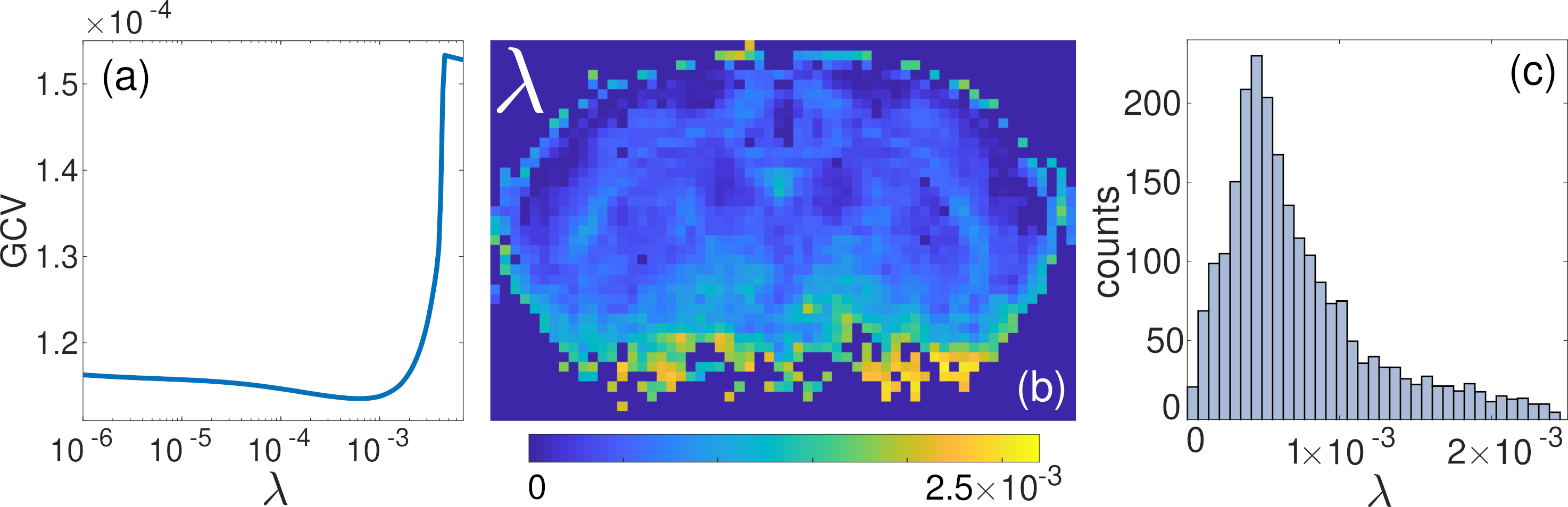
The generalised cross-validation (GCV) method16,17 was used to find the optimal regularisation parameter, $$$\lambda_\mathrm{opt}$$$. The minimum of the GCV function was found via linear search in the range $$$\lambda\in\left[10^{-8},10^{-2}\right]$$$, and a map of the optimal $$$\lambda$$$ was created. Finally, a histogram was built, and $$$\lambda_\mathrm{opt}$$$ was chosen as its peak value.
Results and discussion
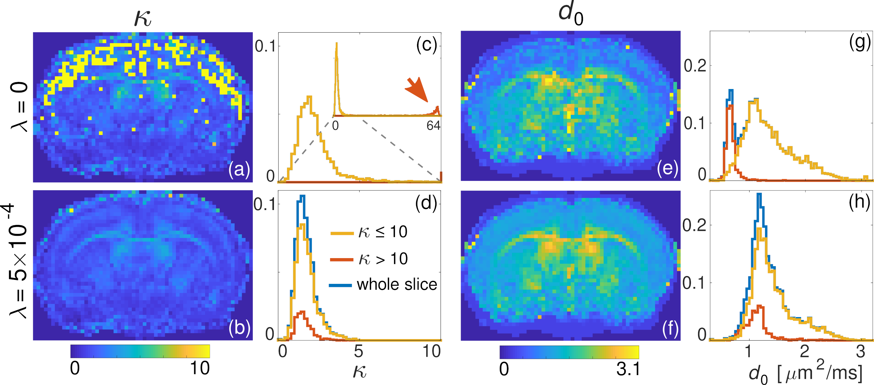
Fig. 1 shows the GCV function for a single voxel, a map of $$$\lambda$$$, and its histogram (peak at $$$\lambda_\mathrm{opt}=5×10^{-4}$$$).The maps of $$$\kappa$$$ and $$$d_0$$$ without ($$$\lambda=0$$$) and with ($$$\lambda=5×10^{-4}$$$) regularisation and their histograms are shown in Fig. 2. Clearly, the estimation of $$$\kappa$$$ for $$$\lambda=0$$$ is unstable, showing two distinct sets of solutions: one characterised by $$$\kappa\lesssim5$$$, and the other having $$$\kappa\sim64$$$ (red arrow, Fig. 2c). The use of the regularisation term helps to penalise the solutions with large $$$\kappa$$$ (Fig. 2b,d).
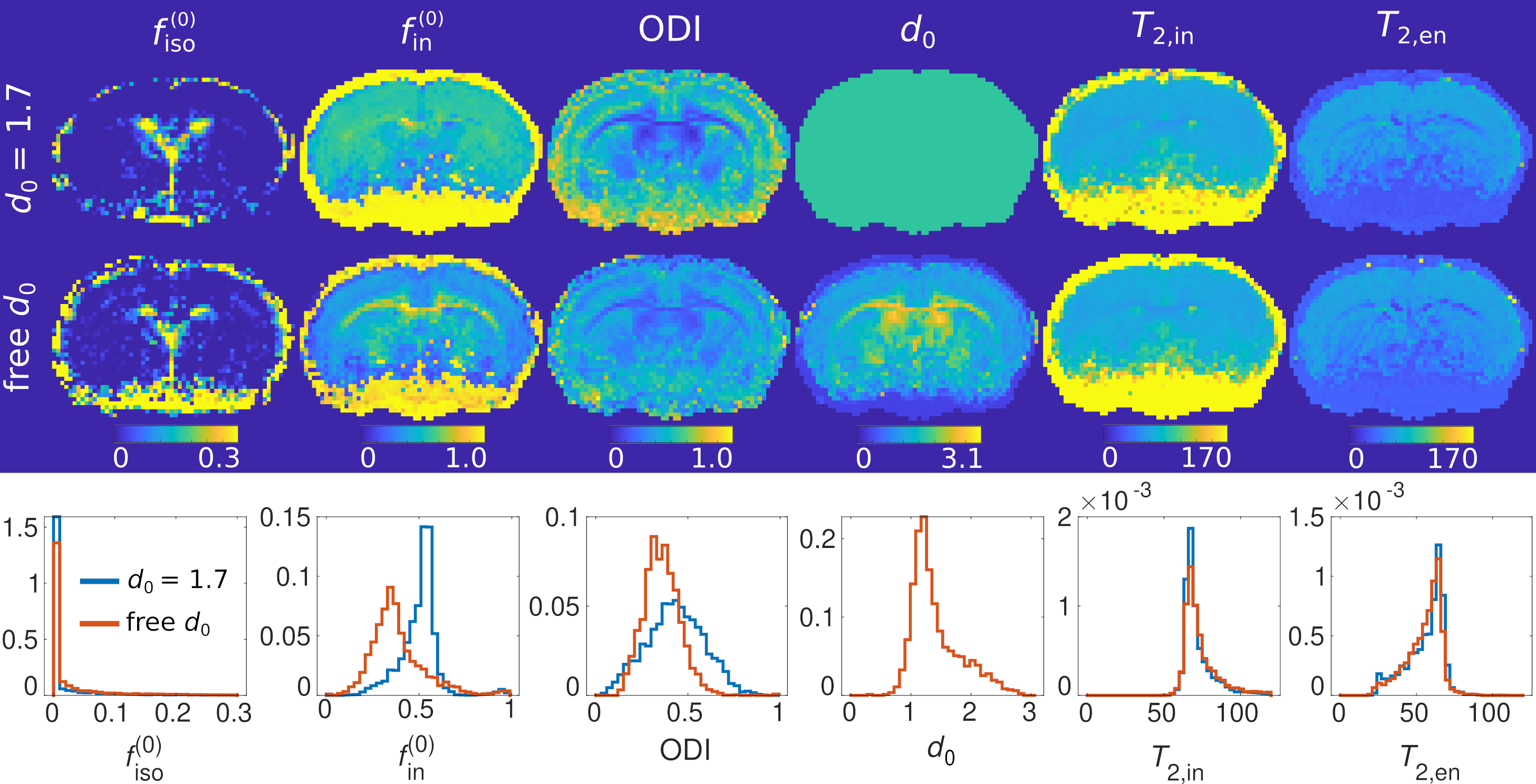
Fig. 3 shows the MTE-NODDI parameters for the conventional ($$$d_0=1.7\;\mathrm{\mu}\mathrm{m}^2/\mathrm{ms}$$$, first row) and the proposed approaches (second row), with the histograms demonstrated in the third row. The map of $$$d_0$$$ shows a strong tissue contrast, with $$$d_0\approx1.1\;\mathrm{\mu m}^2/\mathrm{ms}$$$ in grey matter and $$$d_0\approx2.4\;\mathrm{\mu m}^2/\mathrm{ms}$$$ in white matter4,18. Conversely, the relaxation times $$$T_\mathrm{2,in}$$$ and $$$T_\mathrm{2,en}$$$ do not show noteworthy differences.
Conclusions and outlook
We demonstrated that the spurious solutions in NODDI estimation with released $$$d_0$$$, characterised by large $$$\kappa$$$ values, can be overcome with the addition of an l2-norm regularisation term to the cost function. Crucially, we observed that $$$d_0$$$ is spatially dependent4 and, therefore, fixing it to a global value may lead to bias in the other model parameters. In future work, we intend to evaluate the bias in the parameters induced by the use of the regularisation term via simulations. Finally, the present approach will be used to study tissue microstructure in stroke animal models, a pathology where the release of $$$d_0$$$ is indispensable19.Acknowledgements
We thank Ms Claire Rick for proofreading the abstract.References
1. Zhang, H., et al. NODDI: Practical in vivo neurite orientation dispersion and density imaging of the human brain. Neuroimage 2012;61:1000-1016.
2. Kamiya, K., et al. NODDI in clinical research. Journal of Neuroscience Methods 2020;346:108908.
3. Jelescu, I.O., et al. Degeneracy in model parameter estimation for mulci-compartmental diffusion in neuronal tissue. NMR in Biomedicine 2016;29:33-47.
4. Guerrero, J.M., et al. Optimizing the intrinsic parallel diffusivity in NODDI: An extensive empirical evaluation. PLoS ONE 2019;14(9):e0217118.
5. Peled, S., et al. Water Diffusion, T2, and Compartmentation in Frog Sciatic Nerve. Magnetic Resonance in Medicine 1999;42:911-918.
6. Veraart, J., et al. TE dependent Diffusion Imaging (TEdDI) distinguishes between compartmental T2 relaxation times. Neuroimage 2018;182:360-369.
7. Gong, T., et al. MTE-NODDI: Multi-TE NODDI for disentangling non-T2-weighted signal fractions from compartment-specific T2 relaxation times. Neuroimage 2020;217:116906.
8. Wu K.J., et al., Transplantation of Human Placenta-Derived Multipotent Stem Cells Reduces Ischemic Brain Injury in Adult Rats. Cell Transplantation. 2015; 24: 459–470.
9. Farrher, E., et al. Spatiotemporal characterisation of ischaemic lesions in transient stroke animal models using diffusion free water elimination and mapping MRI with echo time dependence. Neuroimage 2021;244:118605.
10. Cho, K.-H., et al. Development, integration and use of an ultrahigh-strength gradient system on a human size 3T magnet for small animal MRI. PLoS ONE 14:e0217916 (2019).
11. Veraart, J., et al. Denoising of diffusion MRI using random matrix theory. Neuroimage 2016;142:394-406.
12. Andersson, J. L., et al. An integrated approach to correction for off-resonance effects and subject movement in diffusion MR imaging. Neuroimage 2015;125:1063-1078.
13. Andersson, J., et al. How to correct susceptibility distortions in spin-echo echo-planar images: application to diffusion tensor imaging. Neuroimage 2003;20:870-888.
14. Kellner, E., et al. Gibbs-ringing artefact removal based on local subvoxel-shifts. Magnetic Resonance in Medicine, 2016;76:1574-1581.
15. Tournier, J.-D., et al. MRtrix3: A fast, flexible and open software framework for medical image processing and visualisation. Neuroimage 2019;202:116-137.
16. Golub, G.H., et al. Generalized Cross-Validation as a Method for Choosing a Good Ridge Parameter. Technometrics 1979;21(2):215-223.
17. O’Sullivan, F., et al. A Cross Validated Bayesian Retrieval Algorithm for Nonlinear Remote Sensing Experiments. Journal of Computational Physics 1985;59:441-455.
18. Howard A.F.D., et al. Estimating axial diffusivity in the NODDI model. Neuroimage 2022;262:119535.
19. Budde, M. D., et al. Neurite beading is sufficient to decrease the apparent diffusion coefficient after ischemic stroke. Proceedings of the National Academy of Sciences 2010;107:14472:14477.
Figures