3478
Submillimeter diffusion MRI using in-plane segmented 3D multi-slab EPI and a denoiser-regularized reconstruction1Wellcome Centre for Integrative Neuroimaging, FMRIB, Nuffield Department of Clinical Neurosciences, University of Oxford, Oxford, United Kingdom
Synopsis
Keywords: Diffusion Acquisition, Image Reconstruction, 3D multi-slab imaging, Submillimeter, Diffusion acquisition, Diffusion reconstruction
Motivation: Submillimeter diffusion MRI is desirable for neuroscientific research but suffers from intrinsically low SNR.
Goal(s): To achieve submillimeter isotropic resolution in-vivo diffusion MRI with superior SNR and minimal blurring and distortion.
Approach: We quantify the effective resolution and SNR for high-resolution diffusion-weighted EPI, based on which a 3D Fourier encoding-based acquisition with in-plane segmented multi-slab EPI at 7T and a denoiser-regularized reconstruction framework are developed.
Results: In-vivo whole-brain experiments at 0.61 mm isotropic resolution reveal more detailed microstructure compared to 1.05 mm data, which also demonstrates great consistency with a previous post-mortem study. The acquired data also exhibit great anatomical fidelity.
Impact: Our study achieves 3D Fourier-encoded 0.61 mm isotropic resolution whole-brain in-vivo diffusion MRI with minimal blurring and distortion and superior SNR, showing great potential to reveal more detailed depiction of microstructure of the living human brain and advance neuroscientific research.
Introduction
Achieving high resolution diffusion MRI in-vivo is challenging due to the intrinsically limited SNR and T2* blurring associated with large matrix sizes1. We integrate in-plane segmented 3D multi-slab EPI with a denoiser-regularized reconstruction to achieve 0.61mm isotropic resolution dMRI at 7T. The use of orthogonal Fourier bases for 3D EPI provides improved SNR and sharp details2-5 compared to previous super-resolution-based approaches6-9 relying on conditioning which might introduce blurring. We also adopt in-plane segmented EPI to shorten the TE (improving SNR) and readout duration (reducing T2* blurring). The denoiser-regularized reconstruction further enhances SNR.Methods
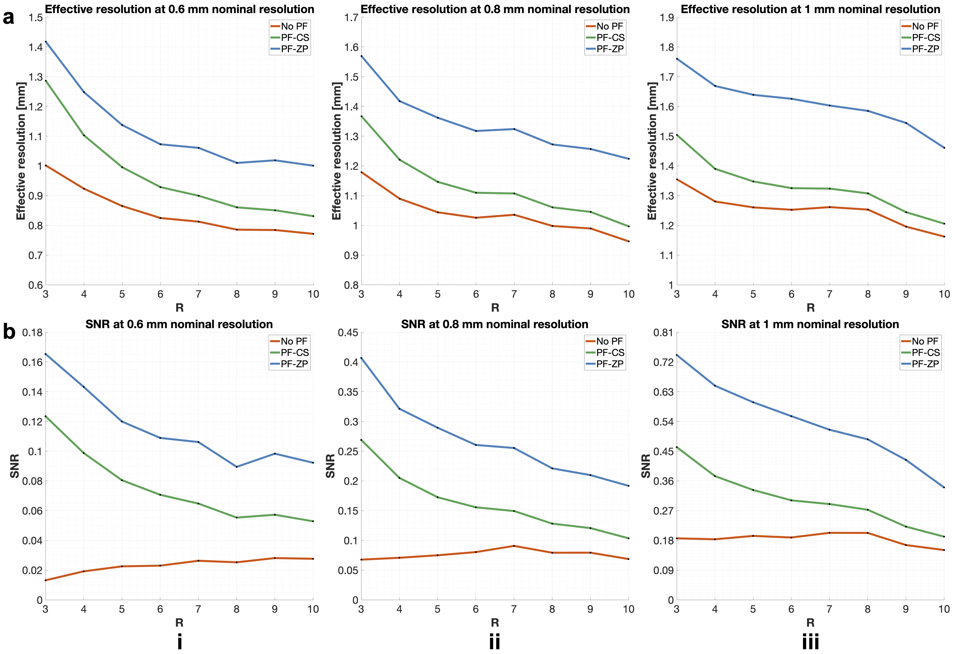
Acquisition simulationWe simulated the effective in-plane resolution and SNR of dMRI at 0.6, 0.8, and 1mm nominal resolutions using EPI trajectories with and without partial Fourier (PF), employing various acceleration factors along ky. The simulation adopted the framework developed by Feizollah and Tardif1 with modified parameters for realistic 3D multi-slab dMRI acquisition (Fig.1). The effective resolution was quantified by the full-width-half-maximum (FWHM) of the point spread functions simulated under various acquisition parameters. For the PF sampling, conjugate symmetric filling (assuming no phase) and zero-padding were investigated. The SNR was quantified based on:
$$SNR\propto\frac{(\Delta x)^3e^{-TE/T2}}{\sqrt{R}},$$
where $$$(\Delta x)^3$$$ is the effective voxel size determined by the FWHM, TE is simulated under different acquisition parameters, and R is the acceleration factor along ky.
In-vivo acquisition
A pilot dataset at 0.61mm isotropic resolution was acquired at 7T. The acquisition encodes each 3D slab using kz phase encoding and R interleaved EPI segments within the kx-ky plane. Based on the simulation (Fig.1), we used R=8 to balance acquisition time, effective resolution, and SNR. To minimize blurring, PF was not used.
Other acquisition parameters: 9 slabs, 20 slices per slab, 1.1× kz oversampling, 2-slice overlap (FOV:220×220×106mm3, matrix:360×360×174), TE1(imaging)/TE2(navigator)/TR=87/153/2600ms, echo spacing=1.22ms (effective: 0.15ms), bandwidth: 992Hz/pixel, anterior-posterior phase encoding, 6 directions (b=1000 s/mm2) and 2 b=0. Total scan time ~60min (7.6min/volume). To validate the feasibility for a shortened acquisition time, each volume was retrospectively under-sampled by 2- and 4-fold along ky (3.8 and 1.9min/volume, respectively). CAIPI shifts were introduced for 4-fold under-sampling to improve the reconstruction10,11.
A 48-direction dataset at 1.05mm from the same subject using 3D multi-slab imaging5 was used for comparison. An MPRAGE image at 0.86mm resolution was acquired for anatomical reference.
Image reconstruction
We developed an iterative SPIRiT12-based plug-and-play13 denoiser-regularized reconstruction (DnSPIRiT) to further enhance the SNR. At the kth iteration:
$$x^k=\arg\min_{x}{\sum_i{||D_iFP_i^HF^{-1}x-y_i||_2^2+\lambda_1||(G-I)x||_2^2+\lambda_2||x-Θ(x^{k-1})||_2^2}},$$
where:
- $$$D_i$$$: shot-sampling mask;
- $$$P_i^H$$$: conjugate of ith navigator phase (motion-induced phase correction);
- $$$y_i$$$: acquired data for ith shot;
- G: SPIRiT kernel;
- Θ: denoiser;
- F: Fourier transform;
- $$$\lambda_1,\lambda_2$$$: regularization weights (10 and 2, respectively).
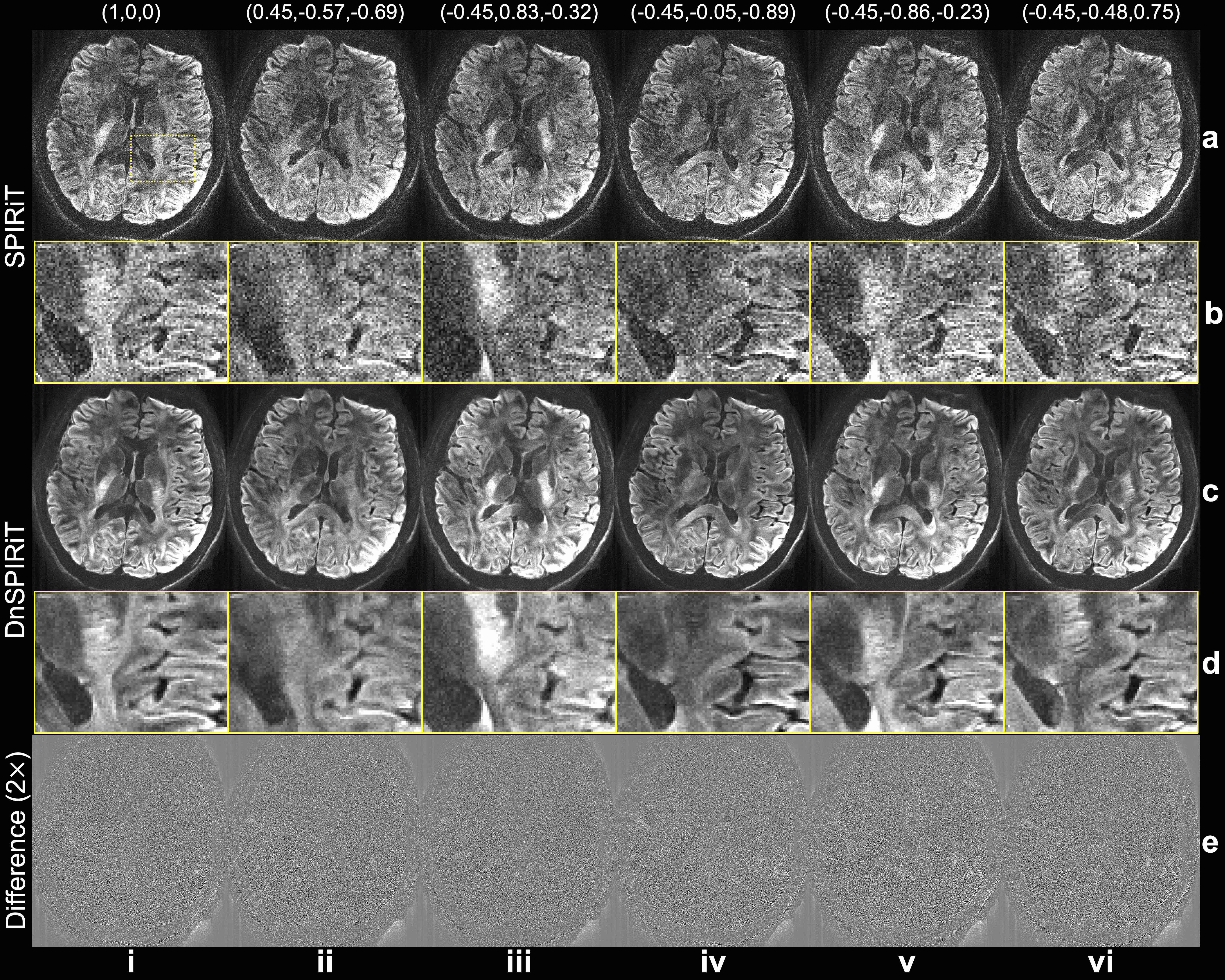
BM4D14 with adaptive noise level estimation was adopted as the denoiser, which acts on the coil-combined magnitude data with Rician noise distribution specified. The denoised magnitude image was multiplied with coil sensitivity estimated using ESPIRiT15 to obtain the multi-coil complex data. Data were also reconstructed using standard SPIRiT with $$$\lambda_2=0$$$ for comparison.
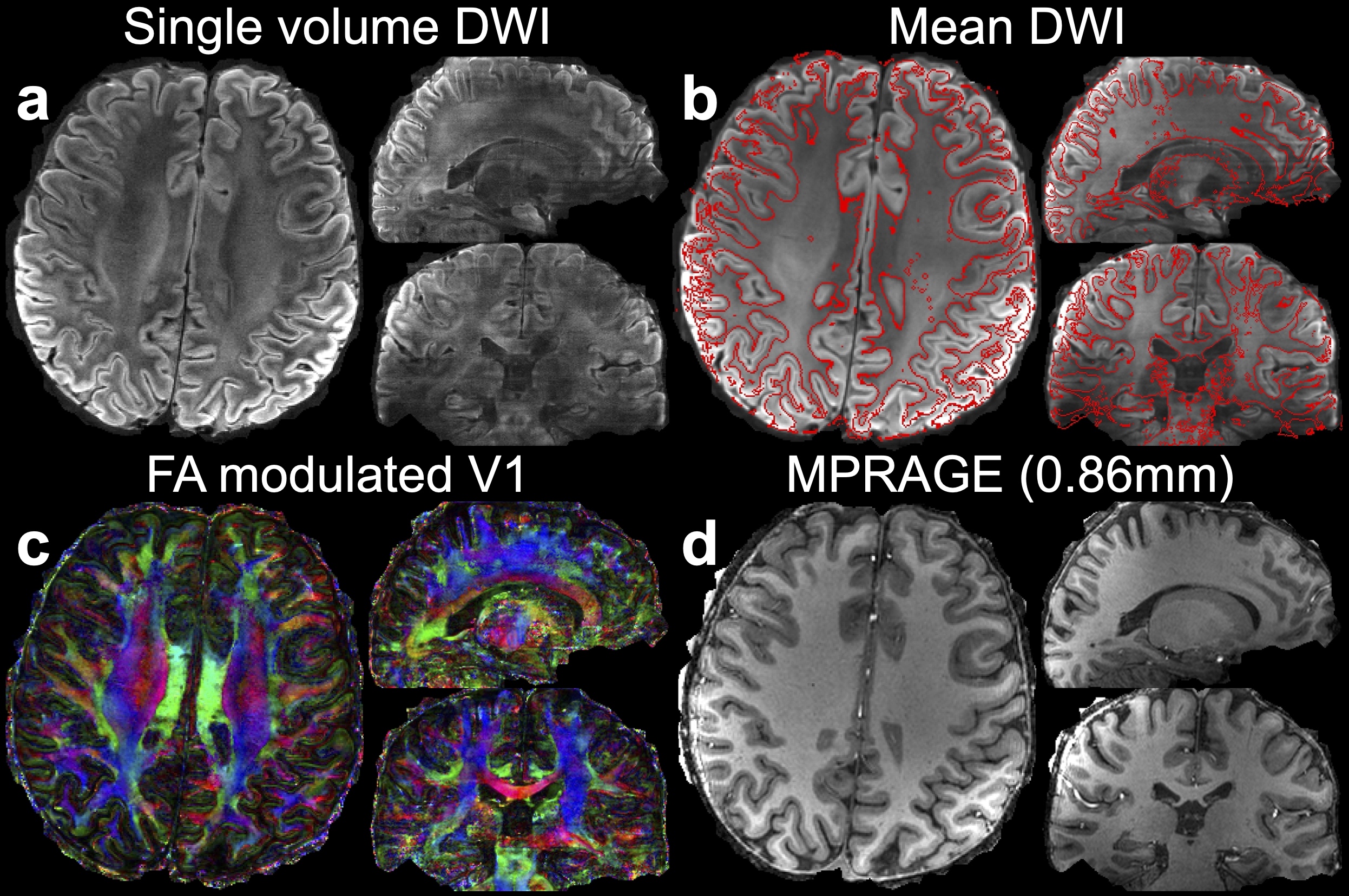
“NPEN”16 was used to combine the multi-slab data. The diffusion data were processed by FSL’s “eddy_correct”17 to align different volumes. DTI fitting was conducted using “dtifit”17. The MPRAGE image was co-registered to diffusion space using “epi_reg”18,19.
Results
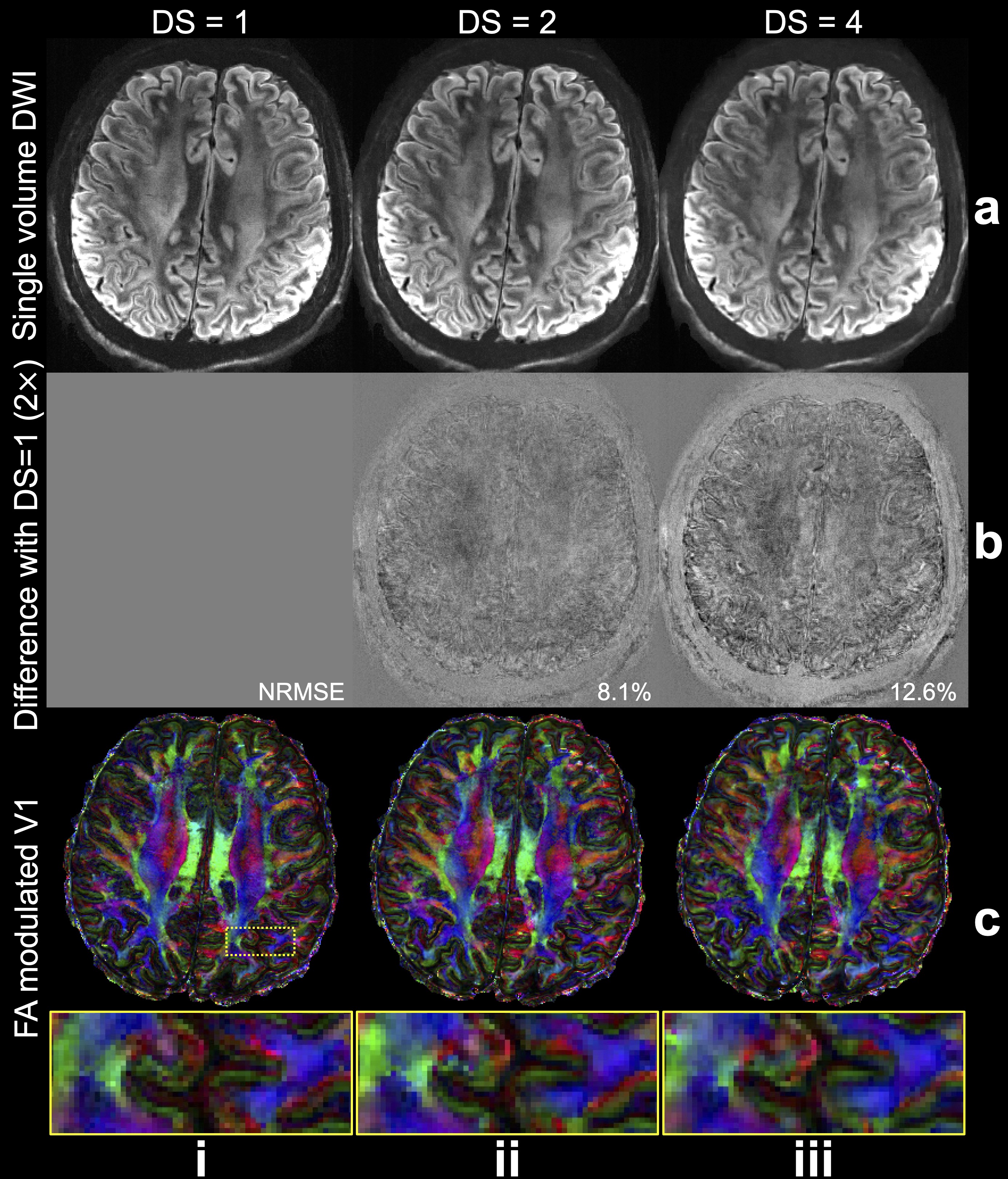
T2* blurring reduces the effective resolution in all cases (Fig.1), which is mitigated by increasing R. For 0.6mm nominal resolution, the effective resolution reaches 0.8mm at R=8, with marginal improvement from further increasing R. PF can improve SNR due to shorter TE and increased blurring, particularly when used with zero-padding. In practice, phase errors cause imperfections in conjugate symmetry for PF-acquisition mean that the simple PF-CS simulated here are infeasible, further compromising effective resolution. Due to the short T2 at 7T, acquisitions without PF can gain SNR at a higher R due to shortened TE. But for acquisitions with PF, the SNR generally reduces as R increases.DnSPIRiT achieves superior SNR with negligible biases or blurring (Fig.2). The whole-brain diffusion data resolve rich details in brain microstructure (Fig.3). Remarkably, even without distortion correction, the diffusion data exhibit great anatomical fidelity thanks to the short effective echo spacing (0.15ms) from in-plane segmentation.
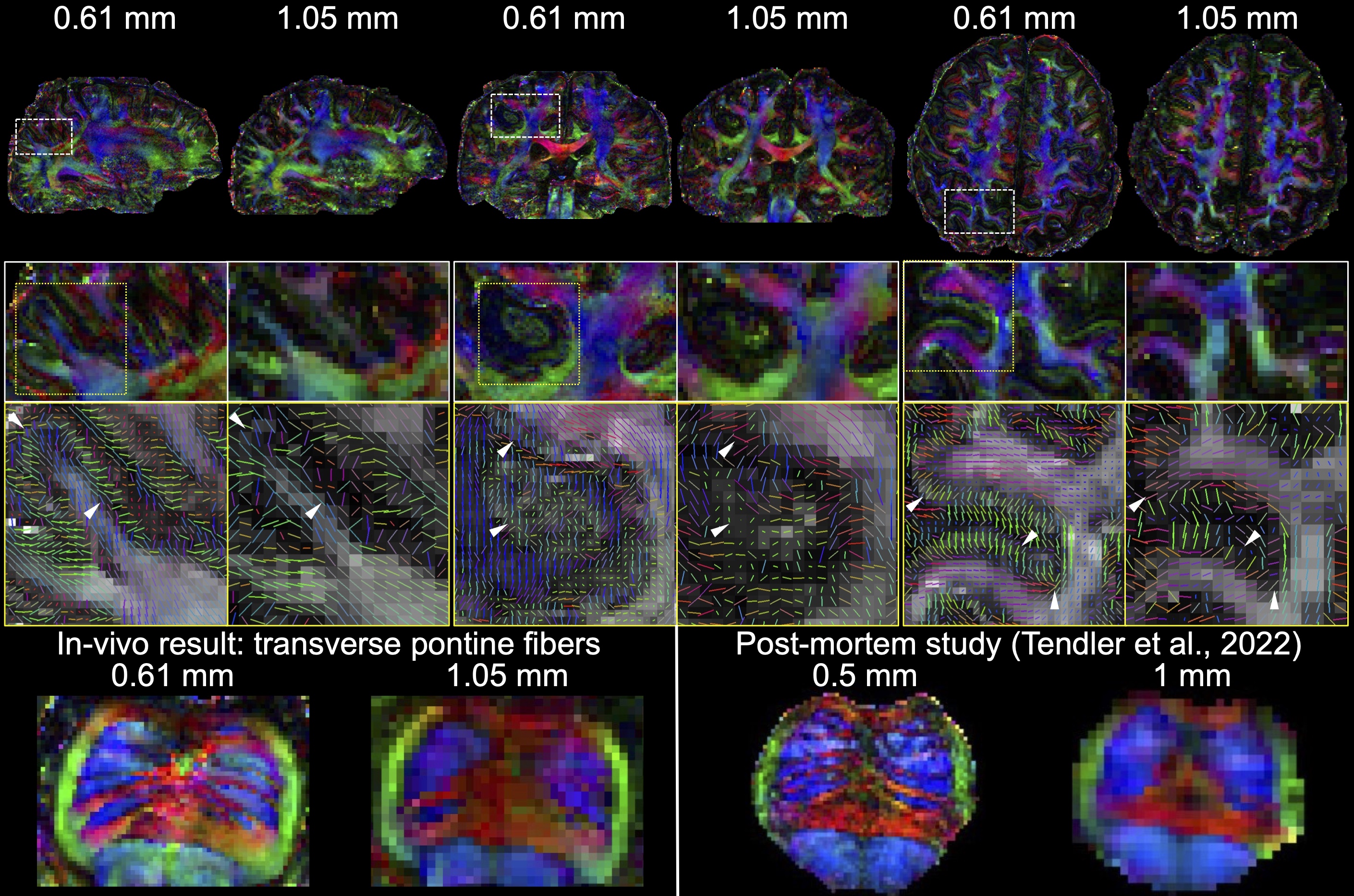
Compared to diffusion data at 1.05mm isotropic resolution, our ultrahigh-resolution dataset reveals substantially more detailed microstructure (Fig.4). Notably, our in-vivo results for resolving transverse pontine fibers demonstrate excellent agreement with post-mortem data20. Figure 5 demonstrates the potential for further accelerated acquisition, enabling collection of more diffusion-encoding directions with the same scan time. This may enable very high-resolution tractography and/or microstructure modelling21.
Discussion and Conclusion
This preliminary work demonstrates the potential of 3D Fourier-encoding-based method to achieve high-quality submillimeter in-vivo dMRI of the living human brains within a one-hour session.Acknowledgements
W.W. is supported by the Royal Academy of Engineering (RF\201819\18\92). K.L.M. is supported by the Wellcome Trust (WT202788/Z/16/A). This study is supported by the NIHR Oxford Health Biomedical Research Centre (NIHR203316). The views expressed are those of the author(s) and not necessarily those of the NIHR or the Department of Health and Social Care. The Wellcome Centre for Integrative Neuroimaging is supported by core funding from the Wellcome Trust (203139/Z/16/Z and 203139/A/16/Z).References
1. Feizollah S, Tardif CL. High-resolution diffusion-weighted imaging at 7 Tesla: single-shot readout trajectories and their impact on signal-to-noise ratio, spatial resolution and accuracy. NeuroImage. 2023;274:120159.
2. Chang H-C, Sundman M, Petit L, et al. Human brain diffusion tensor imaging at submillimeter isotropic resolution on a 3 Tesla clinical MRI scanner. Neuroimage. 2015;118:667-675.
3. Frost R, Miller KL, Tijssen RH, Porter DA, Jezzard P. 3D Multi‐slab diffusion‐weighted readout‐segmented EPI with real‐time cardiac‐reordered k‐space acquisition. Magnetic resonance in medicine. 2014;72(6):1565-1579.
4. Wu W, Poser BA, Douaud G, et al. High-resolution diffusion MRI at 7T using a three-dimensional multi-slab acquisition. NeuroImage. 2016;143:1-14.
5. Li Z, Miller KL, Andersson JL, et al. Sampling strategies and integrated reconstruction for reducing distortion and boundary slice aliasing in high‐resolution 3D diffusion MRI. Magnetic Resonance in Medicine. 2023;90(4):1484-1501.
6. Setsompop K, Fan Q, Stockmann J, et al. High‐resolution in vivo diffusion imaging of the human brain with generalized slice dithered enhanced resolution: Simultaneous multislice (g S lider‐SMS). Magnetic resonance in medicine. 2018;79(1):141-151.
7. Liao C, Bilgic B, Tian Q, et al. Distortion‐free, high‐isotropic‐resolution diffusion MRI with gSlider BUDA‐EPI and multicoil dynamic B0 shimming. Magnetic resonance in medicine. 2021;86(2):791-803.
8. Liao C, Yarach U, Cao X, et al. High-fidelity mesoscale in-vivo diffusion MRI through gSlider-BUDA and circular EPI with S-LORAKS reconstruction. NeuroImage. 2023;275:120168.
9. Dong Z, Polimeni JR, Wald LL, Wang F. SuperRes-EPTI: in-vivo mesoscale distortion-free dMRI at 500μm-isotropic resolution using short-TE EPTI with rotating-view super resolution. In Proceedings of the International Society for Magnetic Resonance in Medicine (ISMRM), London, 2022:3488.
10. Breuer FA, Blaimer M, Mueller MF, et al. Controlled aliasing in volumetric parallel imaging (2D CAIPIRINHA). Magnetic Resonance in Medicine: An Official Journal of the International Society for Magnetic Resonance in Medicine. 2006;55(3):549-556.
11. Setsompop K, Gagoski BA, Polimeni JR, Witzel T, Wedeen VJ, Wald LL. Blipped‐controlled aliasing in parallel imaging for simultaneous multislice echo planar imaging with reduced g‐factor penalty. Magnetic resonance in medicine. 2012;67(5):1210-1224.
12. Lustig M, Pauly JM. SPIRiT: iterative self‐consistent parallel imaging reconstruction from arbitrary k‐space. Magnetic resonance in medicine. 2010;64(2):457-471.
13. Ahmad R, Bouman CA, Buzzard GT, et al. Plug-and-play methods for magnetic resonance imaging: Using denoisers for image recovery. IEEE signal processing magazine. 2020;37(1):105-116.
14. Maggioni M, Katkovnik V, Egiazarian K, Foi A. Nonlocal transform-domain filter for volumetric data denoising and reconstruction. IEEE transactions on image processing. 2012;22(1):119-133.
15. Uecker M, Lai P, Murphy MJ, et al. ESPIRiT—an eigenvalue approach to autocalibrating parallel MRI: where SENSE meets GRAPPA. Magnetic resonance in medicine. 2014;71(3):990-1001.
16. Wu W, Koopmans PJ, Frost R, Miller KL. Reducing slab boundary artifacts in three‐dimensional multislab diffusion MRI using nonlinear inversion for slab profile encoding (NPEN). Magnetic resonance in medicine. 2016;76(4):1183-1195.
17. Smith SM, Jenkinson M, Woolrich MW, et al. Advances in functional and structural MR image analysis and implementation as FSL. Neuroimage. 2004;23:S208-S219.
18. Jenkinson M, Smith S. A global optimisation method for robust affine registration of brain images. Medical image analysis. 2001;5(2):143-156.
19. Greve DN, Fischl B. Accurate and robust brain image alignment using boundary-based registration. Neuroimage. 2009;48(1):63-72.
20. Tendler BC, Hanayik T, Ansorge O, et al. The Digital Brain Bank, an open access platform for post-mortem imaging datasets. Elife. 2022;11:e73153.
21. Zhang H, Schneider T, Wheeler-Kingshott CA, Alexander DC. NODDI: practical in vivo neurite orientation dispersion and density imaging of the human brain. Neuroimage. 2012;61(4):1000-1016.
Figures